Introduction
Aphidius colemani Viereck is a solitary endoparasitoid of aphids, which is commercially produced as a common biocontrol agent against numerous pest aphids in greenhouse and open field settings (Hågvar & Hofsvang, Reference Hågvar and Hofsvang1991; Fernandez & Nentwig, Reference Fernandez and Nentwig1997; Starý, Reference Starý2002). Despite its commercial importance, the biological, ecological and taxonomical status of A. colemani populations is still surprizingly understudied. Originally, A. colemani was said to be distributed over the Mediterranean and Central Asia (Starý, Reference Starý1975), where it was recorded to parasitize over 100 small-sized aphid host species, mainly from the genera Aphis L., Myzus Passerini, Capitophorus van der Goot, Brachycaudus van der Goot and Dysaphis Börner (Starý, Reference Starý1975, Reference Starý1995; Takada, Reference Takada1998; Kavallieratos et al., Reference Kavallieratos, Tomanović, Starý, Athanassiou, Sarlis, Petrović, Niketić and Veroniki2004). The A. colemani group is easily recognizable and is characterized by the costate anterolateral part of the petiole (Eady, Reference Eady1969). Due to the great biological and ecological heterogeneity and host specificity of many A. colemani populations, there is a long list of synonyms for this species in the literature (Starý, Reference Starý1972, Reference Starý1973, Reference Starý1975; Takada, Reference Takada1998; Kavallieratos & Lykouressis, Reference Kavallieratos and Lykouressis1999). Based on literature, we recognized two additional taxa related to A. colemani: Aphidius transcaspicus Telenga and Aphidius platensis Brethes. A. transcaspicus was synonymized with A. colemani due to the large overlap in morphological characteristics and its distribution in the Mediterranean and Central Asia (Starý, Reference Starý1975). However, the biological specificity of A. transcaspicus populations associated with the Hyalopterus spp. and Melanaphis donacis Passerini hosts has been supported by previous studies, raising the questions about the possible morphological (Takada, Reference Takada1998; Kavallieratos & Lykouressis, Reference Kavallieratos and Lykouressis1999) and genetic differentiation of A. colemani and A. transcaspicus (Jafari-Ahmadabadi et al., Reference Jafari-Ahmadabadi, Karimi, Awal and Rakhshani2011). Lozier et al. (Reference Lozier and Mills2009a, Reference Lozier, Roderick and Millsb) addressed one of these questions by studying the genetic structure of A. transcaspicus populations in the Mediterranean and revealed several regional clusters. This finding was useful for further investigation of A. transcaspicus as a biological control agent against the invasive mealy plum aphid, Hyalopterus pruni (Geoffroy), after its unsuccessful releases from 2000 to 2008 in the USA (California and Hawaii) (Wang & Messing, Reference Wang and Messing2006; Latham & Mills, Reference Latham and Mills2012). Aphidius platensis is classified as a member of the Neotropical Faunistic Complex and described as a native species for South America (Starý, Reference Starý1970). However, Starý (Reference Starý1972, Reference Starý1975) discussed its Indian origin with, hypothetically, an accidental introduction to South America, Africa and Australia along with a list of 37 aphid taxa. Based on the original description of A. platensis, we have recognized these phenotypes from South American and Iranian material during our preliminary research, which is tested here for its validity.
The aim of the present study was to explore the genetic structure and morphological variability of the A. colemani group from different aphid host/plant associations, covering a wide area of distribution, using the partial sequences of the mitochondrial cytochrome oxidase subunit I (mtCOI) gene and geometric morphometric analysis of the forewing shape. Furthermore, we tested the congruence between the diversification of the A. colemani group detected by mtCOI sequence analysis and geometric morphometrics. The genetic and morphological heterogeneity of the A. colemani group is also discussed. Aphidius platensis is diagnostified, redescribed, and illustrated; and a key for the A. colemani group is presented.
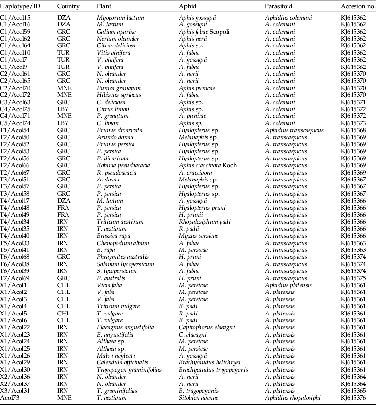
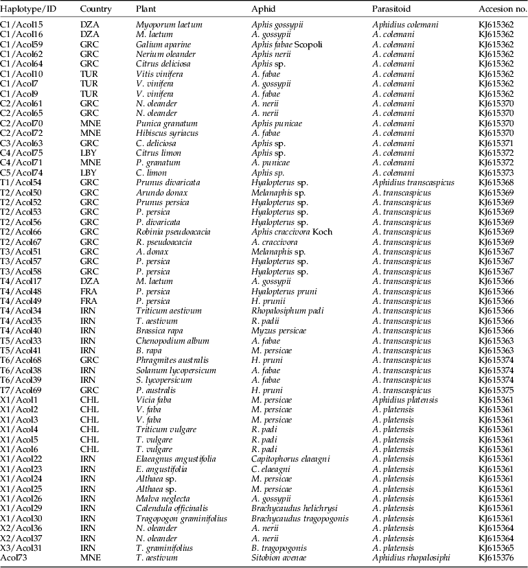
List of specimens belonging to Aphidius colemani group submitted to molecular analysis with designated geographic origin and aphid host/plant associations.
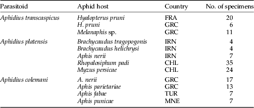
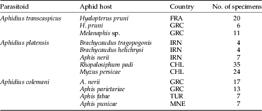
List of specimens belonging to the Aphidius colemani group used for geometric morphometrics with designated geographic origin and aphid hosts.
Material and methods
Material used for DNA study
The parasitoid specimens used in the DNA study were collected from Algeria (DZA), Chile (CHL), France (FRA), Greece (GRC), Iran (IRN), Libya (LBY), Montenegro (MNE) and Turkey (TUR) (Table 1). A total of 54 specimens belonging to the A. colemani group were collected after emergence from mummies. Prior to DNA extraction, all specimens were stored in 96% ethanol at −20 °C. Our approach follows the Meyer & Paulay (Reference Meyer and Paulay2005) technique, according to which the specimens have been accepted a priori as well-identified individuals and assigned into different groups. The nomenclature of parasitoids follows Sharkey & Wharton (Reference Sharkey, Wharton, Wharton, Marsh and Sharkey1997). The nomenclature of aphids is based on Remaudière & Remaudière (Reference Remaudière and Remaudière1997).
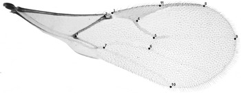
Landmarks scored on a right forewing of an Aphidius colemani female.

DNA extraction, PCR amplification and sequencing
DNA was extracted from each individual wasp using the KAPA Express Extract kit (Kapa Biosystems, Inc. Boston, USA) following the manufacturer's instructions. A region of approximately 710 bp of the barcoding region of the mtCOI gene was amplified using the primers LCO1490 and HCO2198 (Folmer et al., Reference Folmer, Black, Hoeh, Lutz and Vrijenhoek1994). DNA amplification was performed in a final volume of 25 μl. The reaction mixture contained 1 μl of the extracted DNA as the template, 1×KAPA2G Robust HotStart ReadyMix (containing 2 mM MgCl2 at 1X) (Kapa Biosystems) and 0.5 μM of each primer. All PCR reactions were conducted in an Eppendorf Mastercycler® (Hamburg, Germany)® using the following thermal profile: initial denaturation at 95 °C for 5 min, followed by 35 cycles of 94 °C for 60 s, 54 °C for 60 s, 72 °C for 90 s and a final extension at 72 °C for 7 min. The PCR products were purified using the QIAquick PCR Purification Kit (Qiagen, Valencia, CA, USA) according to the manufacturer's instructions. DNA sequencing in both directions was performed by Macrogen Inc. (Seoul, Korea). The nucleotide sequence data were deposited in the GenBank database under accession numbers KJ615361–KJ615376.
Genetic analysis
The sequences were edited using FinchTV (www.geospiza.com), and then, aligned by CLUSTAL W that had been integrated in MEGA5 software (Tamura et al., Reference Tamura, Peterson, Peterson, Stecher, Nei and Kumar2011). Kimura's two-parameter method (K2P) of base substitution (Kimura, Reference Kimura1980) was used to calculate an average genetic distance between the sequences within each group and between the groups of species using a bootstrap procedure. Maximum parsimony (MP) and maximum likelihood (ML) trees were also obtained using the MEGA5 software, with 500 bootstrap replicates performed to assess the branch support. Aphidius rhopalosiphi De Stefani Perez was used as an outgroup for the phylogenetic analysis. For ML, the best-fitting model of sequence evolution based on Bayesian Information Criterion (BIC) and Akaike Information Criterion corrected (AICc) (Nei & Kumar, Reference Nei and Kumar2000) was the Hasegawa–Kishino–Yano model (Hasegawa et al., Reference Hasegawa, Kishino and Yano1985). A haplotype network using statistical parsimony with a confidence limit of 95% was created using the program TCS ver. 1.21 (Clement et al., Reference Clement, Posada and Crandall2000) (Table 3).
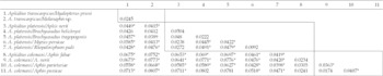
Mean K2P genetic distances between (italic) and within the groups of parasitoids belonging to the Aphidius colemani group.

Geometric morphometrics
A total of 155 females belonging to the A. colemani group were submitted to a geometric morphometric analysis (Zelditch et al., Reference Zelditch, Swiderski and Sheets2012) to explore and quantify the variation of the wing size and shape; a constellation of 11 homologous landmarks of the right wing was scored on each individual (fig. 1). Specimens were a priori classified into three different groups: A. colemani (44 specimens from Montenegro, Greece and Turkey), A. platensis (74 specimens from Chile and Iran) and A. transcaspicus (37 specimens from France and Greece) (Table 2). The groups selected for geometric morphometric analysis correspond with the samples selected for genetic analysis in terms of their geographic origin and aphid hosts.
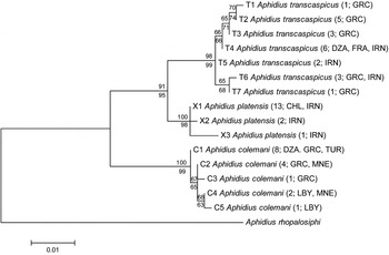
Phylogenetic tree based on mtCOI obtained using maximum likelihood (ML) and maximum parsimony (MP) methods. Bootstrap values for ML are indicated above branches and for MP below branches. Scale bar indicates the number of substitutions per site. Numbers and letters between parentheses refer to the number of sequences for each haplotype and geographic origin of sequences, respectively.

Generalized Procrustes analysis (GPA) (Zelditch et al., Reference Zelditch, Swiderski and Sheets2012) was applied to obtain a matrix of the wing shape coordinates (Procrustes coordinates) from which the differences due to position, scale and orientation had been discarded. The wing size was computed as the centroid size (CS), which is the measure of the size in geometric morphometrics and reflects the amount of dispersion around the centroid of the landmark configuration. To explore the variation in wing shape, we carried out a multivariate ordination by principal component analysis (PCA) based on the covariance matrix. To reduce the dimensionality of the wing shape dataset, the PCA scores were used as the shape variables. The effects of the phylogenetic lineage (species) and the host effects within the lineage on the size (CS) and a wing shape (PCA scores for the first ten principal components).The statistical significance of the difference in wing size between the lineages was tested using a post-hoc test (Tukey's studentized range test). To quantify the shape differences, the Procrustes distances were calculated for each pair of lineages (or between the specimens that emerged from different hosts within the lineages). The statistical significance of the differences between the mean shapes was estimated using a permutation test with 10,000 iterations using the MorphoJ software (Klingenberg, Reference Klingenberg2011), and these results were interpreted following a Bonferroni correction (Table 4).
Divergence in the wing shape of Aphidius that emerged from different host species expressed as Procrustes distances. The statistically significant differences between the populations (P<0.05) after Holm–Bonferroni correction are marked by *. Intra-lineage comparisons are boxed.

Results
Genetic relationships
The sequences of the mtCOI gene were obtained from 54 specimens of the A. colemani group (A. colemani – 16, A. platensis – 16, A. transcaspicus – 22). The aligned sequences were indel-free and were trimmed to 626 bp. Within the 16 analysed specimens of A. colemani collected in Algeria, Greece, Turkey, Montenegro and Libya, a total of five haplotypes were identified (C1, C2, C3, C4 and C5). The most numerous haplotype was C1, determined within eight specimens originating from Algeria, Greece and Turkey, and found attacking five different aphid host/plant associations (Table 1). Haplotype C2 was detected within four A. colemani specimens sampled from three different aphid/plant associations in Greece and Montenegro, whereas the other three haplotypes were identified within only one (C3, C5) or two A. colemani individuals (C4) (Table 1). The mean K2P genetic distance between these haplotypes was 0.3% (Table 3). A total of 22 A. transcaspicus were submitted to molecular analysis, which identified seven haplotypes (T1, T2, T3, T4, T5, T6 and T7) with a mean divergence rate of 0.5%. These haplotypes originate from a wide geographic area including Greece, France, Iran and Algeria where the parasitoid specimens were sampled from 12 different aphid host/plant associations. The most dominant haplotypes within the analysed material were T2 and T4, present with six specimens each, whereas the other five were identified in three (T3, T6), two (T5) or one A. transcaspicus specimens (T1, T7) (Table 1). Three mtCOI haplotypes were detected within 16 analysed A. platensis (X1, X2, X3) with an average distance of 0.5% (Table 3). These specimens were collected from eight different aphid host/plant associations in Chile and Iran (Table 1).
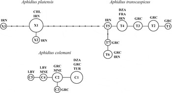
Haplotype networks obtained from 54 Aphidius specimens using a statistical parsimony (TCS). Circles represent specific haplotypes, size of circle reflects the number of individuals with that haplotype (not to scale). Smaller filled circles represent missing haplotypes; lines between circles are mutational steps. The broken line represents a separation place for Aphidius platensis and Aphidius transcaspicus with a confidence limit ≥96%. Geographical distribution of the sequenced specimens is abbreviated next to the haplotype circles.

While the mean nucleotide distances between haplotypes within each group were low (K2P≤0.5%), between-group comparison determined that A. colemani diverged from A. platensis and A. transcaspicus by 4.7 and 4.2%, respectively, while the distance between the latter two groups was 2.2% (Table 3).
The topology of the MP and ML trees was identical with 98–100% bootstrap support, clustering the following three groups as separate taxa: (1) A. colemani, (2) A. platensis and (3) A. transcaspicus (fig. 2). Moreover, A. platensis clustered with A. transcaspicus with 91% (MP), i.e., 95% (ML) bootstrap supports, which corresponds with a substantially lower genetic distance between these two taxa in comparison with A. colemani (Table 3).
Estimation of the haplotype network with a confidence limit ≤95% produced two networks with no ambiguities (fig. 3). One network was built only of the A. colemani haplotypes. The second network consists of the A. platensis and A. transcaspicus haplotypes connected with the haplotype T5 from Iran. However, when the haplotype network was estimated with a confidence limit ≥96%, three separate networks were recognized, one for each of the analysed groups (fig. 3).
Geometric morphometrics
We found significant variation in the wing size among the three lineages and among the specimens that emerged from different hosts within the lineages (ANOVA: factor lineage, SS=3,338,248.739, model df=2, error df=173, F=84.16, P<0.0001; factor host nested within the lineage, SS=1,814,639.57, model df=8, error df=173, F=11.44, P<0.0001). The post-hoc analysis of the wing size differences between the lineages revealed that A. colemani (with a mean CS value and standard error of 1636.52±27.95) has significantly smaller wings than A. platensis (1980.67±13.45) and A. transcaspicus (2054.66±28.26), whereas the latter two do not differ in wing size.
The PC axes 1–10 together described 94.8% of the observed variation in the wing shape (for details see Supplementary Table S1). There were highly significant differences in the wing shape between the lineages (MANOVA, Wilks’ Lambda=0.10626945, F=33.91, df1=20, df2=328, P<0.0001). Additionally, a significant variation in the wing size and a shape was found among the specimens that emerged from different hosts within the lineages (MANOVA, Wilks’ λ=0.21071542, F=3.65 df1=80, df2=1048.7, P<0.0001).
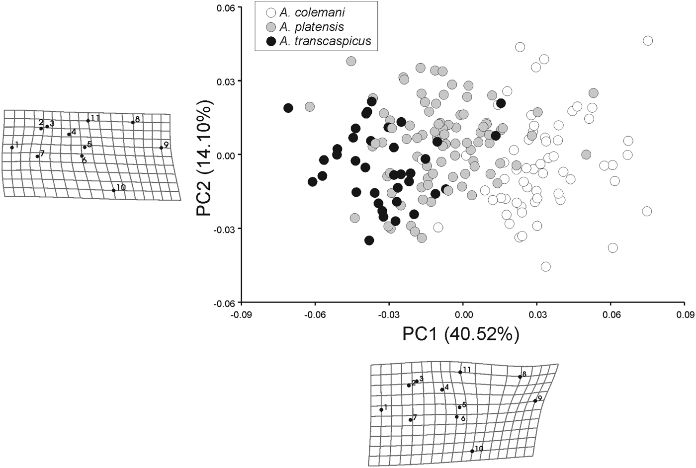
The positions of the specimens in the morphospace defined by the two principal components that describe 54.6% of the total variation in the wing shape are presented in fig. 4. The lineages gradually differentiate along the first PC axis, which describes over 40% of the total variation in wing shape. The shape changes along the second PC axis, which describes 14% of the total variation, are mostly related to the narrowing/widening of the wing, and describe the shape changes within the lineages.
Morphospace of the forewing shape of A. colemani group obtained by a principal components analysis (PCA). The deformation grids illustrate the shape changes along the PC1 and PC2 in a direction of increasing scores.
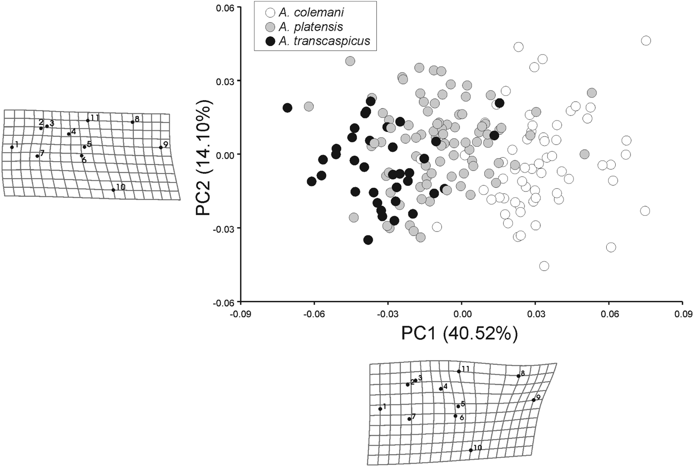
The shape changes that discriminate the lineages are related to the changes in the distal part of the wing (landmarks 8, 9, 10, 11). Individuals of A. transcaspicus have shorter R1 veins (landmarks 11 and 8) and relatively wider distal parts of the wing (demarcated by landmarks 6, 8, 9 and 10) compared to A. colemani, which has a longer R1 vein and a narrower and relatively smaller distal wing area. Regarding the forewing shape, individuals of A. platensis are positioned between A. transcaspicus and A. colemani.
The Procrustes distance (D P) between A. colemani and A. platensis is D P=0.0443, between A. colemani and A. transcaspicus D P=0.0656 and between A. platensis and A. transcaspicus D P=0.0375. The three lineages significantly diverge in mean wing shape (P<0.001 in all comparisons).
Statistically significant differences in mean wing shape among the specimens that emerged from different hosts within the lineages were also found (Table 4). Within A. platensis, the specimens that emerged from Brachycaudus spp. host species, i.e., Brachycaudus helichrysi (Kaltenbach) and Brachycaudus tragopogonis (Kaltenbach) diverged in wing shape from specimens that emerged from Myzus persicae (Sulzer) and Rhopalosiphum padi (L.) but not from each other, while within A. colemani, specimens that emerged from Aphis parietariae Theobald diverged in wing shape from those that emerged from Aphis nerii Boyer de Fonscolombe and A. punicae Shinji.
Redescription of Aphidius platensis Brethes
Material examined
Chile, lab culture reared from M. persicae on Vicia faba, 20 females and 11 males, slide mounted, leg. P. Starý; laboratory culture reared from R. padi on Triticum vulgare, 26 females and 18 males, leg. P. Starý; Chile, Rinconada, 09 October 1992 nr. St. Teresa, reared from mixed colonies Diuraphis noxia (Kudjumov), Metopolophium dirhodum (Walker) and R. padi on Triticum sp., 58 females and 24 males, leg. P. Starý; Chile, Santiago, 30 October 1992, reared from M. persicae on Callendula officinalis, 15 females and 16 males, leg. P. Starý; Chile, La Cruz, 01 October 1992, reared from M. persicae on Solanum muricatum, two females leg. P. Starý; Chile, El Sauce, 02 October 1992, reared from R. padi and Rhopalosiphum maidis (Fitch) on Hordeum vulgare, 26 females and 14 males, leg. P. Starý.
Additional material
Iran, Tehran-Peykanshahr, 26 April 2002, reared from Aphis gossyspii Glover on Malva neglecta, two females slide mounted, leg. E. Rakhshani (Collection of the Institute of Zoology, University of Belgrade); Iran, Kerman – Jiroft, 24 February 2009, reared from M. persicae on Althaea sp., three females and four males, slide mounted, leg. H. Barahoei (Collection of the Institute of Zoology, University of Belgrade); Iran, Kerman, 18 May 2009, reared from B. helichrysi (Kaltenbach) on C. officinalis, four females and one male, leg. A. Alipour; Iran, Zahedan, 24 March 2003, reared from A. nerii Boyer de Fonscolombe on Nerium oleander Boyer de Foncolombe, nine females, three males, leg. E. Rakhshani; Iran, Zahedan, 14 April 2003, reared from Capitophorus elaeagni (del Guercio) on Elaeagnus angustifolia, two females, slide mounted, leg. E. Rakhshani (Collection of the Institute of Zoology, University of Belgrade); Iran, reared from B. tragopogonis on Tragopogon graminifolius, two females, slide mounted, leg. E. Rakhshani (Collection of the Institute of Zoology, University of Belgrade).
Diagnosis
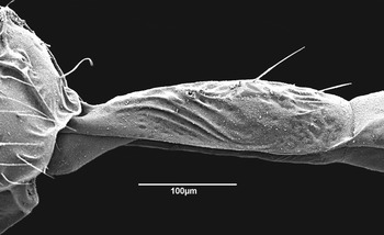
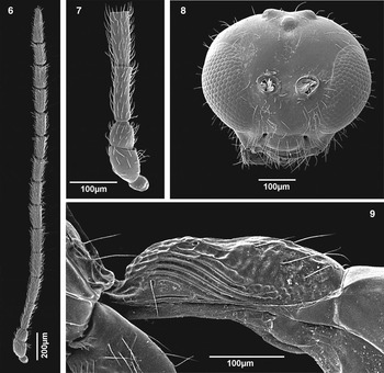
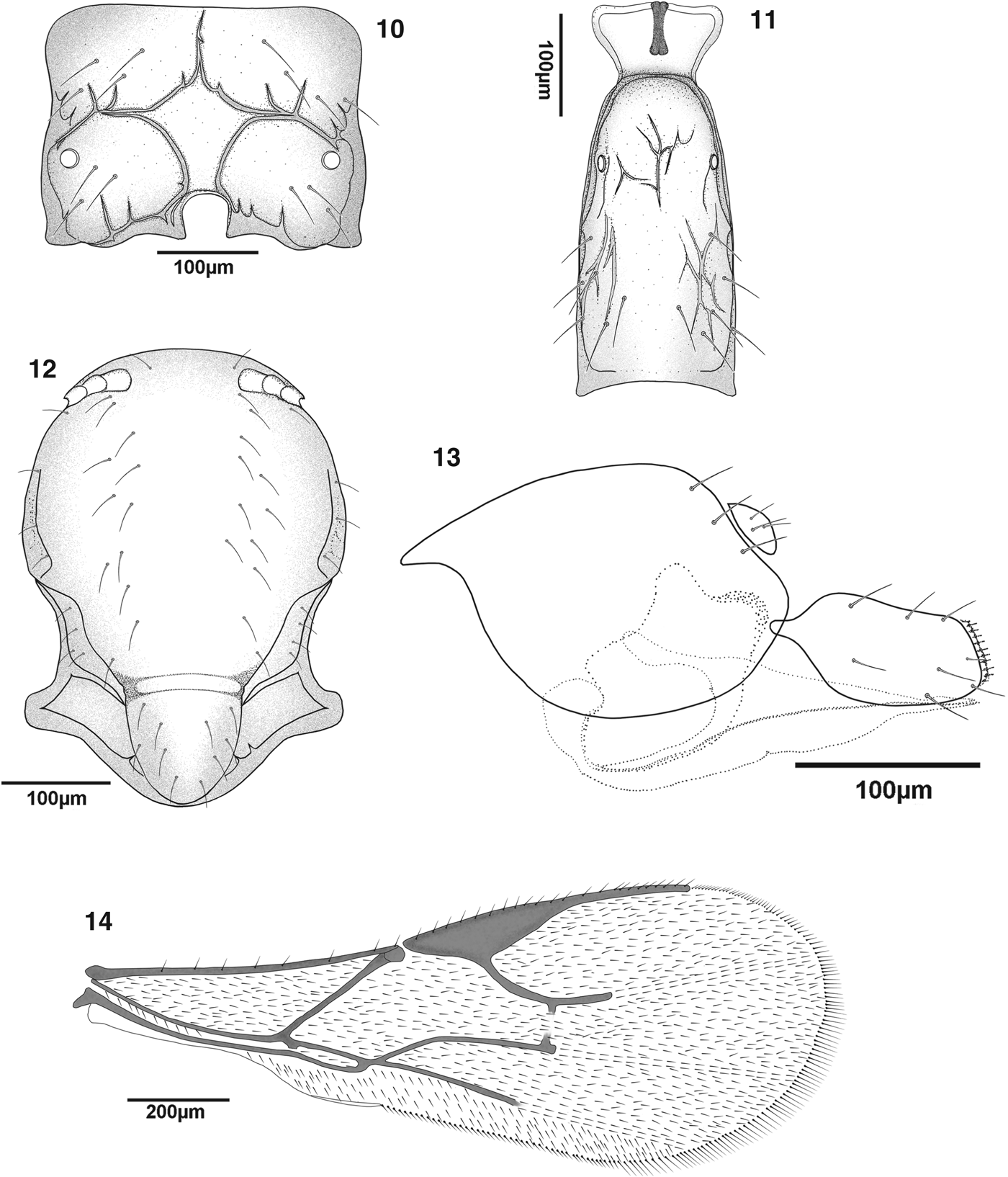
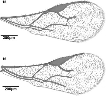
Aphidius platensis belongs to the A. colemani group, having costae on the anterolateral area of the petiole. A. platensis differs from A. colemani by having a shorter R1, which is approximately one-third shorter than the stigma length (fig. 14) (proportion between the length of R1 and the stigma in A. platensis is 0.77–0.89) and a narrower stigma than A. colemani (fig. 15). Costae on the anterolateral area of the petiole in A. platensis are sharper than in A. colemani (figs 5 and 9). Aphidius platensis differs from A. trancaspicus by having longer R1 (figs 14 and 16) and labial palps with two palpomeres (A. transcaspicus has three palpomeres).
Aphidius colemani, anterolateral area of petiole.

Description
Female: Head (fig. 8) wider than mesosoma at the tegulae (proportion between width of head and width of mesoscutum, 1.35–1.45). Frons, vertex and occipital area with dense setae. Face moderately setose (fig. 8). Tentorial index 0.40–0.50. Malar space equal to 0.30–0.35 of longitudinal eye diameter. Eyes oval, converging towards clypeus. Clypeus rounded, with 6–11 long setae. Antennae 14–15-segmented, moderately thickened at the apex (fig. 6), with semi-erected and adpressed setae, which are as long approximately as half of segment diameter. Scape and pedicel subglobular. Flagellar segments stout, with sparse semi-erected setae. Flagellar segment 1 (=F1), 3.00–3.40 times as long as its maximum width (fig. 7). F2, 2.50–3.00 times as long as its maximum width. F1 equal to F2. F1 and F2 with 0–1 and 2 longitudinal placodes, respectively (fig. 7). Maxillary palps with four palpomeres. Labial palps with two palpomeres.
Aphidius platensis, female. (6) Antenna, (7) first antennal segments, (8) frontal view of head, (9) anterolateral area of petiole.

Mesosoma: Mesonotum with notaulices in the ascendant portion of its anterolateral area, erased dorsally and outlined by one or two rows of long sparse setae, which extend near to the scutellum (fig. 12). Scutellum with 5–9 long setae, mostly in lateral parts. Forewing (fig. 14) stigma moderately elongated, 3.50–4.00 times as long as its width, slightly longer than R1 (the proportion between R1 and stigma length is 0.77–0.89). Propodeum (fig. 10) areolated with a wide pentagonal central areola, clearly carinated. Upper areolae with seven long setae laterally and lower areolae with five setae. Hind femur and tibia with semi-erected sparse setae, more dense near the tibial spur.
Aphidius platensis, female. (10) Propodeum, (11) dorsal aspect of petiole, (12) dorsal aspect of mesonotum, (13) last genital segment and ovipositor sheath, (14) forewing.
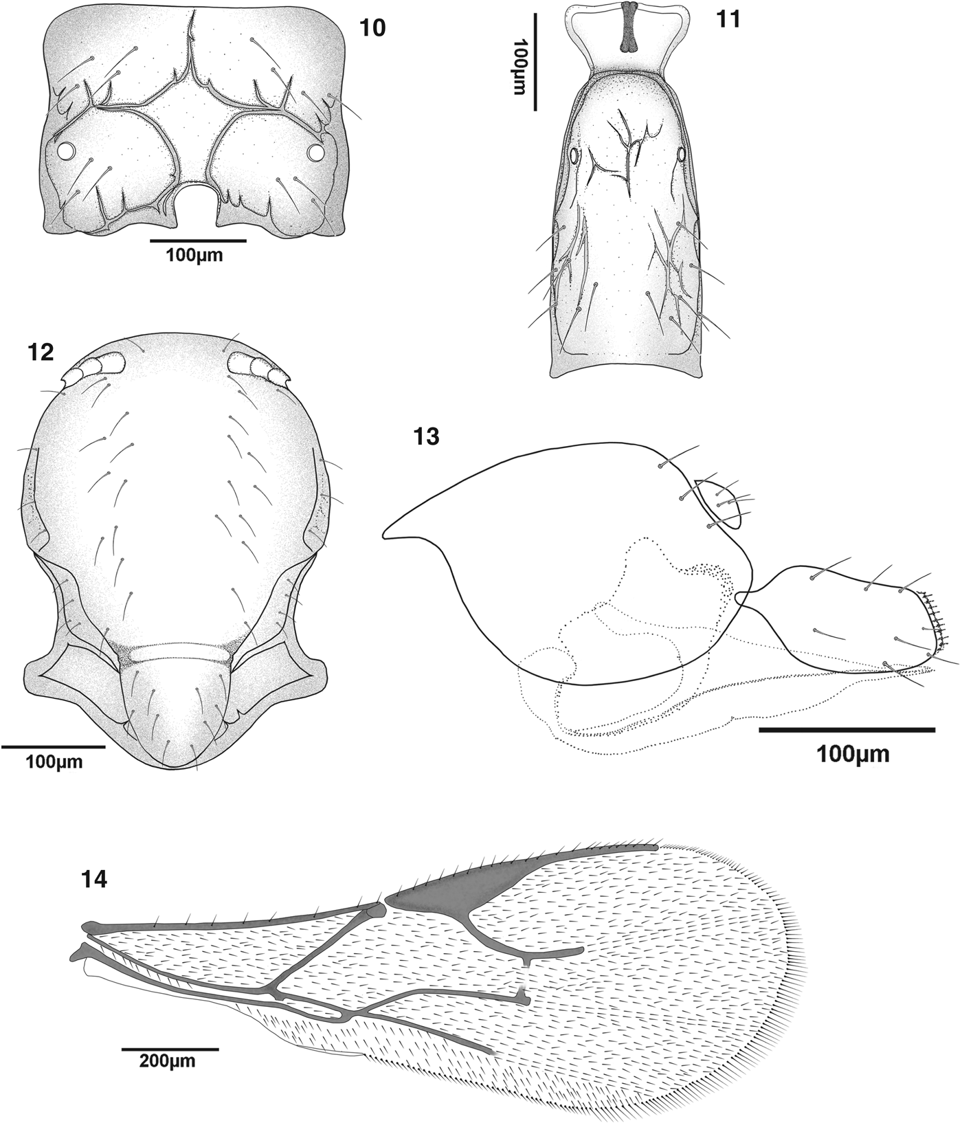
Metasoma: Petiole almost parallel-sided, 3.00–3.50 times as long as its width at the spiracles, anterolateral area with 5–7 costulae (fig. 9). Dorsal surface of the petiole with fine rugosities and six long semi-erected lateromedial setae on its lower half (fig. 11).
Genitalia: Ovipositor sheath (fig. 13) moderately concave at the dorsal margin.
Coloration: Head brown with black eyes, face and genae yellow to light brown, mouthparts yellow; scapus, pedicel and annellus yellow; except for a narrow yellow ring at the base of F1, remaining parts of flagellum uniformly brown. Pronotum yellow. Mesonotum brown with a light brown metapleuron. Legs yellow with dark apices. Wings hyaline. Petiole yellow, the rest of the metasoma light brown with a black ovipositor sheath.
Body length: 1.7–2.3 mm.
Male: Antennae 18–19-segmented. Generally darker than the female. Scapus and pedicel yellow to light brown. Face and mouthparts light brown. Pronotum yellow to light brown. Legs yellow with dark apices. Remaining body parts brown.
Hosts
Aphis gossypii Glover, A. nerii, M. persicae, R. padi, C. elaeagni, B. helichrysi, B. tragopogonis. In addition to the above host aphids, Starý (Reference Starý1972) reviewed 37 aphid hosts for A. platensis.
Distribution
On the basis of our research and available material, A. platensis is distributed in South America (Chile) and the Middle East (Iran). According to Starý (Reference Starý1972), this species has also been recorded in Australia and Africa.
Key for adult female Aphidius colemani species group:
1 forewing R1 vein subequal to half of the stigma length (fig. 16); F1 equal to 3.50–4.00 times as long as centrally wide. … … … …Aphidius transcaspicus
2 forewing R1 vein almost equal to stigma length (fig. 15); anterolateral area of the petiole bluntly costated (fig. 5); antennae (14)15–16-segmented. … … … … Aphidius colemani
Forewings. (15) Aphidius colemani, (16) Aphidius transcaspicus.

Discussion
Aphid parasitoids are characterized by a high level of host specialization, but a low level of morphological diversification, which results in the existence of many cryptic species within Aphidiinae (Starý, Reference Starý, Minks and Harrewijn1988; Mitrovski-Bogdanović et al., Reference Mitrovski-Bogdanović, Petrović, Mitrović, Ivanović, Žikić, Starý, Vorburger and Tomanović2013). However, an integrative approach will substantially contribute to applied biological research. This approach was gradually established using the taxonomic studies of aphidiine parasitoids during the last decade, and combines the molecular characterization, geometric morphometrics and aphid host specificity that resolved several taxonomic problems within this subfamily (Žikić et al., Reference Žikić, Tomanović, Ivanović, Kavallieratos, Starý, Stanisavljević and Rakhshani2009; Kos et al., Reference Kos, Petrović, Starý, Kavallieratos, Ivanović, Toševski and Tomanović2011; Mitrovski-Bogdanović et al., Reference Mitrovski-Bogdanović, Petrović, Mitrović, Ivanović, Žikić, Starý, Vorburger and Tomanović2013).
Taxonomic studies within the A. colemani group were based mainly on a few morphological characteristics and samples with restricted distribution records (Starý, Reference Starý1972, Reference Starý1975; Takada, Reference Takada1998; Kavallieratos & Lykouressis, Reference Kavallieratos and Lykouressis1999; Jafari-Ahmadabadi et al., Reference Jafari-Ahmadabadi, Karimi, Awal and Rakhshani2011). Our phylogenetic analyses of mtDNA COI sequences revealed a clear genetic structure, recognizing A. colemani, A. transcaspicus and A. platensis as separate taxa with distances ranging from 2.2 to 4.7%. Lower divergence rate determined between A. transcaspicus and A. platensis has been reported in recent years for other Aphidiinae as well (Sandrock et al., Reference Sandrock, Schirrmeister and Vorburger2011; Derocles et al., Reference Derocles, Le Ralec, Plantegenest, Chaubet, Cruaud, Cruaud and Rasplus2012; Petrović et al., Reference Petrović, Mitrović, Starý, Petrović-Obradović, Žikić, Tomanović and Vorburger2013). It clearly indicates a close relatedness of the two species, which is also supported by their haplotypes being clustered as a separate taxa at a confidence level exceeding 96%. A low intraspecific genetic variation and no evident association with aphid hosts or region of origin were determined for haplotypes of all three species from the A. colemani group. A. transcaspicus and A. platensis clustered together on both phylogenetic trees and diverged half as much compared to A. colemani. A TCS network analysis also confirmed the closer relationship between A. transcaspicus and A. platensis, indicating that these two parasitoids share the same ancestor, while the absence of the network connection for the A. colemani haplotypes could be interpreted as possibly extant but not as sampled haplotypes or extinct ancestral sequences. Additionally, the analysed A. colemani specimens had a Mediterranean distribution, while A. transcaspicus were more widely distributed, encompassing an area of the Mediterranean and Central Asia. Lozier et al. (Reference Lozier, Roderick and Mills2008) recognized haplotypes of A. transcaspicus geographically structured into two main clades (Western Mediterranean+the Middle East and Eastern Mediterranean). Although we recognized these two clades of A. transcaspicus in our results as well, some eastern Mediterranean haplotypes are closely connected with the Western Mediterranean+Middle East. It is known that A. transcaspicus in the Eastern Mediterranean accepts more diversified aphid hosts than in the Western Mediterranean, which can possibly be explained by a different recent evolutionary history of these populations (Lozier et al., Reference Lozier, Roderick and Mills2008).
Our research confirmed significant morphological differences in the forewing shape among A. colemani, A. transcaspicus and A. platensis. Although we detected statistically significant differences in the forewing shape and wing-venation pattern, some overlap is evident. The variability of the taxonomic characters commonly used in distinguishing these species (Starý, Reference Starý1972, Reference Starý1973, Reference Starý1975; Pennacchio, Reference Pennacchio1989; Takada, Reference Takada1998; Kavallieratos & Lykouressis, Reference Kavallieratos and Lykouressis1999; Tomanović et al., Reference Tomanović, Kavallieratos, Starý, Athanassiou, Žikić, Petrović-Obradović and Sarlis2003; Kavallieratos et al., Reference Kavallieratos, Tomanović, Starý and Mitrovski-Bogdanović2008; Rakhshani et al., Reference Rakhshani, Talebi, Starý, Tomanović, Kavallieratos and Manzari2008), such as the number of antennal segments and the forewing venation pattern, led to a long list of synomizations within the A. colemani group (Starý, Reference Starý1975). Differences in the shape of the forewing investigated within the biotypes of the A. colemani group are congruent with their genetic diversification on the basis of the barcoding region of mtCOI. Although clearly separated, A. platensis is genetically closer to A. transcaspicus than to A. colemani. The same pattern was observed for the forewing shape. Aphidius platensis and A. transcaspicus are more similar to each other than to A. colemani. Our study and redescription support the existence of A. platensis as a separate species with clustered South American and Central Asian analysed specimens.
We have found some differences related to the original description of A. platensis (Brethes, Reference Brethes1913) and redescription on the basis of the paratypes from the British Natural History Museum (Eady, Reference Eady1969; Starý, Reference Starý1972). We found clear differences among all examined samples/specimens of A. platensis in the sharpness of costae on the anterolateral area of the petiole (figs 5 and 9) and the length of R1. Most likely, records of A. platensis in the literature are occasionally mixed with A. colemani. Unfortunately, the holotype in the Natural History Museum of Buenos Aires was not available to us for re-examination. However, in our opinion, the examination of the holotype is necessary in light of the new diagnostic characters we have proposed to confirm the identity as A. platensis. The prevalence of A. platensis phenotypes in many areas of South America (Starý, Reference Starý1995) and the absence of other A. colemani phenotypes support the holotype's value to the identification of A. platensis because the holotype was derived also from field samples.
The taxonomic history of the colemani–platensis–transcaspicus group can be briefly presented as follows: Starý (Reference Starý1975) synonymized A. platensis and A. transcaspicus under the ‘almost forgotten’ A. colemani. Prior to this synonymization, A. platensis, which was originally described in South America, was the only known species from the A. colemani group in South America and Central Asia (India). Fifty years ago, A. platensis was intentionally introduced from Brazil to the USA (California) and the British Isles (Starý, Reference Starý1972), but without reliable reports on the establishment of the species thereafter. Starý (Reference Starý1995) and Starý et al. (Reference Starý, Sampaio and Bueno2007) listed many records of A. colemani in South America, which should be re-examined because they may belong to A. platensis. Although some researchers asserted the neotropical origin of A. platensis, Starý (Reference Starý1972) argued for the Indian origin of A. platensis, with accidental introduction to South America, Africa and Australia, based on the known species distribution and aphid host origin.
In summary, there are three species, A. colemani, A. platensis and A. transcaspicus that have most likely originated in the Mediterranean and subsequently expanded to nearby areas with subtropical/tropical climates. However, the range of their expansion is different and needs to be revisited. Preliminarily, it is most probable that A. platensis has expanded over the Sub-Saharan area and from there was accidentally introduced to South America. On the basis of our preliminary screening, these three species also occur at least in a part of Sub-Saharan Africa, South-Eastern Asia and Australia.
The occurrence of the costate anterolateral area was introduced in the identification of Aphidius species groups by Eady (Reference Eady1969). However, it has been determined in a small number of congeners in the world, as follows: Aphidius avenae Hal. (West Palearctic), Aphidius betrandi Benoit (sub-Saharan Africa), Aphidius pseudopicipes Starý (Mexico), and the A. colemani species group.
Our results open questions regarding the origin of A. platensis, which can be resolved by including samples from the whole area of this species in appropriate phylogeographic analysis. Although A. platensis and A. colemani share a common host range pattern, it would be interesting to estimate and compare the role of these two species in future biological control strategies against aphids of economic importance. Currently, aphid parasitoids within global commercially distributed materials tend to be a mixture of A. colemani, A. platensis and possibly even A. transcaspicus.
Our results indicate that ‘genetic screening’ using mtCOI gene barcoding is a reliable method for identification within the A. colemani group. The high variation in wing shape between species, including significant divergences in wing shape among specimens that emerged from different hosts, make the forewing shape and wing venation less reliable for species determination. However, further investigation is required on that issue inside the subfamily Aphidiinae.
The supplementary material for this article can be found at http://www.journals.cambridge.org/BER
Acknowledgements
This study was supported by the Grant no. III43001 (The Ministry of Education, Science and Technological Development of the Republic of Serbia) and by the programs and 10 TUR/3-7-2 ‘Aphid parasitoids (Hymenoptera: Braconidae: Aphidiinae): diversity of trophic associations and their role in agroecosystems’ (General Secretariat for Research and Technology, Ministry of Development of the Hellenic Republic). Participation by P. Starý was conducted with institutional suppoprt RVO: 60077344.