Introduction
Sialendoscopy is a minimally invasive procedure that is effective for both the diagnosis and management of obstructive salivary gland disorders, including the removal of salivary gland stones. Sialendoscopy has been shown to be a superior diagnostic technique for salivary gland disorders compared with conventional radiology, ultrasound, computed tomography (CT) or magnetic resonance imaging (MRI), all of which are traditional methods for evaluating salivary duct system problems.Reference Harrison1 Here, we report the value of sialendoscopy during transoral resection of the sublingual gland for a plunging ranula to prevent iatrogenic injury to the submandibular duct.
Case report
A 20-year-old healthy male presented with an 8-month history of swelling (of an unknown provocative event) in the right submandibular region. The patient reported that the swelling in the right side of his neck was soft, painless and gradually enlarging. He had noticed that the right, upper neck prominence become more apparent when ‘clenching his neck’ (contraction of upper neck musculature) along with downward pressure with his tongue toward the floor of his mouth.
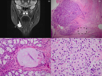
Examination revealed a soft, mobile, non-tender, 3–4 cm mass underlying normal mucosa in the floor of the mouth. The MRI revealed a 4.1 cm ovoid cystic structure adjacent to the right submandibular gland, with extension through the mylohyoid muscle (Figure 1a). Ultrasonography demonstrated a well-defined, lobulated fluid collection. A fine-needle aspiration (FNA) biopsy showed histiocytes. Repeat FNA targeted for amylase analysis revealed mucoid fluid that could not be assessed because of thickness. The clinical presentation and imaging findings were suggestive of a plunging ranula.
(a) Short-tau inversion-recovery magnetic resonance imaging coronal slice, showing enhancing cystic lesion extending from right submandibular space through the plane of the mylohyoid. (b–d) Photomicrographs of the sublingual gland, showing: (b) area of fibrosis and granulation tissue with abundant foamy macrophages (dotted box indicates region of interest magnified in part d) (H&E; ×4); (c) dilated duct filled with mucin (H&E; ×20); and (d) foamy, mucin-filled macrophages (H&E; ×40).

The patient underwent right submandibular sialendoscopy and ductoplasty (sialodochoplasty) with transoral resection of the sublingual gland. The right submandibular duct was identified and a 0.381 mm guidewire was passed without difficulty over the duct, which was then dilated with a Marchal dilator. The diagnostic sialoendoscope was passed into the right submandibular duct with no gross abnormalities. The sialoendoscope was then removed and the Marchal dilator was replaced to guide dissection, permitting the identification and preservation of the duct. The lingual nerve was identified and preserved. Intra-operatively, thick yellow mucoid material was encountered within the pseudocyst at the mid lower border of the sublingual gland. The sublingual gland was resected in its entirety. The partial resection of the pseudocyst included the upper portion where it attached to the sublingual gland. The submandibular gland was exposed but not violated. The sublingual gland was sent for permanent section pathological analysis.
Microscopically, the sublingual gland showed an area of fibrosis and granulation tissue, with abundant foamy macrophages (Figure 1b), and an area of ductal dilatation (Figure 1c). Higher magnification of the fibrotic area shows the foamy macrophages more clearly (Figure 1d).
The patient's clinical history and pre-operative imaging findings suggested a plunging ranula (extravasating pseudocyst of the sublingual gland) on the right side of the neck, which was consistent with the intra-operative findings and pathology.
On follow-up examination, the patient's submandibular duct was intact, his neck was palpably negative and there was no swelling of the submandibular area. He did have slight numbness to the tongue tip, which resolved after 2–3 days.
Discussion
A ranula is a salivary cyst that is thought to arise from the sublingual gland as a result of mucus extravasation.Reference Harrison1, Reference Magadevan and Vasan2 A ranula can be classified based on its extent or location: the intraoral or simple ranula is contained entirely within the sublingual space; the less common plunging ranula (also known as a diving or cervical ranula) extends into the submandibular space.Reference Harrison1–Reference Zhao, Jia, Chen and Zhang3
The intraoral ranula characteristically presents as a bluish, painless, soft swelling in the floor of the mouth. There is often a history of spontaneous rupture and leakage of viscous mucus into the oral cavity. This drainage is frequently followed by healing and reformation of the cyst. The mass may displace the tongue and interfere with oral function. Ranulas that are large or located in the area of the submandibular duct orifice (caruncula sublingualis) may lead to partial obstruction of the submandibular duct and result in submandibular swelling during eating.Reference Yulin, Yifang and Xinming4
Intraoral ranulas have been managed by various surgical methods, including: excision of the ranula via an intraoral or cervical approach; marsupialisation; intraoral excision of the sublingual gland and drainage of the lesion; or combined excision of the ranula and sublingual gland.Reference Yulin, Yifang and Xinming4 Other, less commonly applied treatment modalities include: intra-cystic injection with OK-432;Reference Rho, Kim, Kwon, Lee, Sung and Song5 hydrodissection;Reference Choi and Oh6 cryosurgery;Reference Mortellaro, Dall'Oca, Lucchina, Castiglia, Farronato and Fenini7 erbium, chromium: yttrium-scandium-gallium-garnet (‘Er,Cr:YSGG’) laser treatment;Reference Baurmash8 carbon dioxide laser treatment;Reference Niccoli-Filho and Morosolli9 and, in rare cases, radiation therapy.Reference Shimm, Berk, Tilsner and Coulthard10 Untreated or recurring oral ranulas may progress into the more complicated plunging ranula.Reference Zhao, Jia, Chen and Zhang3 Removal of the sublingual gland is considered the definitive therapy; management with marsupialisation is associated with a higher recurrence rate.
A plunging ranula usually manifests as a painless, non-mobile, anterior neck mass,Reference Rho, Kim, Kwon, Lee, Sung and Song5 occurring in conjunction with, or independent of, a floor-of-mouth component. The differential diagnosis of cervical swelling with a ranula-type appearance should also include lymphangiomas, which present similarly. Less common masses (or causes of masses) in this region include the thyroglossal duct cyst, parathyroid cyst, dermoid or epidermoid cysts, cervical thymic cyst, intramuscular haemangioma, cystic or neoplastic thyroid disease, benign teratoma, infectious cervical lymphadenopathy (e.g. Epstein–Barr virus, cat scratch disease and tuberculosis), hematoma, lipoma, laryngocele, branchial cleft cyst, anterior cystic hygroma, and submandibular sialoadenitis.Reference Yulin, Yifang and Xinming4
Intraoral and plunging ranulas may involve pseudocyst formation, the latter of which is lined with a connective tissue wall instead of an epithelial lining.Reference Harrison1, Reference Yulin, Yifang and Xinming4 One cause of ranula formation is thought to be local trauma or previous surgery. This can lead to acinar rupture or obstruction of the sublingual ducts, resulting in mucus extravasation. That fluid accumulation produces an inflammatory reaction and causes fibrous encapsulation. The macrophages break down the organic component, allowing the water and inorganic component to drain away in the lymphatics, with granulation tissue forming fibrous tissue, which restricts the extravasation and sometimes seals the leak. The plunging ranula may develop as a result of recurrent rupture, fluid extravasation, and re-encapsulation that may gradually extend the pseudocyst further down the neck as a result of gravity. This path of extension has been reported to travel either through the mylohyoid muscle or posterior to the muscle and anterior to the submandibular gland.Reference Harrison1 Another possible aetiology for ranula development is the interruption of drainage through Bartholin's duct; the variable anatomical location of this duct occasionally makes it susceptible to injury from surgical manipulation of the floor of the mouth.Reference Mun, Choi, Kim, Park, Jung and Sung11
A pre-operative diagnosis of a plunging ranula can be difficult to make because of the absence of specific clinical findings, including an often-undetectable intraoral component. Besides a mylohyoid defect, the key to diagnosing a plunging ranula radiographically is the identification of a connection to the sublingual space (Figure 1a) with CT or MRI. This may be no more than a thin tail of fluid or a significant local fluid collection.Reference Zhao, Jia, Chen and Zhang3 This tail is due to extension behind the mylohyoid muscle; it confirms that the ranula arises from the sublingual gland. A cystic fluid aspirate positive for mucin and macrophages can be helpful for the pre-operative diagnosis, as can biochemical positivity for salivary amylase and high protein content.Reference Harrison1–Reference Zhao, Jia, Chen and Zhang3 Sialography of the submandibular duct typically shows no communication with the cyst.Reference Mortellaro, Dall'Oca, Lucchina, Castiglia, Farronato and Fenini7
Histopathologically, most simple and diving or plunging ranulas are pseudocysts, formed in response to the salivary secretions.Reference Magadevan and Vasan2, Reference Rho, Kim, Kwon, Lee, Sung and Song5, Reference Mortellaro, Dall'Oca, Lucchina, Castiglia, Farronato and Fenini7 Our pathological specimen revealed normal appearing salivary tissue surrounding an area of fibrosis, inflammation and histiocytes.
Conservative therapy for a plunging ranula, such as aspiration, injection of sclerosing agents and marsupialisation, tends to result in high recurrence rates. Following a review of 864 ranula cases in the literature, Patel et al. concluded that the lowest complication and recurrence rates are achieved with excision of the offending sublingual gland via an intraoral approach, in conjunction with draining or partial removal of the ranula.Reference Patel, Deal and Shockley12 Complete excision of the pseudocyst is not necessary if the gland itself is excised, as the sublingual gland, not the pseudocyst, is thought to be the source of mucus formation.Reference Lesperance13 The extraoral approach has also been utilised to remove the ranula and sublingual gland, in order to avoid injury to the submandibular duct, Wharton's duct and lingual nerve; however, the recurrence and complication rates are higher.
• A ranula presents as a mass either in the mouth floor, where it elevates mucosa, often with a bluish tinge, or in the neck
• The key to ranula diagnosis is identification of a connection to sublingual space (e.g. thin tail of fluid or local fluid collection)
• Biopsy of lateral part of the neck may reveal only amorphous material, with rare inflammatory cells and predominant histiocytes, which stains positive for mucin
• Identification of submandibular duct and cannulation with a dilator may prevent ductal injury during an intraoral approach to a plunging ranula
In the case described, we used an intraoral approach with sialendoscopic assistance. Sialendoscopy permits endoscopic transluminal visualisation of the major salivary gland ductal system, and offers a mechanism for diagnosing and treating both inflammatory and obstructive pathology related to the ductal system. It is regarded as an acceptable and often preferred diagnostic and treatment tool for chronic sialadenitis and non-neoplastic obstruction of the salivary system, including sialolithiasis. In our case, sialendoscopy provided a minimally invasive means of evaluating the submandibular duct lumen and ensuring its preservation when placing the Marchal dilator. The presence of this dilator within the submandibular duct provides a palpable landmark during surgery to aid in skeletonisation and preservation of the duct. This may help prevent iatrogenic injury to the submandibular duct at the time of primary surgery, potentially avoiding post-obstructive sialadenitis of the submandibular gland.
The intraoral approach with use of a dilator can be an effective means of treating a plunging ranula; this technique can minimise injury to the submandibular duct and other local structures.