Introduction
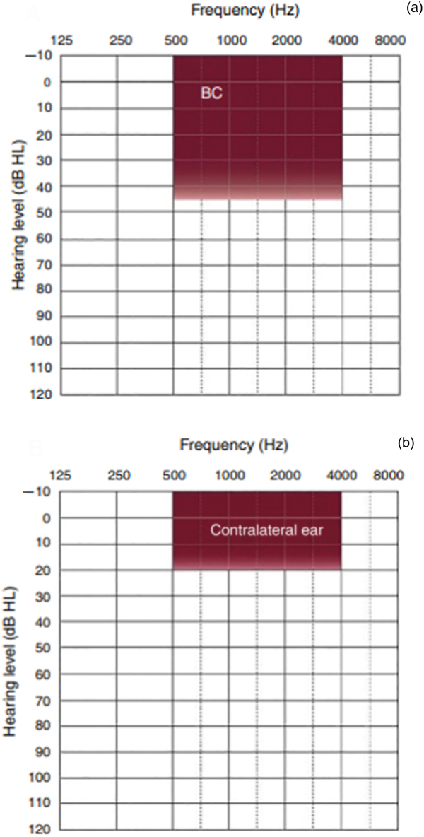
Bone conduction implants are indicated for patients with conductive or mixed hearing loss who are not good candidates for a conventional acoustic prosthesis, and for patients with severe to profound unilateral sensorineural hearing loss (single-sided deafness) with normal hearing in the contralateral ear (Figure 1).Reference Zernotti and Sarasty1
Audiological criteria for the Bonebridge active bone conduction system, for: (a) conductive or mixed hearing loss, and (b) unilateral sensorineural hearing loss. BC = bone conduction

Several types of bone conduction implants exist and are classified according to the modality of conduction. In cutaneous implants, vibration is transmitted through the skin (passive transcutaneous bone conduction implant systems, such as the Sophono® device and bone-anchored hearing aid (BAHA) Attract system (Cochlear, Sydney, Australia)). In direct implants, vibration is transmitted directly to the bone. They are classified into percutaneous devices (e.g. BAHA and Oticon Medical Ponto) and active transcutaneous systems (e.g. Bonebridge, by Med-El, Innsbruck, Austria).
Bonebridge, the first active transcutaneous bone conduction system, is a partially implantable device. It includes an external part, the audio processor, and an implanted part, the bone conduction implant. The audio processor is worn on the head, and contains two microphones to receive the sounds, a digital signal processor and a battery. The bone conduction implant (BCI601) consists of a magnet surrounded by a receiving coil, a demodulator and an electromagnetic floating mass transducer.Reference Sprinzl, Lenarz, Ernst and Hagen2,Reference Manrique, Sanhueza, Manrique and de Abajo3 The implant is placed completely under the skin. It connects to the processor through magnetic attraction, and transforms the received signal into mechanical vibrations that are transmitted through the temporal bone to the inner ear. The first Bonebridge device was implanted in June 2011. In 2014, it was approved for patients aged over five years.Reference Sprinzl and Wolf-Magele4
A pre-operative computed tomography (CT) scan is needed to acquire information about bone thickness, and height of dura mater and sigmoid sinus, to determine where best to place the bone conduction floating mass transducer and the screws.Reference Canis, Ihler, Blum and Matthias5–Reference Law, Bhatia, Tsang, Tong and Shi7 The bone conduction device can be implanted in three different locations.Reference Barbara, Perotti, Gioia, Volpini and Monini8 In cases of normal anatomy, it is placed in the sinodural angle. If the mastoid is already drilled out or if there is little space in the sinodural angle, the bone conduction floating mass transducer can be placed either behind the sinusReference Zernotti, di Gregorio, Galeazzi and Tabernero9,Reference Lassaletta, Sanchez-Cuadrado, Muñoz and Gavilan10 or above the temporal line in the middle fossa region.Reference Zernotti and Sarasty1
In our department, when an anatomical variant exists or a previous surgery such as a petrosectomy or a radical mastoid cavity has been performed, we prefer the middle fossa approach, placing the bone conduction floating mass transducer at squamous portion of the temporal bone to avoid exploring the previously operated ear. Furthermore, we use a surgical technique that makes the procedure fast and safe, with no risk of damaging the dura mater. This article describes our preferred technique to perform the middle fossa approach as a second option when the classic pre-sigmoid approach is not feasible.
Materials and methods
A total of 40 patients were implanted with the Bonebridge active bone conduction system in our hospital between 2012 and 2018. The classic transmastoid pre-sigmoid approach was utilised in 22 patients. In the remaining 18 patients, in whom this approach was not possible because of anatomical variants or previous surgical procedures, a retrosigmoid and a middle fossa media approach were used, in 4 and 14 cases respectively (Figure 2). We have now practically abandoned the retrosigmoid approach; in cases where the pre-sigmoid approach is not possible, we employ the middle fossa media approach, using the surgical technique described below.
Distribution of the three different approaches used in our series of patients.

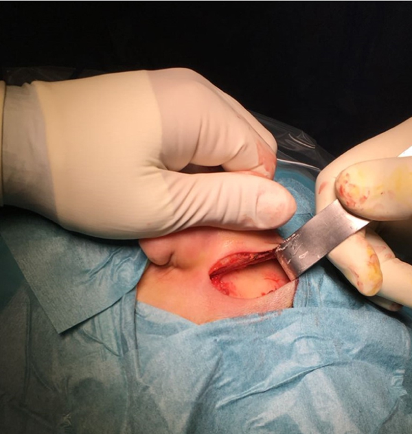
The surgery is conducted under general anaesthesia and using an orotracheal tube. The patient is placed in a supine position with their head turned to the contralateral side. Local infiltration with lidocaine and adrenaline (1:100 000) is used for the anaesthesia. A 3 cm post-auricular incision, above the level of the pinna, is performed (Figure 3).
A post-auricular 3 cm incision is performed on the left ear. The squamous portion of temporal bone is exposed.

Two flaps are prepared: a superficial flap that includes the skin and subcutaneous tissue, and a deeper fasciomuscular flap to reduce the risk of device extrusion. Haemostasis is achieved with monopolar or bipolar electrocautery. However, once the bone conduction implant is in the surgical field or if the patient already has an implant on the other side, bipolar electrocautery is mandatory.
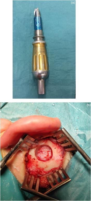
The next step is to prepare the bone bed for the bone conduction implant floating mass transducer, below the temporal muscle. We use a 14 mm drill head (Neuro Drill) to create the bed at the squamous portion of the temporal bone, superior to the supramastoid crest or temporal line, between the inferior temporal line and the middle temporal artery sulcus (Figure 4). The time needed to create a 14 mm bone bed is around 14 seconds (see the short video, available on The Journal of Laryngology & Otology website; Appendix 1), which is much faster than if using a standard otological drill. Afterwards, we enlarge the bone bed with a Kerrison rongeur until a 16 mm diameter bed is achieved to place the bone conduction floating mass transducer.
(a) The 14 mm Neuro Drill used to create the bone bed at the squamous portion of the temporal bone. (b) A 14 mm bone bed is created.

The next steps are the same as described for the classic pre-sigmoid approach. The periosteal pocket is prepared to accommodate the coil and demodulator of the bone conduction implant, and the bone conduction implant is fixed in the correct position. Finally, the incision is closed by suturing the two flaps previously described and applying a pressure dressing to the wound.
Patients who underwent a middle fossa approach were divided into two groups according to the technique used to create the bone bed for the bone conduction implant. In group A, a classic otological drill was used; in group B, the 14 mm Neuro Drill (described above) was employed. The rate of intra-operative and post-operative complications was analysed. The surgical time was compared between the two groups and with the retrosigmoid approach group. The student's t-test was used, and the statistical significance was defined as p < 0.05. Follow-up duration ranged from 6 to 45 months.
Results
A total of 14 out of 40 Bonebridge devices were implanted through a middle fossa approach (35 per cent). In the first four cases (group A), a classic otological drill was used to create the bone bed for the bone conduction implant. In the other 10 cases (group B), the technique described in this article was performed.
Regarding intra-operative complications, no cases of dura mater damage were observed in the two groups. In group A, a mild haemorrhage following rupture of the sigmoid sinus wall was observed in two patients.
In group A, the average surgical time was 47 minutes. Surgical time was shorter than 30 minutes in group B, with an average surgical time of 28 minutes. There was a statistically significant difference between the two groups (student's t-test, p < 0.05). Furthermore, only 14 seconds were needed to create a 14 mm bone bed, before it was enlarged with a Kerrison rongeur to obtain a 16 mm diameter bed.
With respect to the retrosigmoid approach, performed in 4 out of 40 patients (10 per cent), the average surgical time was 120 minutes, which is much longer than the surgical time needed for the middle fossa approach (student's t-test, p < 0.05).
In all cases, we used the two-flap technique (skin and subcutaneous tissue, and fasciomuscular flap) to close the incision. There were no cases of device exposure, cerebrospinal fluid leak, wound haematoma, wound infection or wound dehiscence. There were no complications associated with the bone conduction floating mass transducer contacting the dura mater. The follow-up duration ranged from 6 to 45 months, with an average of 28 months.
Discussion
The Bonebridge active bone conduction system is used for patients with bilateral conductive or mixed hearing loss where the bone conduction thresholds do not exceed 45 dB (average of 0.5, 1, 2 and 3 kHz), or those with a single-sided deafness and normal hearing in the contralateral ear (bone conduction thresholds better than 20 dB).
Three different surgical approaches have been described: pre-sigmoid, retrosigmoid and middle fossa approaches. The pre-sigmoid approach is the preferred technique if normal anatomy is demonstrated on the pre-operative CT scan. In the presence of anatomical variants, such as a low middle fossa dura plate or anterior procident sigmoid sinus, or in cases of radical cavities, the pre-sigmoid placement of the implant is not possible. In these cases, we can choose a retrosigmoid or middle fossa approach.
In our hospital, we began to use the Bonebridge system in 2012, and we have now implanted a total of 40 patients. In all cases where the pre-sigmoid approach was not possible, we utilised the middle fossa approach, using the technique described above. We consider that the use of the 14 mm Neuro Drill to create the bone bed makes this approach safe and easy. In fact, its mechanism of action is based on the automatic interruption of its movement when it does not find bone resistance, which guarantees the integrity of the dura mater. Compared with a classic otological drill, the Neuro Drill significantly shortens the operative time, and reduces the risk of damage to the dura mater and sigmoid sinus.
When compared with a retrosigmoid approach, our technique is easier, the surgical time is significantly shorter (30 minutes vs 120 minutes), and there is no risk of damaging the sigmoid sinus or dura mater, which are principal risks of the retrosigmoid approach. For all these reasons, in 2014 we stopped performing the retrosigmoid approach when the pre-sigmoid approach was not feasible.
Our technique for the middle fossa approach has not previously been described in the literature. We consider it an excellent alternative to the classic pre-sigmoid approach when it cannot be performed. It is easy, safe, fast and reproducible, and does not require special surgical skills. We strongly recommend it in all cases where a classic approach cannot be employed.
Supplementary material
The supplementary material for this article can be found at https://doi.org/10.1017/S0022215119000501.
Competing interests
None declared