Introduction
Skull base paragangliomas may invade the facial nerve,Reference Makek, Franklin, Zhao and Fisch1, Reference Mafee, Raofi, Kumar and Muscato2 however, a primary paraganglioma of the facial nerve (glomus faciale) is a rare entity. There have been seven previous reports of histologically confirmed paragangliomas arising in the facial nerve canal.Reference Bartels, Pennington, Kamerer and Browarsky3–Reference Wippold, Neely and Haughey7 We present a rare case of a predominantly extracranial glomus faciale tumour.
Case report
A 54-year-old woman presented with a six-month history of right sided facial twitching and weakness of her lower lip. She had also experienced pain and tenderness in the right post-auricular region. Clinical examination confirmed grade 2 (House-Brackmann) right sided facial nerve function and a right parotid mass. Otoscopy was normal. Ultrasound scanning at the referring hospital revealed a parotid mass of mixed echogenicity with colour Doppler indicating a hypervascular lesion. A subsequent ultrasound guided 20-gauge core biopsy demonstrated histological features of a paraganglioma.
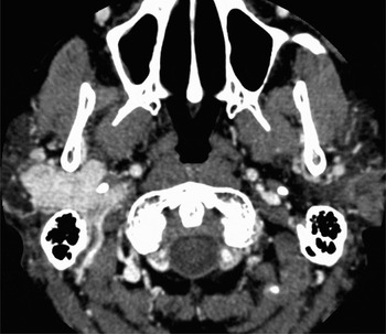
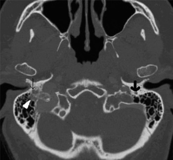
Computed tomography (CT) and CT angiography showed a 4.3 × 2.7 × 4.2 cm mass centred within the deep lobe of the right parotid gland (Figure 1). The mass underwent avid contrast enhancement in both the arterial (Hounsfield units ranging from 200–240) and delayed phases. It extended laterally to infiltrate the superficial lobe of the parotid, medially to abut the lateral aspect of the internal jugular vein and posteriorly into the retrostyloid region. There were multiple enhancing vessels at the periphery of the mass. Superiorly, the mass extended to the inferior mastoid where there was irregular enlargement of the descending portion of the facial nerve canal, a small area of enhancing soft tissue within adjacent mastoid air cells and permeative lucency extending towards the jugular foramen (Figure 2). The patient was unable to tolerate magnetic resonance imaging. Fluoro-deoxy-glucose positron emission tomography (FDG-PET) revealed no additional lesions and 24-hour vanillylmandelic acid urinary estimation was not elevated. At exploration it was apparent that the tumour arose from the facial nerve from the second genu to the proximal divisions within the parotid gland. There was medial extension to the internal jugular vein immediately inferior to the jugular bulb but it was apparent that the tumour did not arise from it. The tumour was removed completely though a cervico-temporal approach and the facial nerve repaired by placing a sural nerve interposition graft from the mastoid to its superior division and anastomosing the hypoglossal nerve to the lower division.
Axial image from a CT angiography study through the mid parotid with soft tissue algorithm. The markedly enhancing parotid mass is centred on the deep lobe with posterior extension to the retrostyloid region. The internal jugular vein is difficult to separate from the mass and lies adjacent to its medial aspect.

Axial image from a CT scan through the inferior mastoid with bone algorithm. There is irregular expansion of the descending portion of the facial nerve canal (thin black arrow). The normal calibre left facial nerve canal (thick black arrow) may be compared. There is adjacent permeative lucency extending towards the dominant right sided jugular foramen. The soft tissue opacity within inferior mastoid air cells (white arrowhead) underwent marked enhancement and represents tumour.

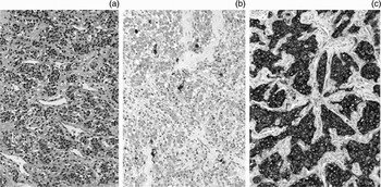
Histological examination of the operative specimen confirmed a paraganglioma of the facial nerve (Figure 3). There was improved facial nerve function at eight-month follow up.
Representative areas showing typical paraganglioma comprising nests of small round or polygonal cells in a highly vascular and sclerotic stroma (a). Immunostaining reveals scattered sustentacular cells with S100 (b) and there is strong uniform positivity for the neuroendocrine marker chromogranin (c). Original objective magnification ×10.

Discussion
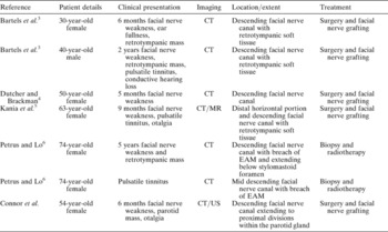
Paragangliomas are neoplasms arising from glomus bodies (paraganglia). The majority of skull base glomus bodies are found in the adventitia of the jugular bulb, along the inferior tympanic canaliculus and over the cochlear promontory,Reference Guild8 hence these are the most frequent locations for skull base paragangliomas. A small number of glomus bodies may be found within the descending portion of the facial nerve canal explaining the rare occurrence of paragangliomas at this site. Previous reports of glomus faciale tumoursReference Bartels, Pennington, Kamerer and Browarsky3–Reference Wippold, Neely and Haughey7 have described lesions confined to the temporal bone or demonstrate minimal extension inferior to the stylomastoid foramen.Reference Bartels, Pennington, Kamerer and Browarsky3, Reference Dutcher and Brackman4, Reference Petrus and Lo6 Table I documents the clinical presentation, macroscopic findings and treatment of pathologically confirmed glomus faciale tumours. Such tumours have occasionally grown proximally along the course of the facial nerve to the tympanic portionReference Kania, Bouccara, Colombani, Molas and Sterkers5 and have expanded radially into the external auditory meatus or posterior tympanum. This appears to be the first case of a glomus faciale tumour presenting as a predominantly extracranial lesion. Therefore, this entity should be considered as a rare differential diagnosis of a hypervascular parotid mass.
Clinical presentation, imaging investigations, location/extent and treatment of pathologically confirmed glomus faciale tumours

CT = computed tomography; EAM = external auditory meatus; US = ultrasound; MR = magnetic resonance
The salient imaging features in this patient were of a hypervascular parotid mass (marked colour flow on Doppler ultrasound and marked contrast enhancement during the arterial phase of a CT study) together with irregular widening of the descending portion of the facial nerve canal and adjacent permeative lucency. The differential diagnosis to consider would include facial nerve schwannoma, malignant parotid tumour, haemangioma, haemangiopericytoma and glomus jugulare tumour. A schwannoma only rarely demonstrates hypervascularityReference Aygenc, Selcuk and Ozdem9 and the facial nerve canal expansion would be well defined without permeative lucency. Malignant parotid tumours are usually not hypervascular although irregular widening of the facial nerve canal and adjacent ill-defined lucency may result from peri-neural extension. Benign vascular tumours such as rarer ‘capillary type’ facial nerve or parotid haemangiomas would be unlikely to demonstrate ill-defined bony changes although these may be present with more aggressive vascular tumours such as haemangiopericytomas. The pattern of invasion would be unusual for a glomus jugulare tumour, which would typically erode the margins of the jugular fossa and then extend superolaterally towards the middle ear, although extension to the parotid region has been described.Reference Brandrick, Das Gupta and Singh10
• Paraganglioma of the facial nerve (glomus faciale) may present in a predominantly extracranial location
• Paraganglioma of the facial nerve (glomus faciale) should be considered as a rare differential diagnosis of a hypervascular parotid mass
The diagnosis of a glomus faciale tumour requires confirmation that the tumour arose from glomus bodies along the facial nerve. This tumour did extend medially to the jugular foramen and it is difficult to exclude an origin from paraganglia along the mastoid canaliculus with subsequent extension along the facial nerve. However, the extensive facial nerve involvement with generally intact bony margins to the jugular foramen would argue against this possibility. Also, in view of the large extracranial mass, an origin from paraganglia within the parotid gland cannot be completely discounted. If this were the case, it is still likely that they would still be located along the course of the facial nerve. It has been postulated that paragangliomas originating in unusual locations within the head and neckReference Nielson, Sejean and Onerheim11 are derived from glomus bodies along distal cranial nerves.
In conclusion, a facial nerve paraganglioma or glomus faciale may be predominantly extracranial and should be considered in the differential diagnosis of a hypervascular parotid mass. Computed tomography will prove helpful in such a case in order to limit the differential diagnosis and to define the extent of skull base involvement.