Introduction
Entomopathogenic nematodes in the family Heterorhabditidae are obligate pathogens of a wide range of insect pests (revised by Shapiro-Ilan et al., Reference Shapiro-Ilan, Gouge, Koppenhöfer and Gaugler2002) and have been recovered from diverse climates and geographic regions of the world (revised by Adams et al., Reference Adams, Fodor, Klein, Smith, Stackenbrandt and Stock2006). These nematodes are mutualistically associated with Photorhabdus bacteria (γ-Proteobacteria: Enterobacteriaceae). The bacterial symbionts are harboured in the anterior portion of the intestine of the only free-living stage of these nematodes, also known as the infective juvenile or third-stage infective juvenile (IJ). The nematodes regurgitate the bacteria, which kill the insects and provide a food source for the nematodes to grow and multiply. At present more than a dozen species of Heterorhabditis nematodes have been described, and only three Photorhabdus species have been identified. Of them, P. asymbiotica is the only species known to be a human pathogen. Indeed, this species invades soft tissues and disseminates bacteraemic infections in human patients both in the United States and Australia (Akhurst & Smith, Reference Akhurst, Smith and Gaugler2002; Gerrard et al., Reference Gerrard, Joyce, Clarke, ffrench-Constant, Nimmo, Looke, Feil, Pearce and Waterfield2006). Originally, this bacterium was not found in association with nematodes, and because of this it was given the species name ‘asymbiotica’ ( = not a symbiont). Gerrard et al. (Reference Gerrard, Joyce, Clarke, ffrench-Constant, Nimmo, Looke, Feil, Pearce and Waterfield2006) isolated a Heterorhabditis nematode from sandy soil samples taken at Kingscliff beach, New South Wales, Australia, a site where a patient with P. asymbiotica infection had been reported. Preliminary observations depicted this nematode as an undescribed Heterorhabditis species. In this study we provide a thorough morphological description of this new species, which is supplemented with internal transcribed spacer (ITS) rDNA sequence data and cross-hybridization assays. Evolutionary relationships of this nematode with other members of Heterorhabditis are presented and discussed. Moreover, the bacterial symbiont of this new Heterorhabidtis species was phenotypically characterized and compared with other existing P. asymbiotica strains.
Materials and methods
Nematode isolation and rearing
Soil samples (650 g) were collected from the property where the reported patient contracted the bacterial infection, in Kingscliff, Australia. Five mealworm larvae, Tenebrio mollitor L. (Coleoptera: Tenebrionidae) were placed in each container as baits according to procedures described by Kaya & Stock (Reference Kaya, Stock and Lacey1997). Dead insects were removed from the containers 5 days later and placed in modified White traps to recover the nematodes (Kaya & Stock, Reference Kaya, Stock and Lacey1997). Third-stage infective juveniles (IJs) began to emerge 15 days after exposure of mealworm larvae to nematodes in the soil samples. Emerging IJs were surface sterilized in 0.4% Hyamine (Sigma, St. Louis, Missouri, USA) for 15 min and washed three times in sterile phosphate-buffered saline (PBS). Surface sterilized nematodes were used to infect Galleria mellonella larvae (100 IJs per insect) to confirm Koch's postulates for pathogenicity (Kaya & Stock, Reference Kaya, Stock and Lacey1997).
Morphological characterization
Differential interference contrast microscopy
For morphological studies, first-generation hermaphrodites and second-generation adults were obtained by dissecting infested cadavers on days 4–5 and 7–8 after initial infection. Third-stage infective juveniles were obtained upon emergence and during the first 2 days. A total of 20 specimens from each life stage were considered for evaluation of qualitative and quantitative data. Nematodes were examined live and/or heat-killed in 60°C Ringer's solution. Heat-killed nematodes were placed in triethanolamine–formalin (TAF) fixative (Kaya & Stock, Reference Kaya, Stock and Lacey1997) and processed to anhydrous glycerine for mounting (Seinhorst, Reference Seinhorst1959). Measurements and digital light microscopy images were made from live and mounted specimens using an Olympus IX51 microscope equipped with differential interference contrast (DIC) optics and a digital video image system. Specimen measurements were made using AnalySIS Image software (Soft Imaging System Corp., California, USA).
The following abbreviations for morphological features are used in the text and tables: ABW = anal body width, D% = (EP/ES) × 100, E% = (EP/TL) × 100, EP = distance from anterior end to excretory pore, ES = distance from anterior end to oesophagus base, GL = gonad length, GS = GuL/SpL, GuL = gubernaculum length, MBW = maximum body width, NR = nerve ring position, StL = stoma length, StW = stoma width, SpL = spicule length, SW = SpL/GuL, TBL = total body length, TL = tail length, TRF = testis reflexion, V% = (TBL/length to vagina) × 100, WA = width at anus/cloaca.
Scanning electron microscopy (SEM)
Adult males and third-stage infective juveniles were rinsed three times in M9 buffer for 5 min per rinse. All nematodes were relaxed and heat-killed as above, then fixed in 8% glutaraldehyde buffered in cacodylate at pH 7.33 overnight. Fixed nematodes were rinsed in distilled water three times, post-fixed in OsO4 for 1 h, rinsed again in distilled water and serially dehydrated at 15-min intervals in ethanol (McClure & Stowel, Reference McClure and Stowel1978). Specimens were then critical point dried in liquid CO2, mounted on SEM stubs, coated twice with gold and scanned using a Philips XL series microscope at 20 kV accelerating voltage.
Molecular characterization
Nucleic acid preparations used for polymerase chain reaction (PCR) amplifications of the new Heterorhabditis sp. were extracted from pools of 10–50 third-stage infective juveniles. Nucleic acids were extracted from the digestion supernatant using phenol–chloroform enrichment, ethanol/ammonium acetate precipitation (Ausbel, 1989). The resulting pellet was washed with 70% ethanol, resuspended in Tris-EDTA (TE) buffer (pH 8.0), treated with 50 μg of RNAse A (1 h at 37°C), and DNA recovered following reprecipitation with ethanol. DNA was quantified by spectrophotometry, and 100–200 ng used per PCR reaction.
PCR was used to amplify a region within the 5′-end of the nuclear internal transcribed spacer (ITS) region. Typical PCR reactions included 0.5 μm of each primer, 200 μm deoxynucleoside triphosphates, and MgCl2 concentration of 2 mm in a total reaction volume of 25 μl. PCR conditions, e.g. annealing temperature and MgCl2 concentration, were adjusted empirically as needed to optimize reaction specificity for individual isolates. Typical conditions included denaturation at 94°C for 3 min, followed by 33 cycles of 94°C for 30 s, 60°C for 30 s, and 72°C for 1.15 min, followed by a post-amplification extension at 72°C for 5 min (Stock et al., Reference Stock, Campbell and Nadler2001).
PCR primers considered were 94, forward (5′–TTGAACCGGGTAAAAGTCG) and 93, reverse (5′–TTAGTTTCTTTTCCTCCGCT), as described by Stock et al. (Reference Stock, Campbell and Nadler2001). One microlitre of each PCR product was used for agarose gel electrophoresis (1.3% agarose in 1 × Tris-borate-EDTA (TBE) buffer) to confirm amplicon size and yield. PCR products were prepared for direct sequencing using enzymatic treatment with EXOSAP-IT (USB Corp., Cleveland, Ohio, USA). Sequencing reactions were performed using dye-terminator cycle sequencing chemistry, and reaction products were separated and detected using an ABI 3730 automated DNA sequencer. Sequences for each species were completely double-stranded for accuracy, using the PCR primers and two internal sequencing primers. The forward internal primer was #264 (5′-GGTTTTTCATCGATACGCG), and the reverse internal primer #389 (5′-TGCAGACGCTTAGAGTGGTG).
Sequence analysis and phylogenetic analysis
Contig assembly and sequence conflict resolution was performed with the aid of EditSeq (DNAStar software, Lasergene Corp., Madison, Wisconsin, USA). Sequence regions corresponding to the PCR amplification primers were removed prior to multiple sequence alignment and phylogenetic analysis, because primer incorporation during amplification masks potential mismatches (substitutions) that may occur in PCR priming sites. Sequences were aligned initially using CLUSTAL X v.1.53b (Thompson et al., Reference Thompson, Gibson, Plewniak, Jeanmougin and Higgins1997), and the resulting output was adjusted manually to improve homology statements using MacClade software v.4.0 (Maddison & Maddison, Reference Maddison and Maddison2002). ITS sequences of H. gerrardi n. sp. were deposited in GenBank under the accession number FJ1525445.
Sequence data were analysed by unweighted maximum parsimony (MP) using PAUP* v. 4.0b3a (Swofford, Reference Swofford2002). Unrecoded gaps were treated as missing data. Tree searches for these ITS datasets were performed using heuristic methods with TBR (tree-bisection-reconnection) branch swapping, and a minimum of 1000 replicates of random stepwise addition. Reported consistency indices (CI) do not include uninformative characters. Bootstrap parsimony analyses were performed using heuristic searches (simple stepwise addition, TBR branch-swapping, MULPARS) and 2000 pseudoreplicates.
Cross-hybridization tests
Cross-breeding tests were carried out on lipid agar plates according to Kaya & Stock (Reference Kaya, Stock and Lacey1997). The Kingscliff isolate of the new Heterorhabditis species was crossed with two H. indica strains T1 (Thailand) and CR4 (Costa Rica). One female and one male of the appropriate strains were placed on the agar plates (35 mm diameter) and incubated at 25°C. Controls consisted of one male and one female of the same isolate/species. Ten plates per isolate were tested. Progeny production was checked daily and for a period of three consecutive days according to Stock & Kaya (Reference Stock and Kaya1996). Hybridization assays were repeated three times.
Isolation and characterization of bacteria
A motorized pestle was used to crush five individual IJs in 100 μl of Luria broth (LB). Thirty-five microlitres were plated on to LB and neutral bromothymol (NBT) agars and incubated at 30°C for 48 h. Single colonies were replicated on to LB agar for incubation at 30 and 37°C and allowed to grow for an additional 48 h and then examined for bioluminescence. Nematode-borne bacteria were analysed by MLST (multi-locus sequence typing) (Ed Feil, pers. comm.). Bacteria were also characterized phenotypically following procedures described by Joyce & Clarke (Reference Joyce and Clarke2003).
Mutualistic exclusivity
Members of all three characterized P. asymbiotica strains were assessed for their ability to support the growth and development of different nematode species. Bacteria, representative of all three known strains, were seeded separately on to lipid agar plates in triplicate and incubated at 28°C for 48 h. Surface sterilized Kingscliff infective juveniles (IJs) were added (day 1) and plates were examined and scored for nematode growth and development from day 5 to 18 using a stereomicroscope with a magnification of × 50. When infective juveniles appeared on plates, they were collected, surface sterilized and crushed individually, as described above, to determine whether repackaging of that particular bacterial species had occurred, thereby completing mutualism with the Kingscliff nematode.
Results and discussion
Description of Heterorhabditis gerrardi n. sp.*
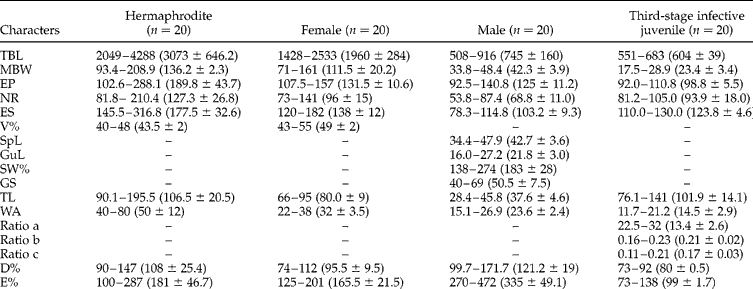
The morphological traits of adult stages and third-stage infected juveniles of H. gerrardi are given in table 1.
Morphometric traits of adult stages and third-stage infective juveniles of H. gerrardi n. sp. Measurements – range and average (in parentheses) – are in micrometres.
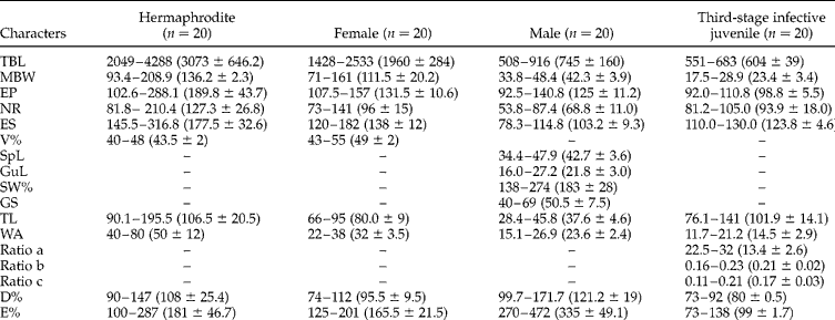
ABW, anal body width; D%, (EP/ES) × 100; E%, (EP/TL) × 100; EP, distance from anterior end to excretory pore; ES, distance from anterior end to oesophagus base; GS, GuL/SpL; GuL, gubernaculum length; MBW, maximum body width; NR, nerve ring position; ratio a, TBL/MBW; ratio b, TBL/ES; ratio c, TBL/TL; SpL, spicule length; SW%, (SpL/GuL) × 100; TBL, total body length; TL, tail length; V%, (TBL/length to vagina) × 100; WA, width at anus/cloaca.
Hermaphrodite
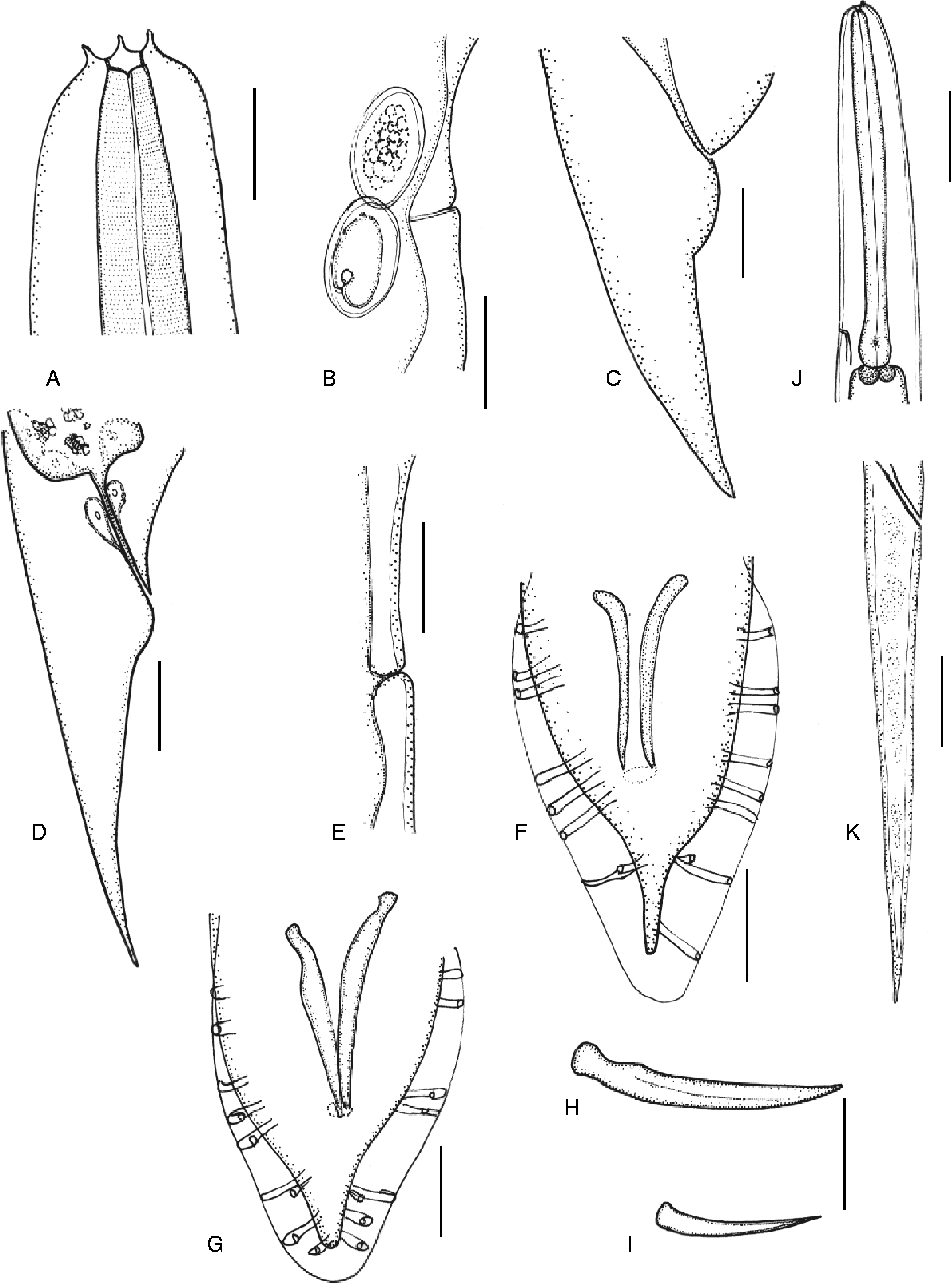
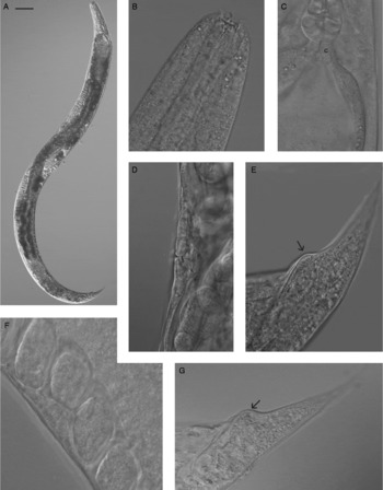
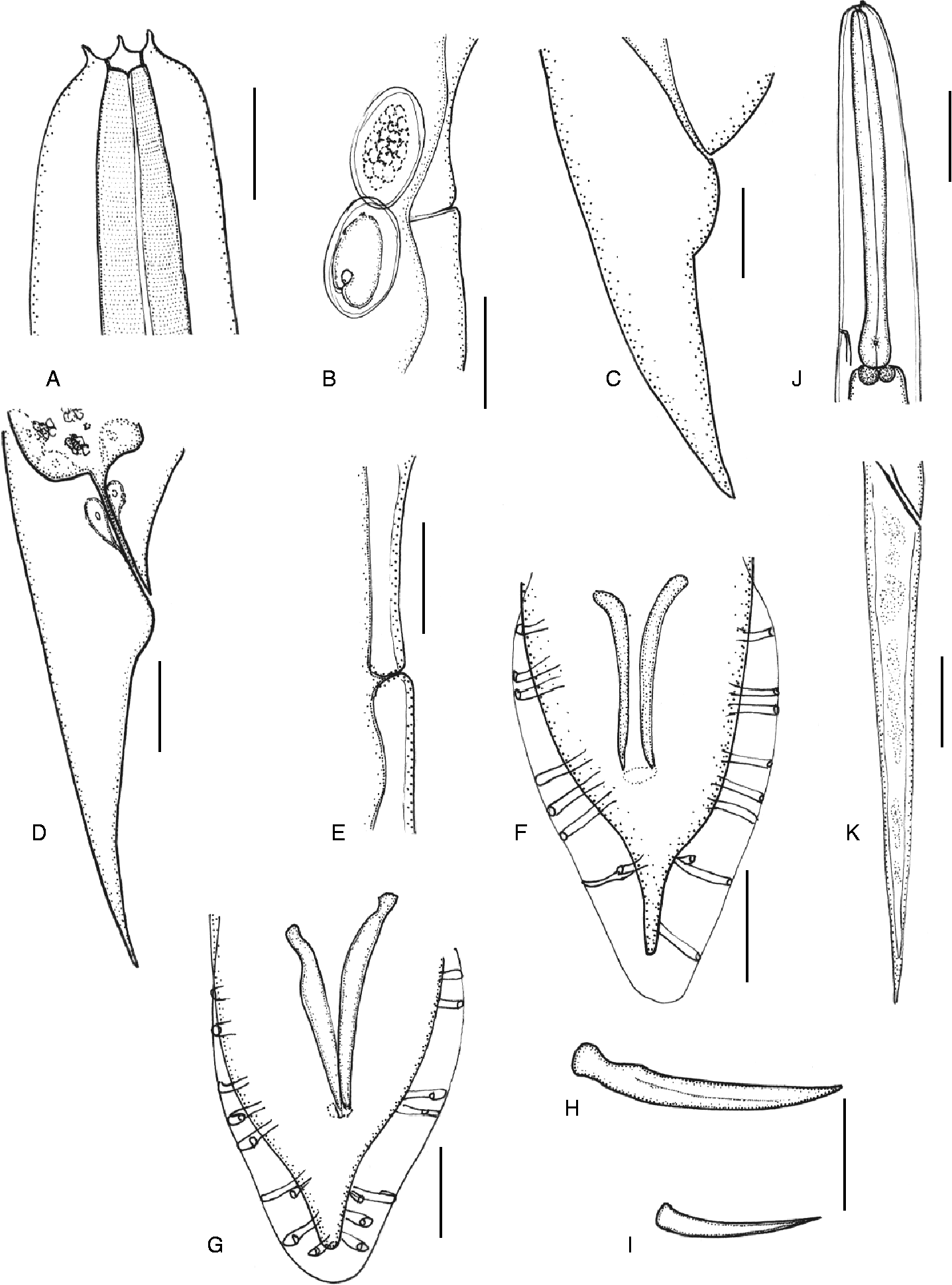
C-shaped after heat killed. Cuticle with fine transverse striae. Head region slightly truncated (figs 1A and 2B). Labial region with six prominent lips, each bearing one papilla. Cephalic papillae not observed with SEM. Amphidial opening small, pore-like. Stoma funnel-shaped, with refractile cheilorhabdions (figs 1A and 2B). Oesophagus with a cylindrical corpus. Isthmus present. Basal bulb pyriform or slightly rounded with small valve. Cardia present (fig. 2C). Nerve ring located in the middle of the isthmus. Gonad didelphic, amphidelphic. Vulva located near the mid-body region (fig. 2A). Vulval lips slightly protruding or non-protruding and symmetrical (figs 1A and 2D). Copulatory plug not observed. Tail conoid with slightly protruding post-anal swelling (figs 1C and 2E). Phasmids inconspicuous.
Heterorhabditis gerrardi n. sp. line drawings. (A) hermaphrodite, anterior end, lateral view; (B) hermaphrodite vulval region, lateral view; (C) hermaphrodite tail region, lateral view; (D) female tail region, lateral view; (E) female vulval region, lateral view; (F, G) male tail ventral view showing numerical variation of bursal rays; (H) spicule, lateral view; (I) gubernaculum lateral view; (J, K) third-stage infective juvenile: (J) anterior end (lateral view), (K) tail region (lateral view). Scale bars: (A) 27 μm; (B) 43 μm; (C) 28.5 μm; (D, F, G) 23 μm; (E) 33 μm; (H) 16 μm; (I) 12.5 μm; (J) 30 μm; (K) 20 μm.
Heterorhabditis gerrardi n. sp. DIC and SEM micrographs of hermaphrodite and second-generation female. (A) Female, second generation, in toto; (B) hermaphrodite, anterior end, lateral view; (C) hermaphrodite, lateral view showing cardias (c) and anterior portion of intestine; (D) vulva opening of hermaphrodite, lateral view; (E) tail of hermaphrodite, lateral view, showing post-anal swelling (arrow); (F) vulva opening of second-generation female, lateral view; (G) tail of second-generation female, lateral view, showing post-anal swelling (arrow). Scale bars: (A) 186 μm; (B) 12 μm; (C, F) 14 μm; (D, E) 15 μm; (G) 32 μm.

Male
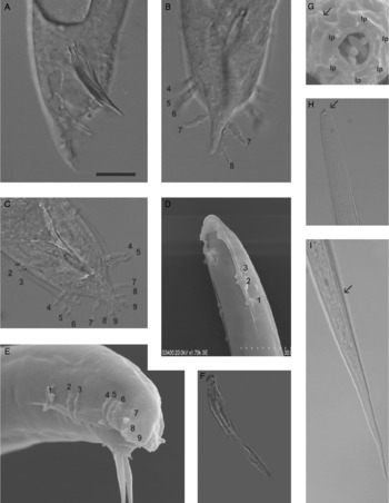
Body curved ventrally when heat killed. Cuticle with fine longitudinal striae. Head truncated or slightly rounded. Six labial papillae. Amphidial opening conspicuous. Mouth opening funnel-shaped, stoma short. Oesophagus with cylindrical procorpus and metacarpus. Isthmus distinct. Basal bulb pyriform, with reduced valve. Nerve ring surrounding isthmus or just above the basal bulb. Excretory pore usually posterior to basal bulb. Testis single, reflexed anteriorly. Vas deferens well developed. Spicules paired, separate, with rostrum (figs 1H and 3A). Gubernaculum thin, straight or slightly curved ventrally. Tail conoid with peloderan bursa, slightly curved ventrally. Nine pairs of genital papillae (bursal rays) with the typical Heterorhabditis formula: 1 2 () 3 3 (fig. 3B–E). Pairs 4 and 7 curved outward (laterally) (fig. 2E). Pairs 7, 8 and 9 form a terminal group at the end of the bursa. Approximately 30% of the examined specimens (total 40 males) showed variable number of papillae in the two terminal sets (i.e. pairs 4–6 and 7–9). In some instances pair 6 was missing on one side of the bursa, whereas in other specimens pairs 8 and 9 or 9 were missing on both sides or only one side of the bursa (fig. 3B and C).
Heterorhabditis gerrardi n. sp. DIC and SEM micrographs of male and third-stage infective juvenile. (A–C) Light microscopy images of tail of male, showing spicules and arrangement and number of bursal rays (A, lateral view; B,C, ventral views); (D, E) scanning electron micrographs of male tail showing bursal rays; (F) spicule, lateral view; (G) En face view of male head showing stomal opening, labial papillae (lp) and amphid (arrow), also Photorhabdus asymbiotica bacteria are shown inside the stoma; (H) third-stage infective juvenile showing dorsal cuticular tooth (arrow); (I) tail of third-stage infective juvenile showing anal opening (arrow). Scale bars: (A–C) 14 μm; (D) 30 μm; (E) 20 μm; (F) 13 μm; (G) 5 μm; (H, I) 19.5 μm.

Female
Body C-shaped after heat-killed. Cuticle with fine transverse striae. Head region, lips and oesophageal region similar to that of the hermaphrodites, but smaller. Gonads didelphic, amphidelphic. Vulva, a transverse slit, located in the mid-body region. Copulatory plug not observed. Vulval lips non-protruding. Vagina short. Tail conical with or without post-anal swelling (figs 1D and 2G).
Infective juvenile
Body long and slender, enclosed in the second-stage juvenile cuticle. Cuticle of exsheathed IJ with fine annulations. Lateral field with two longitudinal ridges. Anterior end rounded. Labial region with a conspicuous dorsal cuticular tooth (fig. 3H). Amphidial aperture inconspicuous. Mouth and anus are obliterated. Oesophagus narrow. Isthmus long and narrow. Basal bulb pyriform, with small valve (fig. 1J). Intestinal cardia visible and wide, harbouring symbiotic bacteria. Hemizonid, when observed, located 2–3 annules anterior to excretory pore. Nerve ring located at the isthmus level. Excretory pore located at the level of the basal bulb (fig. 1J). Phasmids inconspicuous. Tail long and pointed terminus (fig. 1K and 3I).
Host
Type host
Unknown.
Type locality
Isolated from sandy soil in Kingscliff, Australia.
Type material
Holotype male deposited in the USDA Nematode Collection, Beltsville, Maryland, USA. Paratypes males (five specimens), females (five specimens), and third-stage infective juveniles (five paratypes) were deposited at the UC Davis Nematode Collection.
Etymology
This species is named after John Gerrard who first isolated and reported the bacterial symbiont of this nematode.
Cross-hybridization tests
No progeny was produced when the Kingscliff isolate was crossed with the two H. indica isolates. Positive controls consisting of second-generation males and females of the Kingscliff isolate yielded fertile progeny.
Diagnosis and relationships
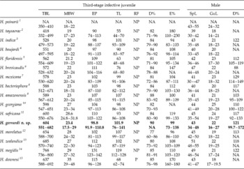
Morphological and molecular evidence suggest that the new Heterorhabditis species is related to species in the indica-group, which currently comprises six species, namely: H. indica Poinar, 1992, H. amazonensis Anadaló et al., 2007, H. baujardi Phan et al., Reference Phan, Subbotin, Nguyen and Moens2003, H. floridensis Nguyen et al., Reference Nguyen, Gozel, Koppenhofer and Adams2006, H. mexicana Nguyen et al., Reference Nguyen, Shapiro-Ilan, Stuart, McCoy, James and Adams2004 and H. taysearae Shamseldean et al., Reference Shamseldean, Abou El-Sooud, Abd-Elgawad and Saleh1996. However, H. gerrardi n. sp. can be differentiated from these species by a combination of morphological and/or morphometric traits of adult and third-stage juvenile stages (table 2). For example, third-stage infective juveniles of H. gerrardi n. sp. have a larger body size (average: 604 μm) than any of the species in the indica-group. Similarly the tail (average: 107 μm) of the third-stage infective juveniles is longer than in other species of the indica-group. Except for H. amazonenis, the excretory pore of the new species is more posteriorly located than in other species members of this clade. As in most Heterorhabditis spp., males of H. gerrardi n. sp. have a peloderan bursa usually supported by nine pairs of bursal rays which are arranged according to the formula: 1 2 3 () 3. However, a reduction of this formula, usually affecting the terminal set of three papillae, with symmetrical or asymmetrical loss of one or two pairs, is characteristic in the new species. Reduction of bursal rays has also been observed in other members of the indica-group, including H. indica, H. amazonensis, H. floridensis, H. georgiana Nguyen et al., Reference Nguyen, Shapiro-Ilan and Mbata2008 and H. mexicana, but usually affecting the terminal set of papillae and not pairs 4–6. Hermaphrodites of H. gerrardi n. sp. can be distinguished from other species in the indica-group by having the vulval opening more anteriorly located (V% average: 43), and with non-protruding or slightly protruding lips. Hermaphrodites of the new species can be separated from H. baujardi, H. floridensis, H. taysearae and H. indica, by having a longer tail (average: 106 μm).
Comparison of morphometric traits (range and mean) of infective juveniles of Heterorhabditis gerrardi n. sp. (in bold) with other Heterorhabditis spp. All measurements in micrometres.

After: 1Kakulia & Mikaia, Reference Kakulia and Mikaia1997; 2Shamseldean et al., Reference Shamseldean, Abou El-Sooud, Abd-Elgawad and Saleh1996; 3Poinar et al., Reference Poinar, Karunakar and David1992; 4Phan et al., Reference Phan, Subbotin, Nguyen and Moens2003; 5Nguyen et al., Reference Nguyen, Gozel, Koppenhofer and Adams2006; 6Liu, Reference Liu1994; 7Nguyen et al., Reference Nguyen, Shapiro-Ilan, Stuart, McCoy, James and Adams2004; 8Poinar, Reference Poinar1976; 9Andaló et al., Reference Andaló, Nguyen and Moino2007; 10Nguyen et al., Reference Nguyen, Shapiro-Ilan and Mbata2008; 11Malan et al., Reference Malan, Nguyen, De Waal and Tiedt2008; 12Liu & Berry, Reference Liu and Berry1996; 13Poinar, Reference Poinar, Gaugler and Kaya1990; 14Poinar et al., Reference Poinar, Jackson and Klein1987; 15Stock et al., Reference Stock, Griffin and Burnell2002.D%, (EP/ES) × 100; E%, (EP/TL) × 100; EP, distance from anterior end to excretory pore; GuL, gubernaculum length; MBW, maximum body width; NA, not available; NP, not present; P, present; RF, tail refractile spine; SpL, spicule length; TBL, total body length; TL, tail length.
The new species can also be separated from other Heterorhabditis species by the size of the infective juveniles, which are much shorter than those of H. zealandica, H. downesi Stock et al., Reference Stock, Griffin and Burnell2002 and H. megidis Poinar et al., Reference Poinar, Jackson and Klein1987, H. gerrardi n. sp. can also be differentiated from H. bacteriophora Poinar, Reference Poinar1976 by the value of D% in the males (average: 121 versus 117) and from H. brevicaudis Liu, Reference Liu1994 by having third-stage infective juveniles with a longer tail (average: 107 versus 76 μm).
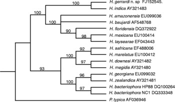
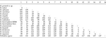
The new species can be further characterized by molecular traits of sequence data from ITS region of ribosomal DNA. Analysis of ITS rDNA sequences yielded three equally parsimonious trees with a tree length of 714 steps and a consistency index (CI), excluding uninformative characters, of 0.83. A total of 321 parsimony informative traits were depicted for this analysis. ITS analysis placed H. gerrardi n. sp. (GenBank accession no. FJ152545) as a member of the indica-clade and as sister taxon of H. indica. Bootstrap re-sampling provided strong support for this topology (100%) and the 50% majority rule consensus tree is given (fig. 4). Results from this analysis indicate H. gerradi n. sp. differs from H. indica by 8 base pairs out of 321 compared base pairs (table 3). These molecular observations provide further evidence for the distinctiveness of this species.
Evidence of internal transcribed spacer region (ITS) of ribosomal DNA lineage independence for Heterorhabditis gerrardi n. sp. based on maximum parsimony analysis. Numbers in bold indicate bootstrap values. Numbers after species names correspond to GenBank accession numbers.

Adjusted character distance matrix based on ITS rDNA sequence comparison between Heterorhabditis spp.

Bionomics
Heterorhabditis gerrardi n. sp. was found in sandy soils in Kingscliff beach, Australia. This new species behaves as other Heterorhabditis spp., its life cycle is completed after 15 days of initial infection of an insect host. First-generation hermaphrodites are fully mature on days 4–5 after initial infection. Second-generation adults can be recovered on days 7–9 after initial infection. Cadavers infected by this new species do not have the typical brick-red/burgundy colouration. Instead cadavers turn grey with blotchy pink spots. Once infection has progressed, cadavers remain with a greyish-pink colouration.
Isolation and characterization of bacteria
Nematodes were observed to contain a monoculture of non-pigmented bacteria. Cultures incubated at 30°C were weakly bioluminescent while those incubated at 37°C were strongly bioluminescent. MLST analysis confirmed the symbiotic bacteria as identical to bacteria isolated from the infected human wound and from the infected insects, and also identified as the Australian group of Photorhabdus asymbiotica (Gerrard et al., Reference Gerrard, Joyce, Clarke, ffrench-Constant, Nimmo, Looke, Feil, Pearce and Waterfield2006).
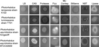
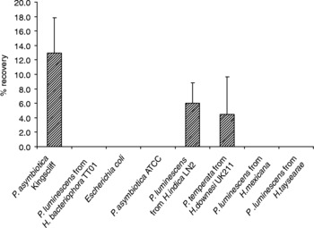
Interestingly, phenotypic tests revealed differences between the human pathogenic strains of this Photorhabdus species. For example, the Kingscliff isolate of P. asymbiotica retained the ability to produce proteases and lipases and is highly bioluminescent when compared to P. asymbiotica USA/ATCC strain, which appears not to produce these factors (table 4, fig. 5). Joyce & Clarke (Reference Joyce and Clarke2003) previously proposed these factors as symbiosis factors that are regulated by the repressor HexA. More interestingly, the USA/ATCC strain produces no detectable siderophores and stilbene production is severely reduced. Both of these factors are required to support normal nematode growth and development (Gerrard et al., 2006; Joyce et al., Reference Joyce, Brachmann, Glazer, Lango, Schwär, Clarke and Bode2008). Indeed P. asymbiotica USA/ATCC did not support nematode growth and development of H. gerrardi n. sp., nor any nematode species when this bacterium was used in growth and development assays. Isolates of P. asymbiotica bacteria from all sources (i.e. nematode, human and insect associated) considered in this study were also tested for their ability to support nematode growth and development in vitro on lipid agar. All three bacterial isolates were capable of supporting recovery and development of the Kingscliff nematodes, and the bacteria were retained by the nematode. Furthermore three members of Photorhabdus luminescens: LN2 (origin H. indica), INAH23 (origin H. indica) and UK211 (origin H. megidis) supported growth and development of the Kingscliff nematode and yielded infective juveniles. However, when nematodes were surface sterilized and individually crushed, bacteria were not retained in any of these nematode species (fig. 6). In similar studies, it has been shown that other Photorhabdus species, such as P. temperata, support growth and development of Heterorhabditis species that naturally harbour P. luminescens; however, bacteria are not retained by non-natural nematode hosts (Joyce, pers. comm.). Furthermore Photorhabdus strains isolated from H. mexicana and H. taysearae (both members of the ‘indica-group’), and Photorhabdus luminescens HP88 and TT01 strains, were toxic to H. gerrardi n. sp., causing death of IJs.
Phenotypic characterization of Photorhabdus asymbiotica isolates recovered from various sources.

CAS, chrome azurol S; LB, Luria broth; EMB, Eosin Methylene Blue (Oxoid); NBT, neutral bromothymol.
Phenotypic characteristics of Photorhabdus asymbiotica (Kingscliff strain) grown in different culture media. LB, Luria broth; CAS, chrome azurol S; Fizz, catalase test using H2O2; NBT, neutral bromothymol.

Recovery of Heterorhabditis gerrardi n. sp. infective juveniles (IJs) grown in lipid agar in the presence of representatives of other Photorhabdus species and/or strains.