Interruption or atresia of the aortic arch is a rare congenital anomaly, associated with poor prognosis if untreated.Reference Collins-Nakai, Dick, Parisi-Buckley, Fyler and Castaneda1 It is characterized by a loss of continuity between the ascending and descending parts of the thoracic aorta, albeit that on occasions it is possible to find an atretic cord joining the disconnected components. Three types are described according to the site of discontinuity in the aortic arch, and usually described alphabetically.Reference Celoria and Patton2 In most patients with such interruption of the aortic arch, there is a ventricular septal defect, and often other intracardiac anomalies that reduce the flow of blood to the ascending aorta during fetal life, typically lesions producing obstruction of the left ventricular outflow tract.Reference Freedom, Bain, Esplugas, Sische and Rowe3
Diagnosis is usually made within the first days of life, with the patients often presenting in shock concomitant with closure of the arterial duct. Nowadays, such neonates are stabilized with an infusion of prostaglandin E1, and surgical repair is performed within the following days after presentation. We report here a patient presenting somewhat later, in whom the ventricular septum was intact, and the descending aorta was supplied exclusively by a voluminous collateral artery arising from the right subclavian artery.
Case report
An infant, aged 2 months, was admitted to our hospital for mild tachypnoea and absence of the femoral pulses. He had been born at full-term after an uneventful pregnancy, albeit that his had received valproate on a chronic basis as a treatment of epilepsy throughout the pregnancy. On admission, he weighed 4.9 kilograms. Physical examination revealed a systolic murmur radiating to the back. Blood pressure was 145 over 95 millimetres of mercury in the right arm, 110 over 70 in the left arm while the pulses in the legs were absent. There was no sign of cardiac failure. The electrocardiogram was normal.
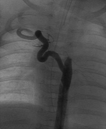
Echocardiography demonstrated concentric left ventricular hypertrophy. There was no ventricular septal defect, nor any other intracardiac anomaly. The ascending aorta was of normal sized, and the aortic arch was left-sided. The aortic isthmus, however, was not seen, and no antegrade flow was revealed by colour-Doppler. We proceeded to cardiac catheterization, which showed systolic pressure in the descending aorta of 35 millimetres of mercury, whereas non-invasive pressures in the right arm had been measured at 140 over 90. The catheter could not be advanced to the descending aorta. Opacification at the expected level of the isthmus showed retrograde filling of a big collateral artery arising from right subclavian artery (Fig. 1). Catheterization in the right heart showed pressures in the right ventricle, and in the pulmonary arteries, within normal limits. A pulmonary angiogram revealed, during the laevophase, interruption of the aortic arch between the left common carotid and subclavian arteries, but the ascending aorta and proximal arch were not precisely imaged. The interatrial septum was intact, and could not be crossed.
Selective angiogram of the collateral vessel arising from the right subclavian artery. Only the descending aorta is opacified.

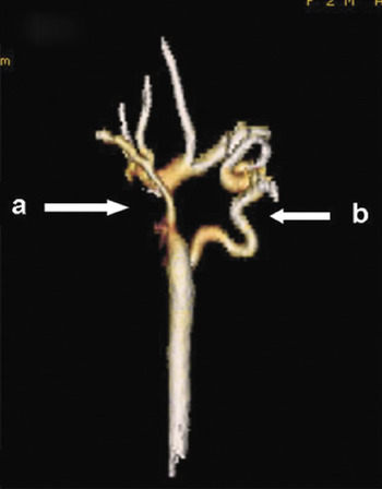
A multi slice computed angiography was performed more accurately to define the structure of the proximal aortic arch. This confirmed interruption of the aortic arch, with a loss of luminal continuity of 18 millimetres between the ascending and descending parts of the aorta, the diameters of the ascending and descending parts being 8, and 7, millimetres, respectively. The flow to the descending aorta was supplied exclusively by the collateral artery arising from the right subclavian artery (Fig. 2). There was no extracardiac anomaly, and a genetic study revealed a 46 XY karyotype without 22q11 microdeletion.
Multi slice computed angiography: posterior view of the aorta revealing the lack of continuity (a) and the collateral artery down from the right subclavian artery (b).

Surgical repair was performed. The anatomy was typical of so-called type B interruption. The ascending aorta gave rise to the brachiocephalic and left common carotid arteries, and had no continuity with the descending aorta. The descending aorta gave rise to the left subclavian artery, and there was a normally located arterial duct. The aortic isthmus, between the left subclavian artery and the descending aorta, was very narrow. A large collateral artery supplying the descending aorta was found, as described, below the arterial duct. The subclavian artery was divided at its junction with the descending aorta, and the hypoplastic isthmus was enlarged with a patch of bovine pericardium. Aortic continuity was then restored by creating a latero-terminal anastomosis between the left side of the left carotid artery and the reconstructed descending aorta.
The postoperative course was uneventful. The patient was discharged home 2 weeks after surgery and remained on captopril, at 3 milligrams per kilogram per day, for moderate residual systemic hypertension, despite no residual aortic obstruction. At follow-up 2 years after surgery, the child is asymptomatic, albeit still treated with captopril for mild systemic hypertension.
Discussion
Interruption or atresia of the aortic arch occurs in 3 per million births.Reference Collins-Nakai, Dick, Parisi-Buckley, Fyler and Castaneda1 It is the most common consequence of abnormal development of the aortic arch involving the neural crest. So-called “Type B” interruption, between the left common carotid and subclavian arteries, results from an abnormal regression by apoptosis of the fourth arch segment,Reference Bergweff, DeRuiter, Hall, Poelmann and Gittenberger-de Groot4 and is often seen in the setting of 22q11 deletion syndrome.Reference Lewin, Lindsay, Jurecic, Goytia, Towbin and Baldini5 In our patient, the karyotype was normal, and the interruption is possibly linked with fetal exposure to Valproic acid, known to be associated with malformations related to abnormal closure of the neural tube.Reference Meador, Baker and Finnell6
During fetal development in patients with interruption of the aortic arch, the descending aortic flow is supplied by the patent arterial duct. At birth, severe signs of cardiac failure usually occur, concomitant with closure of the duct. If untreated, about nine-tenths of the neonates die at a mean age of 4 days.Reference Collins-Nakai, Dick, Parisi-Buckley, Fyler and Castaneda1 In our case, the patient survived thanks to the development of collateral artery filling the descending aorta. Such rapid development of collateral arteries in a neonate with interrupted aortic arch has been previously reported.Reference Starreveld, van Rossum and Hruda7 When supply to the descending aorta is adequate, survival is possible, and late presentation has been reported in an adult with numerous collateral arteries.Reference Kaulitz, Jonas and Van der Velde8
Echocardiography is the best examination in the critically ill neonate for diagnosis and assessment of interrupted aortic arch.Reference Messner, Reul, Flamm, Gregoric and Opfermann9 In our patent, precise delineation of the collateral supply was mandatory for surgery. Cardiac catheterization provided an angiographic description of the collaterals (Fig. 1), and allowed haemodynamic assessment. The proximal aortic arch, however, could not properly be opacified. Less invasively, multislice computed tomography showed accurately the collateral artery and the entire aortic arch. Magnetic resonance angiography would also have optimally imaged the interrupted aorta as well as collateral arteries,Reference Starreveld, van Rossum and Hruda7, Reference Varghese, Gatzoulis and Mohiaddin10 but this would have required longer sedation in a 2 months-old infant.
Acknowledgement
The authors gratefully acknowledge Dr Guillaume Gorincour who realized the multi slice computed tomography and the post processing.