Proteoglycans are expressed on the cell surface, in intracellular compartments, and in the extracellular matrix (ECM) in mammals. The chemical nature of the glycosaminoglycan (GAG) chains attached to core proteins defines proteoglycans as heparan sulfate proteoglycans (HSPGs), chondroitin sulfate proteoglycans (CSPGs), dermatan sulfate proteoglycans (DSPGs), or keratan sulfate proteoglycans (KSPGs). Some are hybrid proteoglycans, carrying both heparan sulfate (HS) and chondroitin sulfate (CS) chains. Studies on GAGs and proteoglycans date back to 1916 when a medical student trying to isolate a procoagulant molecule from liver extracts unexpectedly isolated a potent anticoagulant. This anticoagulant is now known as heparin, a highly sulfated version of HS. HS was first recognised as a mere contaminant in the heparin preparation, but was later distinguished from heparin by the difference in the extent of sulfation and greater structural variability.
For a long time, biological functions of GAGs and proteoglycans were largely speculative. However, studies during the past several decades have revealed critical biological functions of GAGs and proteoglycans in modulating molecular and cellular interactions pertinent to development and disease, including infection. Accumulating evidence indicates that many viral, bacterial and parasitic pathogens subvert proteoglycans at various stages during the course of infection. This review provides an overview of the major mechanisms whereby pathogens exploit proteoglycans to promote infection, using prototypical examples of each. The review also evaluates the potential implications of proteoglycan-based therapies as novel approaches to prevent, attenuate, halt or reverse the course of infectious diseases.
Primer on proteoglycan biology
Structure
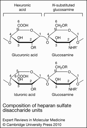
A proteoglycan consists of a core protein and one or several covalently attached GAG chains, which are unbranched polysaccharides composed of repeating disaccharide units. In most proteoglycans, GAGs make up more than 50% of the total molecular mass and mediate the biological functions. GAG biosynthesis is initiated with the formation of a covalent bond between the reducing end of a xylosyl (Xyl) residue and the hydroxyl moiety of certain serine residues within a Ser-Gly dipeptide sequence often repeated two or more times in the core protein. This is followed by formation of the -GlcA-Gal-Gal-Xyl tetrasaccharide linkage domain (where GlcA is glucuronic acid, and Gal is galactose), polymerisation of a characteristic disaccharide unit, and modification of the newly synthesised polysaccharide chain, with each step catalysed by specific enzymes. GAGs are defined by the nature (composition and chemical linkage) of the repeating disaccharide unit, which comprises a hexosamine [e.g. N-acetylglucosamine (GlcNAc), N-acetylgalactosamine (GalNac)] and uronic acid [e.g. GlcA, iduronic acid (IdoA)]. The signature disaccharide unit of HS/heparin is GlcAβ1→4GlcNAcα1→4, CS is GlcAβ1→3GalNAcβ1→4, dermatan sulfate (DS) is IdoAβ1→3GalNAcβ1→4, keratan sulfate (KS) is Galβ1→4GalNAcβ1→3, and hyaluronan (HA) is GlcAβ1→3GlcNAcβ1→4. The chemical structure of HS is shown in Figure 1. Except for HA, GAGs are modified in the Golgi complex by several sulfation and epimerisation reactions. Because the polymerisation and modification reactions do not go to completion, the biosynthetic process generates an exceptionally diverse array of GAG structures.
Composition of heparan sulfate disaccharide units. Heparan sulfate is composed of repeating disaccharide units of hexuronic acid (either glucuronic or iduronic acid) alternating with an N-substituted (R′ = acetate or sulfate) or unsubstituted (R′ = H) glucosamine. Both N-acetylated and N-sulfated glucosamine are commonly found in mature HS chains, whereas N-unsubstituted glucosamine is not. Glucosamine can also be sulfated at the C-6 and C-3 position, although 3-O-sulfated glucosamine is rare in HS. The hexuronic acids can be sulfated at the C-2 position, and this is a common modification in iduronic acid (2-O-sulfated iduronic acid). In general, the unique sulfation pattern dictates the ligand-binding specificity of HS.

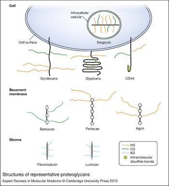
All GAGs, except for HA, are found covalently linked to specific core proteins in vivo as proteoglycans. The spatial and temporal expression patterns of proteoglycans are primarily dictated by the core proteins. Cell-surface proteoglycans include the type I transmembrane syndecans (SDC1–4), NG2 (CSPG4), CD44, betaglycan (TGFBR3) and thrombomodulin (THBD), and glycosylphosphatidylinositol (GPI)-anchored glypicans (GPC1–6) (Refs Reference Bernfield1, Reference Filmus and Selleck2, Reference Hartmann and Maurer3, Reference Park, Reizes and Bernfield4, Reference Taylor and Gallo5). ECM proteoglycans include: perlecan (HSPG2), agrin (AGRN), bamacan (SMC3) and type XVIII collagen in basement membranes; small leucine-rich proteoglycans, such as decorin (DCN), biglycan (BGN), fibromodulin (FMOD), lumican (LUM) and keratocan (KERA); and HA-binding versican (VCAN), aggrecan (ACAN) and brevican (BCAN), among others (Refs Reference Schaefer and Iozzo6, Reference Wight7). Serglycin (SRGN), decorated with highly sulfated heparin and CS chains, is expressed in intracellular vesicles of mast cells (Ref. Reference Kolset and Tveit8). As shown in Figure 2, proteoglycans have distinct structural designs and GAG attachment sites. For example, syndecan core proteins contain an extracellular domain extended in conformation due to a high proline content, whereas glypican extracellular domains are globular due to several intramolecular disulfide bonds formed by the highly conserved cysteine residues. Perlecan structure resembles pearls on a string by rotary shadowing electron microsopy. HS chains are attached distal to the plasma membrane on all syndecans and CS chains are also attached proximal to the cell surface on some syndecans (e.g. syndecan-1). GAG attachment sites in NG2 (CS) and CD44 (HS/CS) are located in the middle portion of the core protein, and those of glypicans (HS), thrombomodulin (CS) and betaglycan (HS/CS) are proximal to the cell surface. HS chains in perlecan are attached at the N-terminus. The cytoplasmic domain of the transmembrane proteoglycans also mediates several important biological functions. The highly conserved, short cytoplasmic domain of syndecans contains several signalling and scaffolding motifs, such as three highly conserved tyrosines and one highly conserved serine, and a C-terminal PDZ-binding domain. The CD44 cytoplasmic domain contains an ezrin–radixin–moesin (ERM) motif that might link surface CD44 to the actin cytoskeleton.
Structures of representative proteoglycans. The structures of several cell-surface (syndecans, glypicans, CD44), intracellular (serglycin), and matrix (bamacan, perlecan, agrin, fibromodulin, lumican) proteoglycans are shown. Serglycin is decorated with highly sulfated HS (i.e. heparin) and CS chains, whereas syndecans and CD44 harbour HS and CS chains. Glypicans, perlecan and agrin contain HS chains, and bamacan contains CS chains. The small leucine-rich proteoglycans fibromodulin and lumican are decorated with KS chains. These proteoglycans interact with various ligands through their glycosaminoglycan chains. Abbreviations: CD, cluster of differentiation; CS, chondroitin sulfate; HS, heparan sulfate; KS, keratan sulfate.

Biological functions and implications for microbial pathogenesis
Proteoglycans, especially those harbouring the structurally diverse HS chains, bind to and regulate a multitude of biological molecules through their GAG chains. The list includes growth factors, cytokines, chemokines, proteinases, antimicrobial factors, ECM components, and many more (Refs Reference Bernfield1, Reference Park, Reizes and Bernfield4, Reference Kreuger9, Reference Rostand and Esko10). Although cell-surface proteoglycans can serve as primary receptors for some ligands, in most cases cell-surface proteoglycans function as coreceptors that capture ligands and facilitate the encounter between ligands and their respective signalling receptors. This also holds for proteoglycan interaction with microbial pathogens, with the majority of HS-binding microbial pathogens using cell-surface HSPGs as coreceptors to facilitate their interaction with secondary internalisation receptors. Proteoglycans can also regulate protein–protein interactions by affecting the stability, conformation and oligomerisation state of either ligand or receptor, and some microbial adhesins and secreted virulence factors subvert these mechanisms to enhance pathogenic activities. Furthermore, proteoglycans can function as soluble molecules because they can be released from the cell surface or ECM by proteolytic cleavage. Once solubilised, proteoglycans show functions similar to or distinct from their immobilised counterparts, and certain bacterial pathogens are known to exploit soluble proteoglycans to inhibit host defence mechanisms. Since GAG fine structures dictate the ligand-binding activities of proteoglycans, regulation of GAG biosynthetic enzymes is also thought to modulate proteoglycan functions; however, it is not known if microbial pathogens possess the means to modulate the expression or function of these host enzymes.
Proteoglycans in microbial pathogenesis and host defence
Proteoglycans in viral attachment and internalisation
Herpes simplex virus
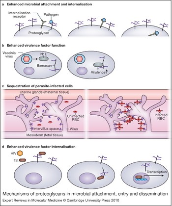
With respect to microbial pathogenesis, proteoglycans were first identified as coreceptors that primarily concentrate pathogens on host cell surfaces, increasing the pathogen's ability to bind to specific secondary receptors (Fig. 3a). Most viruses bind to the HS moiety of cell-surface HSPGs to enhance viral attachment and subsequent internalisation (Refs Reference Shukla and Spear11, Reference Spillmann12). The best-studied example is provided by the alphaherpesviruses herpes simplex virus (HSV) serotypes 1 and 2. HSV glycoproteins gB and gC bind to cell-surface HSPGs and mediate the initial attachment to target host cells (Refs Reference Feyzi13, Reference Laquerre14, Reference Spear15), which are primarily epithelial cells of the skin, cornea, and urogenital system. Although specific HSPGs that mediate HSV attachment have not been identified, the observed target-cell specificity suggests that syndecan-1, the predominant HSPG of epithelial cells, is a likely candidate. Binding to cell-surface HSPGs concentrates HSV virions at the surface and facilitates the interaction of HSV gD with secondary entry receptors, such as herpes virus entry mediator (HVEM/TNFRSF14) and nectin-1 and -2 (PVRL1 and PVRL2) (Refs Reference Feyzi13, Reference Laquerre14, Reference Spear15). Engagement of entry receptors by gD induces fusion of the viral envelope with the host plasma membrane, leading to internalisation of HSV virions. Interestingly, gD can also use a rare 3-O-sulfated form of HS as an entry receptor (Refs Reference O'Donnell16, Reference Shukla17). These findings suggest that cell-surface HSPGs may serve as direct internalisation receptors for HSV in some tissues rich in 3-O-sulfated HS.
Mechanisms of proteoglycans in microbial attachment, entry and dissemination. (a) Enhanced microbial attachment and internalisation. Pathogens use proteoglycans as coreceptors to increase pathogen concentration on the cell surface, facilitating binding to specific secondary receptors. This binding often results in internalisation of the pathogen. (b) Enhanced virulence factor function. Vaccinia virus produces the virulence factor N1L after internalisation. N1L binds to the CSPG bamacan, resulting in improved viral growth in vitro and neurovirulence in vivo. (c) Sequestration of parasite-infected cells. Placental tissue expresses a CS-A (purple), which binds Plasmodium falciparum-infected red blood cells, leading to their sequestration and clinical manifestations including anaemia. (d) Enhanced virulence factor internalisation. HIV Tat binds to cell-surface HSPGs and is then internalised where it can activate transcription. Abbreviations: CS-A, chondroitin sulfate A; CSPG, chondroitin sulfate proteoglycan; HIV, human immunodeficiency virus; HSPG, heparan sulfate proteoglycan; RBC, red blood cell; Tat, transactivator of transcription.

Human immunodeficiency virus
Human immunodeficiency virus (HIV), the aetiologic agent of acquired immune deficiency syndrome (AIDS), binds to cell-surface HSPGs of macrophages, dendritic cells, endothelial cells and epithelial cells through gp120 (Refs Reference Argyris18, Reference Bobardt19, Reference de Witte20, Reference Saphire21). Syndecans are apparently the major cell-surface HSPG receptors because HIV does not attach to K562 cells deficient in syndecans, but does attach when K562 cells are transfected with syndecan-1 (Ref. Reference Saphire21). HIV also shows increased binding to Burkitt lymphoma-derived Namalwa B cells transfected with syndecan-1, -2, -3 or -4 (Ref. Reference Bobardt19). Furthermore, syndecan-3 has been shown to be the major HIV-1 attachment receptor on dendritic cells (Ref. Reference de Witte20). These data suggest that several syndecans expressed in a cell-specific manner may promote HIV attachment. In addition, because target cells of HIV also express other HSPGs, these observations suggest that syndecans harbour HS structures essential for gp120 binding or syndecans are expressed in a cellular niche where gp120 preferentially binds to them. HIV entry receptors include CD4, DC-SIGN (CD209) and mannose receptors, and syndecan-3 has been shown to stabilise the captured virus and enhance viral interaction with DC-SIGN (Ref. Reference de Witte20). Whether syndecans increase the interaction of HIV with other entry receptors remains to be examined. HIV-1 can also bind to the basement membrane HSPG agrin through the envelope glycoprotein gp41 (Ref. Reference Alfsen22). Here, gp41 binds specifically to agrin tethered to the apical epithelial cell surface, reinforcing the interaction of gp41 with its epithelial receptor galactosyl ceramide. Together, these data suggest that HIV possesses multiple mechanisms to bind to HSPGs, and the HSPG interaction is critical for HIV pathogenesis.
Human papillomavirus
Human papillomavirus (HPV) types 11 (Ref. Reference Joyce23), 16, 33 and 39 (Ref. Reference Giroglou24) use cell-surface HSPGs as low-affinity attachment receptors to facilitate their interaction with high-affinity internalisation receptors. HPVs infect skin and mucosal epithelial cells and manipulate the host cell cycle to cause both benign and malignant epithelial tumours. Several studies suggest a mechanism where binding of the major capsid protein L1 to cell-surface HSPGs leads to a conformational change in the minor capsid protein L2, exposing residues in L2 that bind to a secondary internalisation receptor. Consistent with this mechanism, HPV pseudovirions with L1 and L2 have increased infectivity over those with only L1 (Refs Reference Roden25, Reference Unckell, Streeck and Sapp26, Reference Yeager27). In addition, neither anti-L2 neutralising antibody (Refs Reference Roden28, Reference Roden29, Reference Roden30) nor deletion of either the N- or C-terminus of L2 can block initial virion attachment to the host cell surface (Ref. Reference Yang31). Studies using HPV16 demonstrated that the L2 residues responsible for facilitating infection (amino acid residues 13–31) are displayed on the capsid surface only when L1 binds to HSPGs (Refs Reference Kawana32, Reference Kawana33). How this is accomplished is not precisely understood, but a recent study showed that cyclophilin B present on the cell surface in association with HSPGs facilitates conformational changes of L2 (Ref. Reference Bienkowska-Haba, Patel and Sapp34). Whether cell-surface cyclophilin B serves as an internalisation receptor for HPV remains to be determined.
Vaccinia virus
Vaccinia virus, an enveloped, double-stranded DNA virus in the poxvirus family, is able to infect many different cell types. Initial work demonstrated that the vaccinia protein A27L binds to cell-surface HSPGs, and soluble heparin inhibits viral attachment to host cells (Ref. Reference Chung35). Other poxviruses, including cowpox virus, rabbitpox virus, Shope fibroma virus and myxoma virus, also bind to HSPGs, suggesting that HS binding may be a general mechanism of attachment for this group of viruses (Ref. Reference Chung35). Further studies showed that a series of 12 positively charged amino acids in the N-terminal domain of A27L mediate binding to HSPGs, and this interaction allows for subsequent A27L-mediated cell fusion (Ref. Reference Hsiao, Chung and Chang36). Interestingly, vaccinia proteins A34 and B5 have also been shown to mediate virus binding to GAGs on the host cell surface, and these interactions induce nonfusogenic dissolution of the outer viral membrane (Ref. Reference Law37). This is an important mechanism that allows fusion of the inner viral membrane with the plasma membrane and penetration of the virus core into the cytoplasm. Other studies have identified another GAG-binding vaccinia protein, DL8, which binds to CS and has a role in virus entry (Ref. Reference Hsiao, Chung and Chang38). Deletion of DL8 significantly diminished infectivity of virions, confirming the importance of DL8–CS interaction in vaccinia infection (Ref. Reference Hsiao, Chung and Chang38).
In addition, a recent study suggested that binding of vaccinia to the core protein portion of the basement-membrane-associated CS proteoglycan bamacan might be important for its neurovirulence (Ref. Reference Mohan, Zhang and Atreya39) (Fig. 3b). Vaccinia N1L protein binds to the C-terminal 227 amino acids of bamacan (Ref. Reference Mohan, Zhang and Atreya39), whose expression is increased in brain tissues (Ref. Reference Ghiselli, Siracusa and Iozzo40). Increased expression of bamacan in vitro results in improved viral growth (Ref. Reference Mohan, Zhang and Atreya39). Furthermore, N1L, which is produced only after viral infection of a host cell, is responsible for neurovirulence, possibly including neurological complications after vaccination with vaccinia. Thus, N1L binding to bamacan may lead to downstream effects of this virulence factor, such as influencing cytokine (Ref. Reference Zhang41) and ATP levels (Ref. Reference Abrahams42) and inhibiting Toll-like receptor mediated NF-κB signalling (Ref. Reference DiPerna43).
Proteoglycans in bacterial and parasitic attachment and internalisation
Listeria monocytogenes
Several bacterial pathogens similarly bind to the GAG moiety of proteoglycans to promote their attachment and internalisation. Listeria monocytogenes is a Gram-positive, intracellular food-borne pathogen that crosses the intestinal mucosa and enters the systemic circulation where it can induce sepsis and meningitis in immunocompromised hosts. The L. monocytogenes internalin protein A (InlA) binds to E-cadherin (Ref. Reference Mengaud44) and the complex is internalised through caveolin or clathrin-coated pits (Ref. Reference Bonazzi45). In contrast to the monospecific InlA–E-cadherin interaction, internalin B (InlB) is known to bind to three receptors: the receptor for complement factor C1q, the hepatocyte growth factor receptor MET, and cell-surface HSPGs. C1q receptor was identified as an InlB receptor by affinity chromatography (Ref. Reference Braun, Ghebrehiwet and Cossart46). Although the mechanism is not fully understood, C1q receptor mediates the uptake of L. monocytogenes in professional phagocytes. In contrast, the InlB–MET interaction mediates bacterial internalisation in hepatocytes by inducing the mono-ubiquitination of MET and subsequent clathrin-dependent endocytosis (Ref. Reference Veiga and Cossart47). Binding of InlB to cell-surface HSPGs is thought to facilitate internalisation through MET by clustering both InlB and MET at the cell surface (Ref. Reference Jonquieres, Pizarro-Cerda and Cossart48) or by stabilising the InlB–MET complex during bacterial internalisation (Ref. Reference Banerjee49). Specific HSPGs that mediate these processes have not been identified.
Neisseria gonorrhoeae
In the case of the Gram-negative bacterium Neisseria gonorrhoeae, cell-surface HSPGs appear to serve as both coreceptors and direct internalisation receptors. Along with chlamydial infection, gonorrhoeae is one of the two most common bacterial sexually transmitted diseases in the USA. Available data suggest that binding of N. gonorrhoeae Opa protein to syndecan-1 and -4 is important for pathogenesis as overexpression of syndecan-1 or -4 in HeLa cells increases N. gonorrhoeae infection of these cells (Ref. Reference Freissler50). However, N. gonorrhoeae attaches to but does not invade HeLa cells expressing syndecan-1 or -4 constructs lacking the cytoplasmic domain. Furthermore, a syndecan-4 mutant lacking the dimerisation motif in the cytoplasmic domain that binds to protein kinase C (PKC) and phosphatidylinositol 4,5-bisphosphate (PtdIns(4,5)P 2), and a syndecan-4 mutant lacking the invariant C-terminal Glu-Phe-Tyr-Ala PDZ-binding domain does not support gonococcal invasion (Ref. Reference Freissler50). These findings suggest that both syndecan-1 and -4 can serve as direct internalisation receptors for N. gonorrhoeae and that intracellular signalling mediated by the cytoplasmic domain of syndecans is essential. Consistent with this mechanism, N. gonorrhoeae binding to cell-surface HSPGs induces the generation of phosphatidylcholine-specific phospholipase C, diacylglycerol, acidic sphingomyelinase, and ceramide (Ref. Reference Grassmé51). However, how engagement of the HS moiety of syndecans by N. gonorrhoeae leads to core-protein-mediated signalling is not known.
N. gonorrhoeae can also utilise cell-surface HSPGs as coreceptors to facilitate fibronectin-mediated internalisation in HEp-2 human laryngeal carcinoma cells (Ref. Reference van Putten, Duensing and Cole52). Here, gonococci use HSPGs to bind to fibronectin, and use fibronectin as a molecular bridge to bind to β1 integrins, which then mediate N. gonorrhoeae internalisation. Consistent with this mechanism, RGD-containing peptides, fibronectin fragments, and blocking antibodies against α5β1 integrin inhibit fibronectin-mediated uptake of MS11-OpaA, and fibronectin is unable to enhance gonococcal entry into cells that have been treated with heparinase III (Ref. Reference van Putten, Duensing and Cole52).
Borrelia burgdorferi
The spirochaete that causes Lyme disease, Borrelia burgdorferi, expresses two adhesins that bind the DSPG decorin (Ref. Reference Guo53). The physiological significance of this interaction is underscored by the observation that mice deficient in decorin resist Borrelia-induced arthritis, but are as susceptible as wild-type mice to Borrelia infection of other sites including heart and bladder (Ref. Reference Brown54). Longer-term studies suggest that in decorin-rich tissues, such as joint and skin, Borrelia decorin-binding protein A (DbpA) expression and decorin binding may prevent immune-mediated clearance of spirochaetes (Ref. Reference Liang55). The same protection from immune clearance was not seen in tissues with low levels of decorin, such as heart and bladder (Ref. Reference Liang55).
Parasites
Several parasites also bind to proteoglycans to promote their initial attachment and entry. Examples include Plasmodium spp. (Ref. Reference Pancake56), Leishmania spp. (Ref. Reference Love, Esko and Mosser57), and Trypanosoma cruzi (Ref. Reference Ortega-Barria and Pereira58). Plasmodium falciparum, the most virulent parasite that causes malaria, is thought to cause end-organ damage by sequestration of infected red blood cells (IRBCs) in the microvasculature (Ref. Reference Sherman, Eda and Winograd59). P. falciparum binds to multiple adhesion molecules on the host cell surface, and this ability allows the pathogen to switch its mode of attachment as the host develops neutralising antibodies to specific adhesins with repeated infection (Ref. Reference Muthusamy60). A unique case arises in pregnant women, in that the placenta expresses a new receptor for IRBCs: chondroitin sulfate A (CS-A) (Ref. Reference Fried and Duffy61). Studies have shown that IRBCs bind to CS-A in fetal placental tissues (Refs Reference Muthusamy60, Reference Muthusamy62). In the absence of a pre-existing host immune response that can block IRBC binding to the CS-A receptor in the placenta, parasites can multiply at will, leading to IRBC sequestration in the placenta and influx of inflammatory cells (Fig. 3c).
Proteoglycans in virulence factor internalisation
Proteoglycans can also serve as receptors for certain secreted virulence factors. Examples include HIV transactivator of transcription (Tat) and dengue virus nonstructural protein-1 (NS1). Tat is a small cationic polypeptide that is released from HIV-infected cells (Ref. Reference Noonan and Albini63). Tat has important roles in the biological effects of HIV on cells lacking CD4. Tat is thought to be a neurotoxin involved in the pathogenesis of AIDS dementia (Ref. Reference Dewhurst, Gelbard and Fine64) and involved in tumourigenesis by inducing the replication of Kaposi-sarcoma-associated herpes virus (Ref. Reference Zeng65). Four classes of receptors are known to interact with Tat: αvβ3 integrin (Ref. Reference Urbinati66); FLT1 and FLK1/KDR (vascular endothelial growth factor receptors) (Ref. Reference Albini67); chemokine receptors CCR2, CCR3 and CXCR4 (Refs Reference Albini68, Reference Xiao69); and HSPGs (Ref. Reference Rusnati and Presta70). Tat binding to HSPGs is necessary for Tat internalisation (Ref. Reference Tyagi71) and subsequent activation of transcription (Fig. 3d). Overproduction of CS cannot compensate for a lack of HS, indicating that Tat specifically binds to HSPGs (Ref. Reference Tyagi71). The interaction between Tat and HSPGs depends on the size, and degree and type of sulfation, of HS (Refs Reference Rusnati72, Reference Rusnati73). As has been described for interactions of HSPG with growth factors, HSPG binding leads to Tat oligomerisation, which facilitates tyrosine kinase receptor dimerisation and signalling (Ref. Reference Rusnati73). HS/heparin also protects Tat from proteolytic degradation (Ref. Reference Chang74). The cell line WiDr, which lacks all HSPGs except perlecan, is permissive for Tat internalisation (Ref. Reference Argyris75), suggesting that perlecan is one of the HSPG receptors.
Dengue virus NS1 is a secreted glycoprotein that accumulates on the plasma membrane of target host cells. Dengue virus is a mosquito-borne RNA virus of the Flaviviridae family. Immune recognition of NS1 on endothelial cells has been proposed as a mechanism for vascular injury and leakage during severe dengue virus infection. NS1 secreted from infected cells has been shown to bind to cell-surface HS and CS-E of uninfected cells (Ref. Reference Avirutnan76). Consistent with these data, studies with CHO cells established that specific HS and CS structures modified by 2-O- and 3-O-sulfotransferases promote NS1 binding to cell surfaces (Ref. Reference Avirutnan76). Interestingly, NS1 binds preferentially to cultured human microvascular endothelial cells over aortic or umbilical cord vein endothelial cells (Ref. Reference Avirutnan76). Whether this is due to specific HS or CS structures awaits precise comparison of GAG structures on endothelial cells from different tissues.
Proteoglycans in microbial dissemination
The final step of infection where proteoglycans are known to play a role is dissemination. A key role for HSPGs in HIV-1 dissemination has been identified. Dendritic cells are the first cells to encounter HIV-1 at the mucosal surface during sexual transmission (Ref. Reference Patterson77). Dendritic cells bind HIV (Ref. Reference Trumpfheller78) and transfer it to T cells (Ref. Reference Cameron79), where rapid viral expansion occurs. Syndecan-3 has been identified as a dendritic-cell-specific HSPG receptor for HIV-1 (Ref. Reference de Witte20). Syndecan-3 and DC-SIGN together prolong the infectivity of HIV-1, increase infectivity of dendritic cells in cis, and promote transmission to T cells. Thus, syndecan-3 at mucosal surfaces is important for pathogen transmission to T cells in which the virus can explosively replicate and disseminate. Interestingly, syndecan-2 may also function in the transmission of HIV to T cells (Ref. Reference Bobardt19), suggesting that syndecans possess the HS structure required for this activity. Furthermore, cell-surface HSPGs might influence HIV dissemination by facilitating the interaction of infected lymphoid cells with the endothelium through Tat. Here, HSPGs on lymphoid and endothelial cells simultaneously bind to Tat homodimers, leading to the formation of HSPG–Tat–Tat–HSPG quaternary complexes that physically link lymphoid cells to the endothelium, promoting their extravasation (Ref. Reference Urbinati80).
Mycobacterium tuberculosis, the causative agent of tuberculosis, continues to be a major global pathogen infecting an estimated third of the world's population. In pulmonary tuberculosis, the primary target host cell in early infection is the alveolar macrophage. However, the role of alveolar epithelial cells in disseminated tuberculosis is becoming more evident. The heparin-binding haemagglutinin adhesin (HBHA) has been identified as an epithelial cell adhesin for M. tuberculosis and other pathogenic Mycobacteria (Ref. Reference Menozzi81). Deletion of HBHA decreases the ability of M. tuberculosis to attach to type II alveolar epithelial cells in vitro (Ref. Reference Pethe82). Moreover, studies in a mouse model of intranasal tuberculosis demonstrated that HBHA deletion has no effect on the initial pulmonary colonisation, but HBHA deletion significantly decreases the bacteria's ability to disseminate (Ref. Reference Pethe82). This difference was not seen when wild-type and HBHA-deficient M. tuberculosis strains were injected intravenously into mice, suggesting that HBHA does not affect the ability to colonise or replicate in distant organs. Thus, these findings suggest that HBHA is specifically needed for dissemination of primary pulmonary tuberculosis (Ref. Reference Pethe82). How HBHA mediates dissemination is incompletely understood, but HBHA binding to epithelial HSPGs has been shown to lead to reorganisation of the actin cytoskeleton, leaving the tight junctions between cells intact, and trigger endocytosis (Ref. Reference Menozzi83). The resulting vesicles containing M. tuberculosis are transported across the cytoplasm of the epithelial cell to the basal surface, facilitating dissemination (Ref. Reference Menozzi83).
In addition to InlB, the surface protein ActA of L. monocytogenes binds to cell-surface HSPGs (Ref. Reference Alvarez-Dominguez84). ActA is best known for its capacity to manipulate the actin cytoskeleton to allow bacterial migration within and between host cells (Refs Reference Lasa and Cossart85, Reference Portnoy86, Reference Sheehan87). ActA binding to HSPGs is thought to affect the invasion of epithelial cells, possibly through microvilli (Ref. Reference Suarez88). In support of this, coinstillation of heparin during oral inoculation of mice with L. monocytogenes did not alter the bacterial load in the caecum, but did decrease extraintestinal dissemination (Ref. Reference Henry-Stanley89). Although the mechanism is incompletely understood, the ability of ActA to interact with HSPGs may have important ramifications regarding the tissue specificity and dissemination of listerial infection.
The tissue-specific expression and modification of HS may also have important implications in the tissue tropism and dissemination of P. falciparum. Infected mosquitoes inject P. falciparum sporozoites into the skin of a mammalian host, and the sporozoites travel through the bloodstream to the liver where they invade hepatocytes and are transformed into extraerythrocytic forms. Each extraerythrocytic form releases thousands of merozoites, which enter the blood stream and infect erythrocytes, causing the symptoms of malarial disease, such as anaemia, fever, arthralgia and, in severe cases, coma and death. To disseminate from the skin to the liver, P. falciparum apparently detects the different degree of HS sulfation in tissues that it encounters through circumsporozoite protein (CSP), the major surface protein of circumsporozoites. CSP binds to low-sulfated HSPGs on dermal and endothelial cells, but only invades cells with highly sulfated HSPGs, such as those of hepatocytes, allowing P. falciparum sporozoites to migrate through tissues and specifically infect the liver (Ref. Reference Coppi90).
Mechanisms of proteoglycans in pathogen evasion of host defence
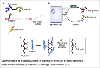
Cationic antimicrobial peptides, including defensins and cathelicidins, are important components of the innate immune response to many pathogens (Ref. Reference Lai and Gallo91). These peptides act by disrupting the lipid membranes of various classes of pathogens including Gram-positive and Gram-negative bacteria, and parasites and fungi (Ref. Reference Lai and Gallo91). Given the highly positive charge of these peptides, it has been hypothesised that pathogens might exploit negatively charged GAGs to neutralise the effect of cationic antimicrobial peptides. Indeed, DS and HS, but not CS, were able to bind to α-defensin and inhibit α-defensin-mediated killing of Pseudomonas aeruginosa, Enterococcus faecalis and Streptococcus pyogenes (Ref. Reference Schmidtchen, Frick and Björck92). Furthermore, P. aeruginosa, E. faecalis and S. pyogenes were found to secrete proteinases that degrade the DSPG decorin and release soluble DS fragments (Ref. Reference Schmidtchen, Frick and Björck92). Together, these data suggest that several Gram-positive and Gram-negative bacteria exploit decorin to promote their survival by secreting proteinases that degrade decorin, releasing soluble DS fragments that inhibit cationic antimicrobial peptides (Fig. 4a).
Mechanisms of proteoglycans in pathogen evasion of host defence. (a) Pathogens including Pseudomonas aeruginosa secrete proteases that cleave decorin, releasing dermatan sulfate. Dermatan sulfate binds to and neutralises cationic antimicrobial peptides such as α-defensin. (b) P. aeruginosa virulence factor LasA usurps the host cell machinery to enhance syndecan-1 shedding into the airspace. Shed syndecan-1 can bind and neutralise antimicrobial peptides such as cathelicidins. (c) Helicobacter pylori binds heparin, enhancing its ability to resist complement-mediated killing by preventing assembly of the membrane attack complex.

Similarly, P. aeruginosa, Staphylococcus aureus and Streptococcus pneumoniae are able to induce the release of syndecan-1 ectodomains from the cell surface, which can inhibit innate immune mechanisms (Refs Reference Chen93, Reference Park94, Reference Park95, Reference Park96). Here, bacterial pathogens secrete virulence factors that stimulate the host cell's shedding mechanism where endogenous metalloproteinases cleave the core protein to release the ectodomain of syndecan-1 as an intact HSPG from the cell surface. Shed syndecan-1 binds to and inhibits antimicrobial peptides, such as cathelicidins and potentially other host defence factors (e.g. collectins), in an HS-dependent manner (Fig. 4b). Consistent with this mechanism, the virulence of P. aeruginosa is significantly reduced in syndecan-1-null mice compared with wild-type mice in models of intranasal lung infection (Ref. Reference Park95) and burn injury infection (Ref. Reference Haynes97). Furthermore, administration of HS or purified shed syndecan-1 increases bacterial virulence in the resistant syndecan-1-null mice, whereas metalloproteinase inhibitors reduce virulence in the susceptible wild-type mice. Together, these findings suggest that subversion of syndecan-1 shedding to inhibit antimicrobial peptides is an important pathogenic mechanism utilised by distinct bacterial pathogens. Interestingly, bacterial pathogens appear to specifically target shed syndecan-1 to promote their pathogenesis. How this is accomplished and whether shed forms of other HSPGs are exploited remain to be determined.
Helicobacter pylori is a spiral-shaped Gram-negative pathogen that colonises the gastric mucosa of approximately half of the human population. H. pylori infection has been associated with diseases including peptic ulcers and some gastric cancers (Ref. Reference Cover and Blaser98). H. pylori binds specifically to heparin and HS even in the presence of pepsin or under low pH conditions (Ref. Reference Ascencio, Fransson and Wadström99). The H. pylori–HSPG interaction has been shown to promote bacterial attachment (Ref. Reference Chmiela100), but other roles for HSPGs have emerged. Heparin/HS enhances the ability of H. pylori to resist phagocytosis in the presence of untreated and heat-treated serum (Ref. Reference Dubreuil101), suggesting that HS interferes with the phagocytosis of H. pylori mediated by the classical and alternative complement pathways (Fig. 4c). This mechanism may partly explain how H. pylori establishes chronic infection because one explanation for the persistence of H. pylori in patients is its ability to resist phagocytosis (Ref. Reference Chmiela, Lelwala-Guruge and Wadstrom102). It remains to be determined whether other bacterial pathogens utilise this HS-dependent mechanism to evade serum-mediated killing mechanisms and cause chronic infections.
Implications of proteoglycan-based antimicrobial therapy
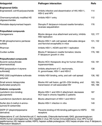
As discussed above, proteoglycans are exploited in a diverse array of pathogenic mechanisms. Moreover, genetically distinct viral, bacterial and parasitic pathogens utilise similar mechanistic approaches to subvert proteoglycans to promote their infection, indicating that subversion of proteoglycans is a broadly used pathogenic strategy. These features suggest that the benefits of developing proteoglycan-based antimicrobial therapies may be great. Several classes of natural and synthetic compounds that interfere with proteoglycan functions have been identified and they include: (1) GAG mimetics that compete with endogenous GAGs; (2) cationic compounds that bind to and inhibit GAGs; (3) inhibitors of GAG biosynthesis; and (4) enzymes that digest the polysaccharide backbone of GAGs or remove sulfate residues of GAGs. Some of these have been tested against proteoglycan–pathogen interactions and have been shown to inhibit pathogenesis both in vitro and in vivo (Table 1).
Proteoglycan-based antagonists of microbial pathogenesis

Abbreviations: E. coli, Escherichia coli; C. trachomatis, Chlamydia trachomatis; GAG, glycosaminoglycan; HCMV, human cytomegalovirus; HCV, hepatitis C virus; HIV, human immunodeficiency virus; HPV, human papillomavirus; HS, heparan sulfate; HSPG, heparan sulfate proteoglycan; HSV, herpes simplex virus; P. falciparum, Plasmodium falciparum.
GAG mimetics
Soluble HS and heparin have been shown to inhibit infection of host cells by HS-binding pathogens, such as HSV (Refs Reference Feyzi13, Reference Laquerre14, Reference Spear15), hepatitis B virus (Refs Reference Schulze, Gripon and Urban103, Reference Leistner, Gruen-Bernhard and Glebe104), HPV (Ref. Reference Giroglou24), L. monocytogenes (Ref. Reference Henry-Stanley89), Staphylococcus spp. (Ref. Reference Henry-Stanley105), Streptococcus spp. (Ref. Reference Henry-Stanley105), and Chlamydia spp. (Refs Reference Davis and Wyrick106, Reference Taraktchoglou107, Reference Yan108), among many others. These findings suggest that along with pharmaceutical heparin, HS and its derivatives are potential candidates for therapy against a wide range of HS-binding pathogens. The major drawbacks of using heparin is that it is a potent anticoagulant that can cause excessive bleeding when misused, and it also has the potential to cause immunopathological manifestations, such as heparin-induced thrombocytopaenia. Relative to heparin, HS is an inefficient anticoagulant and, as a ubiquitous molecule, it is unlikely to be immunogenic. However, because macromolecular HS can bind to and regulate many biological molecules, usage of intact HS as a therapeutic agent could also cause unforeseen adverse effects. These features of HS/heparin have served as a basis for defining the minimum active structure for a given biological function, and this effort has been successful in the development of several HS mimetics specific for a particular function. For instance, based on the finding that induced expression of 3-O-sulfotransferases can convert resistant CHO-K1 cells to susceptibility to HSV-1 entry (Refs Reference O'Donnell16, Reference Xia109, Reference Xu110, Reference Tiwari111), a heparin octasaccharide modified with 3-O-sulfotransferases that inhibits HSV-1 entry was successfully synthesised (Ref. Reference Copeland112). These data suggest that GAG mimetics with selective antimicrobial activities can be synthesised in vitro with a chemoenzymatic approach.
In addition to synthesising defined HS mimetics from HS oligosaccharides and polysaccharides, attempts have been made to synthesise structurally defined K5, a nonsulfated capsular polysaccharide of Escherichia coli with the same structure as that of the HS/heparin precursor N-acetyl heparosan (Ref. Reference Vann113). K5 synthesis can be controlled and modified to enhance binding specificity to a particular ligand. A variety of K5-derived compounds have been studied with regards to their ability to inhibit cellular infection by viruses including HIV, HSV-1 and -2, and HPV (Ref. Reference Rusnati114). Highly sulfated K5 derivatives, with a pI higher than that of heparin, inhibited HIV infection and replication in host cells in culture (Refs Reference Cicala115, Reference Pacciarini116). Three K5 derivatives have also been shown to inhibit HIV Tat binding to cell-surface HSPGs and the subsequent internalisation and transcription activation (Ref. Reference Urbinati117). Further, N- and O-sulfated K5 and O-sulfated and epimerised K5 inhibited infection of Vero cells by HSV-1 and -2 by impairing the attachment and entry into cells and by reducing cell–cell spread of virus particles (Ref. Reference Pinna118). In addition, N- and O-sulfated K5 and highly O-sulfated K5 inhibited infection of host cells by HPV-6, -16 and -18 (Ref. Reference Lembo119). Importantly, all of these K5 derivatives inhibited cellular infection at concentrations that were not cytototoxic to host cells. These data suggest that specifically engineered K5 derivatives are promising therapeutic agents against a broad range of diseases caused by HS-binding pathogens.
Other polysulfated and polysulfonated GAG mimetics also bind to GAG-binding sites in proteins and can function as antagonists of proteoglycan–pathogen interactions (Table 1). The polysulfated compounds with antimicrobial activity include carrageenans (Refs Reference Baba120, Reference Buck121, Reference Talarico and Damonte122, Reference Utt and Wadstrom123, Reference Adams124), fucoidan (Refs Reference Baba120, Reference Romanos125, Reference Fallgren, Andersson and Ljungh126, Reference Ortega-Barria and Boothroyd127), dextran sulfate (Refs Reference Baba120, Reference Fallgren, Andersson and Ljungh126, Reference Ying128, Reference Christensen129, Reference Adams130), phosphomanno pentaose sulfate (PI-88) (Refs Reference Adams130, Reference Nyberg131, Reference Lee132), and pentosan polysulfate (PPS) (Refs Reference Baba120, Reference Fallgren, Andersson and Ljungh126, Reference Adams130). The polysulfonated compounds with antimicrobial activity include suramin (Refs Reference Schulze, Gripon and Urban103, Reference Aguilar, Rice and Wagner133, Reference Clanton134, Reference Fleck135) and poly(sodium 4-styrene sulfonate) (PSS) (Refs Reference Christensen129, Reference Cheshenko136, Reference Taylor137, Reference Herold138). In fact, suramin is the first-line therapy for human African trypanosomiasis, a potentially fatal disease caused by the parasite Trypanosoma brucei. However, the efficacy and toxicity in vivo are not known for most of these compounds. The polysulfated and polysulfonated compounds are thought to interfere with GAG interactions that are required for attachment to host cells and/or entry. Interestingly, some of these compounds may possess additional antimicrobial activities. PI-88 has different effects on infection in vitro than larger compounds such as unfractionated heparin. Heparin-like compounds are known to inhibit HSV-1 and -2 infection by blocking attachment to host cells, but have limited activity against cell–cell spread of infection (Ref. Reference Vaheri139); in contrast, PI-88 has weak activity against preventing HSV-1 or -2 attachment, but can significantly inhibit viral spread from cell to cell (Ref. Reference Nyberg131). Furthermore, in mouse models of dengue virus and flaviviral encephalitis, PI-88, but not suramin or PPS, significantly improved disease outcome (Ref. Reference Lee132), suggesting that in vitro activity does not always predict in vivo therapeutic success. PI-88 is currently under clinical trials in cancer therapy, and results from Phase I studies identified thrombocytopaenia and pulmonary embolism as dose-limiting side effects (Ref. Reference Basche140).
Cationic compounds
Another approach in inhibiting proteoglycan–pathogen interactions is usage of cationic peptides and proteins that bind to the highly negative GAG chains. Examples include lactoferrin, protamine, and Arg- and Lys-rich peptides such as synthetic polylysine and natural cationic antimicrobial peptides. Lactoferrin, an iron-binding glycoprotein of the transferrin family (Ref. Reference Metz-Boutigue141), has multiple cationic domains in its N-terminus, through which it can bind to GAGs (Ref. Reference Wu, Monroe and Church142). Trypsin digestion of lactoferrin yields a cleavage product consisting of the cationic N-terminus of lactoferrin called lactoferricin (Ref. Reference Tomita143). Lactoferricin shares many of the same antiviral properties of lactoferrin. Both were examined for their ability to inhibit HSV-1 and -2 infection and shown to prevent the initial attachment in an HSPG-dependent manner (Ref. Reference Andersen144). In addition, lactoferricin had an inhibitory effect on later stages of infection by a mechanism that is currently unknown (Ref. Reference Andersen144). Initial human studies using lactoferrin in the treatment of hepatitis C infection were promising, with one study showing that high-dose bovine lactoferrin (3.6 g/day) decreased hepatitis C virus RNA load in the blood of patients with chronic hepatitis C, although no improvement in transaminases was seen (Ref. Reference Iwasa145). However, in clinical trials to evaluate the efficacy of lactoferrin in combination therapy with interferon α (Ref. Reference Hirashima146) or with interferon α and ribavirin (Ref. Reference Ishibashi147) in patients with chronic hepatitis C infection, no added benefit was seen with lactoferrin.
Protamine is a cationic peptide that binds to and neutralises GAGs, and is used as a neutralising agent of pharmaceutical heparin (Ref. Reference Guidry, Raschke and Morkunas148). Protamine has been shown to inhibit the attachment of HS-binding viruses to cell-surface HSPGs (Ref. Reference Schulze, Gripon and Urban103). Furthermore, protamine can inhibit pathogenesis by inhibiting the anti-host-defence activity of solubilised HSPGs. As described above, certain bacterial pathogens induce the shedding of syndecan-1 and utilise the anti-host-defence activity of shed syndecan-1 to enhance their virulence. Thus, in a mouse model of intranasal P. aeruginosa infection, coinoculation of protamine significantly reduced the bacterial burden in the lung and spleen (Ref. Reference Park95). Although protamine has antibacterial activities at high concentrations, growth and viability of P. aeruginosa were not affected by the low-dose protamine tested in this study, suggesting that protamine specifically inhibits P. aeruginosa infection by interfering with the anti-host-defence activities of shed syndecan-1.
The aminoquinuride surfen (bis-2-methyl-4-amino-quinolyl-6-carbamide) is another small-compound antagonist of GAGs. Surfen binds to various GAGs in vitro with a preference for binding heparin then DS then HS then CS, an order indicating its preference for highly negatively charged GAGs (Ref. Reference Schuksz149). Although the mechanism is incompletely understood, it is thought that positively charged aminoquinoline moieties of surfen interact with the negatively charged sulfate and carboxyl groups of GAGs (Ref. Reference Schuksz149). Surfen has been shown to inhibit cell attachment and infection of CHO cells by HSV-1 in vitro, suggesting that it inhibits the HSV–HSPG interaction (Ref. Reference Schuksz149).
Derivatives of dispirotripiperazine (DSTP), such as the N,N'-bisheteryl derivative DSTP 27, are another family of small-compound inhibitors of GAG-binding pathogens. DSTP 27 prevents host-cell attachment of a variety of HS-binding pathogens, including pseudorabiesvirus, HSV-1 and -2, HIV and respiratory syncytial virus (RSV) (Ref. Reference Schmidtke150). The inability of DSTP 27 to inhibit infection with varicella–zoster virus or Epstein–Barr virus – herpes viruses that do not use HSPGs as initial attachment receptors – suggests that DSTP 27 specifically interferes with HSPG interactions (Ref. Reference Schmidtke150). The ability of DSTP 27 to prevent attachment persists even after its removal, suggesting a very high affinity of DSTP 27 for HSPGs. Interestingly, DSTP 27 has been shown to have antiviral activities against HPV when applied several hours before or after infection of cells (Ref. Reference Selinka151). Pretreatment of cells with DSTP 27 prevents infection through binding to cell-surface HSPGs and blocking the initial attachment of HPV. However, when administered postinfection, DSTP 27 appears to inhibit infection by blocking the transfer of HPV to a non-HSPG receptor, which induces virion internalisation through a pathway that does not favour infection (Ref. Reference Selinka151).
Inhibitors of GAG biosynthesis
Several chemical inhibitors of GAG synthesis have been tested for their inhibitory effects on infection of host cells in culture. Xylosides containing various hydrophobic aglycone moieties resemble xylose attached to a proteoglycan core protein. Thus, xylosides, such as β-d-xyloside, can function as false acceptors for the formation of the linkage region and subsequent elongation of GAG chains on proteoglycans, and serve as chemical inhibitors of GAG biosynthesis. Xylosides have been used to study mechanisms of GAG synthesis in vitro (Refs Reference Kanwar152, Reference Galligani153) and in vivo (Refs Reference Sobue154, Reference Gibson, Segen and Audhya155, Reference Rada156), but not in the context of infection in vivo. Because xylosides serve as effective primers for CS and DS, and to a lesser extent for HS, toxicity and lack of specificity are a major hindrance for usage in vivo. However, efforts are being made to synthesise selective xyloside inhibitors of GAG synthesis (Ref. Reference Victor157), which may be tested in in vivo models of infection.
By contrast, chlorate inhibits the extent of GAG sulfation by blocking the formation of the universal sulfate donor PAPS (3′-phosphoadenosine-5′-phosphosulfate). Treatment of host cells with sodium chlorate has been shown to inhibit infection by several viruses (Refs Reference Giroglou24, Reference Schulze, Gripon and Urban103, Reference Nyberg131, Reference Barth158), bacteria (Ref. Reference Baron159) and parasites (Ref. Reference Oliveira160), confirming the importance of GAG sulfation in facilitating microbial pathogenesis. However, because of its strong oxidative properties and inhibitory effects on sulfation of not only GAGs but also of other glycans and tyrosines, sodium chlorate is likely toxic in vivo. In fact, one study found that oral administration of sodium chlorate causes nephrotoxicity through formation of methaemoglobins in rabbits (Ref. Reference Steffen and Wetzel161).
Several studies have indicated that proteoglycan–pathogen interactions are mediated by specific GAG modifications. For example, both dengue virus NS1 (Ref. Reference Avirutnan76) and HSV gD (Refs Reference O'Donnell16, Reference Shukla17) bind to 3-O-sulfated HS, which leads to internalisation of ligand. Thus, inhibition of specific GAG modifications may reduce the toxicity associated with therapeutic approaches targeting GAG biosynthesis. For instance, gene knockdown techniques (e.g. RNA interference) against specific GAG biosynthetic enzymes administered in vivo should, in theory, selectively reduce the content of a particular modification and inhibit key steps of pathogenesis. Several function-perturbing antibodies that bind to specific domains in GAGs have also been generated (Refs Reference Ten Dam162, Reference van Kuppevelt163). However, these selective approaches to inhibit GAG biosynthesis have not yet been tested in vivo in the context of infectious diseases.
Enzymes that digest the polysaccharide backbone or remove sulfate residues of GAGs
Enzymes such as bacterial heparinases and mammalian heparanases that digest the polysaccharide backbone of HS, and endosulfatases that remove sulfate residues from GAGs, are potential candidates for proteoglycan-based antimicrobial therapeutics. Flavobacterium heparinases are HS lyases that selectively digest sulfated HS domains (heparinase I), low sulfated domains (heparinase III), or both domains (heparinase II) (Ref. Reference Liu164). In vitro studies showed that treatment of host cells with heparinases inhibits attachment or entry by several HS-binding viral (Refs Reference de Witte20, Reference Giroglou24, Reference Leistner, Gruen-Bernhard and Glebe104, Reference Tiwari111), bacterial (Refs Reference Alvarez-Dominguez84, Reference de Vries165, Reference Moelleken and Hegemann166) and parasitic (Ref. Reference Rathore167) pathogens. In vivo studies of heparinases in infectious diseases are limited, but topical administration of heparinase III prior to genital HPV-16, -31 or -5 pseudovirus infection in mice has been shown to inhibit infection by at least 89% (Ref. Reference Johnson168), suggesting the in vivo feasibility of heparinase therapy.
Mammalian heparanase is an endoglycosidase that degrades HS. Heparanases have been implicated in a variety of pathologies, such as cancer and inflammatory diseases (Refs Reference Reiland169, Reference van den Hoven170, Reference Yang171). Endosulfatases edit the sulfated HS structures by removing a subset of 6-O-sulfate groups within the highly sulfated HS domains (Refs Reference Ai172, Reference Morimoto-Tomita173). By doing so, they regulate the capacity of HSPGs to bind to HS-binding molecules, such as fibroblast growth factor 2 (Ref. Reference Wang174) and hepatocyte growth factor (Ref. Reference Lai175). In light of the inhibitory effects of bacterial heparinases and the requirement of particular sulfate modifications in microbial attachment and entry, it is plausible that heparanases and endosulfatases can also inhibit infection caused by HS-binding pathogens. However, the feasibility of these enzymes as antimicrobial agents has yet to be studied both in vitro and in vivo.
Summary
Proteoglycan–pathogen interactions illustrate the multiple roads taken by pathogens to establish a successful infection. Proteoglycans influence key processes at several steps of pathogenesis, such as adhesion to and invasion of host cells, dissemination into the systemic circulation, and evasion of host defence mechanisms. Given these central roles, it is not surprising that proteoglycans are subverted by a wide variety of viral, bacterial and parasitic pathogens. Studies during the past several decades have revealed that pathogens primarily exploit endogenous functions of proteoglycans, such as serving as a cell-surface coreceptor, enhancing receptor–ligand interactions by inducing a conformational change of ligands or receptors, and mediating the internalisation of ligands. However, proteoglycan roles in transmitting ligands to neighbouring cells (e.g. HIV), mediating dissemination and invasion of distant tissues (e.g. P. falciparum), and inhibiting host defence mechanisms as soluble proteoglycans (e.g. shed syndecan-1) were unexpected. It will be interesting to examine if these previously unknown functions of proteoglycans are important in endogenous processes, such as tumour cell metastasis and protection from inflammatory tissue damage. The subversion of endogenous proteoglycan functions, along with the fact that essential mammalian genes do not evolve as rapidly as those of microbes, suggest that pathogens will not rapidly develop resistance to therapeutic agents that target proteoglycan functions. Thus, in theory, the opportunities to develop antimicrobial agents that are directed against critical propathogenic functions of proteoglycans are great. However, care must be taken in this approach because interfering with a physiological molecule and its mechanisms can potentially have dire consequences. Additional studies directed at defining the key features of proteoglycan–pathogen interactions and evaluating the in vivo efficacy of proteoglycan antagonists should provide a foundation for the development of novel proteoglycan-based antimicrobial therapies.
Acknowledgements and funding
We thank the peer reviewers for their helpful comments. We apologise to many authors for not citing work that may have been relevant, because of space limitations. Our research described in this review was supported by NIH grants HL81474 and HL94613 (P.W.P.), and RR17665 (A.H.B.).