INTRODUCTION
Barnacles (Crustacea: Cirripedia: Thoracica) are among the most widespread and diverse marine crustaceans. They differ from other crustaceans in being irreversibly sessile (non-mobile) as adults, a habit that has been accompanied by dramatic changes in both morphology and biology. These animals have dispensed with the normal crustacean scheme of growing by moulting of the entire exoskeleton. Instead, they are enclosed in armour of mineralized shell plates. They are thoracopodal filter feeders and the majority are hermaphrodites (Anderson, Reference Anderson1994). These drastic adaptations make barnacles excellent model organisms for evolutionary studies (Schram & Høeg, Reference Schram, Høeg, Schram and Høeg1995).
The Azorean cirripedes were first reported by Drouët (Reference Drouët1858) who made the first descriptions. Some decades later, Barrois (Reference Barrois1896) made a collection of exemplars collected at Vila Franca and Mosteiros in São Miguel Island. Pilsbry (Reference Pilsbry1916) included some Azorean barnacles in his monograph on Cirripedia as in the collections of the US National Museum. Gruvel (Reference Gruvel1920) published an exhaustive study about the barnacles sampled in the Príncipe do Mónaco Expedition as did Henry & McLaughlin (Reference Henry and McLaughlin1986). Recently, Southward (Reference Southward1998) increased the number of known Azorean Cirripedia. Presently, there are 57 species recorded from the Azores, most of them from deep waters, contrasting with the low number of intertidal species.
Megabalanus azoricus is well represented in the archipelago, being more abundant in the first 3 m on the low intertidal and infralittoral rocky shores (Morton et al., Reference Morton, Britton and Martins1998; Southward, Reference Southward1998; Young, Reference Young1998). Hawkins et al. (Reference Hawkins, Corte-Real, Pannacciulli, Weber and Bishop2000) described the habitat of this species and its preference for wave exposed locations. This is the largest and third most common shallow water barnacle of the Azores, where it is heavily exploited for food since the colonization of the islands. Consequently, many stocks are currently overfished (Santos et al., Reference Santos, Hawkins, Monteiro, Alves and Isidro1995; Morton et al., 1998). The enormous impact of direct exploitation of resources on rocky shore communities species is a serious concern as target species usually show declining populations (Thompson et al., Reference Thompson, Crowe and Hawkins2002). Therefore, the huge increase (115%) in exploitation levels of M. azoricus in recent years and its absence in places where it used to be present in large populations, has led to a conservation alert. As a result, this species is currently being proposed for urgent action by the OSPAR Convention (1998) and is considered a threatened species by the WWF (2004). Also, more recently, M. azoricus was included in the 100 species for priority management within Macaronesia (Martin et al., Reference Martin, Arechavaleta, Borges and Faria2008).
Pilsbry (Reference Pilsbry1916) made a first description regarding the hard parts of the barnacles, walls and opercular plates, although based on a reduced number of exemplars. Later, Stubbings (Reference Stubbings1967) focused his work on species from Terceira Island and confirmed the description made by Pilsbry. Newman & Ross (Reference Newman and Ross1976) placed this species in a specific rank and restricted its distribution. Henry & McLaughlin (Reference Henry and McLaughlin1986) explained the need to clarify the wide range of intraspecific variation as varieties or subspecies of Megabalanus tintinnabulum. They examined species from different places, including Santa Helena, which present broader terga with shorter spur than the exemplars from the Azores. The features analysed in this study also included the description of the parietes and opercular plates. Young (Reference Young1998) confirmed the description and added two islands where this species could be found (São Miguel and Santa Maria).
A revised diagnosis and description of M. azoricus is provided in the present paper, in anticipation that this will assist in identification, and thus the development of sustainable management protocols for the species. A more detailed description of this species is necessary and essential since we are dealing with an edible species which can be studied for aquaculture and population dynamic purposes. It is also important to study all the diagnostic features to recognize if that species is endemic for Macaronesia. No descriptions of internal morphology have been provided previously for this species. Thoracic and mouth appendages are important for the identification of M. azoricus.
MATERIALS AND METHODS
The study was carried out at São Miguel Island, Azores, where barnacles were sampled in the subtidal zone (0–3 m) by SCUBA diving in several sites along the island's shore.
The morphometric study focused on 215 exemplars of Megabalanus azoricus. Only complete, fresh (unpreserved) specimens representing the full adult size-range were selected for measurements. The size of the exemplars varied between 3.46 and 22.60 of right tergum width with a mean ± standard deviation of 17.10 ± 0.22. The barnacles were dissected and the pattern and colour of the wall plates, and tergo-scutal flaps were recorded (see Southward & Crisp, Reference Southward and Crisp1963). The opercular plates (tergum and scutum) and soft parts of the body were removed from the parietes.
Measurements of shell as operculum maximum diameter, basal maximum diameter, carinal height, rostral height, width and length of right and left tergum and scutum, were taken to the nearest 0.05 mm using digital callipers. In addition, total fresh weight (soft part plus attached opercular plates), gonad weight and edible mass (whole soft body parts determined as fresh weight minus operculum plates weight) of each individual were determined to the nearest 0.1 mg. The soft parts of the body and the opercular plates were removed and the wet weight of each part was recorded. The weight of the gonads (ovaries and testes) was recorded. The features measured were selected based on other studies with barnacles (e.g. Cancino et al., Reference Cancino, Hernandez, Chong, Otaiza, Iriarte and Aviles1998).
The relationship between dimensions or weights of various organs or body parts (y), and dimensions or weights of the entire body or a particular reference organ or part (x) is commonly described by the allometric power function: y = bx a , where b is the intercept at x = 1, and a is the slope (Huxley, Reference Huxley1932). On logarithmic coordinates, this relationship plots as a straight line in the form: log y = a log x + log b. When values for both x and y refer to a length, area or volume, allometry is indicated by a slope value significantly greater or lower than 1. At a = 1 (isometry), proportionality of parts is constant over the size-range specified in the data (Zar, Reference Zar1984).
RESULTS
SYSTEMATICS
Subclass CIRRIPEDIA, Burmeister 1834
Superorder THORACICA, Darwin Reference Darwin1854
Order SESSILA, Lamark 1818
Sub-order BALANOMORPHA, Pilbry 1916
Family BALANIDAE, Leach 1806
Subfamily MEGABALANIDAE, Newman 1979
Genus Megabalanus, Hoek 1913
Megabalanus azoricus, Pilsbry Reference Pilsbry1916
Pilsby (1916)—Balanus tintinnabulum azoricus Pilsbry, United States National Museum Bulletin 93, 62, Pl. 12, Figure 2, 2A, B.
Stubbings (Reference Stubbings1967)—Balanus (Megabalanus) tintinnabulum azoricus Stubbings, Bulletin of the British Museum (Natural History), Zoology 15, 229–319.
Newman & Ross (Reference Newman and Ross1976)—Megabalanus azoricus Newman & Ross, Memoirs of the San Diego Society of Natural History 9, 1–108.
DIAGNOSIS
Megabalanus azoricus (Pilsbry, Reference Pilsbry1916) presents a shell with six wall plates, including paired lateral and carinolateral, carina and rostral plate; the younger individual presents a conical form in juveniles to cylindrical/columnar in the larger specimens.
Parietes present one row of tubes; primary denticles of sutural edges of radii with regular secondary denticles regularly disposed on upper and lower margin of septa. Alae like an internal projection of the parietes being contiguous with the sheath have a small projection covering the alar edge, forming a slot to its attachment. Presence of longitudinal ridge on inner laminae of the radii, holding the ala and not allowing it to attain the sheath border. Plates with spaced and conspicuous growth lines, cuticle persistent only on opercular plates.
The parietes present a strong ribbing, with longitudinal furrows with a smooth and pale pink striped with strong pink. Transverse growth lines with weak longitudinal striae. The opercular aperture is diamond-shaped, with the sides of equal length in the smaller individuals, presenting unequal length in the bigger ones, showing a triangular shape.
The operculum comprises left and right opercular valves with a sand colour. These plates are able to move apart laterally and to close tightly. Each valve is strengthened by two opercular plates, a scutum rostrally and a tergum carinally, locked together to form a single functional unit. The plates are longer than wide and present longitudinal growth lines prominent and well-marked. The opercular valves are attached to the sheath by the chitinous opercular membrane, a circular or oval flap of tissue stretches between the lower part of the sheath deep red with a blue or purple spot on either side.
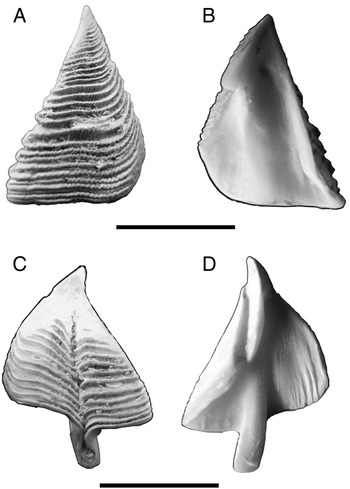
The scutum valve (Figure 1A, B) is triangular, higher than wide, height 1.3 to width. The angle between the 3 sides of the valve is somewhat variable. It is typically about 60°, frequently somewhat greater. Apex is recurved outwards. The extremely narrow tergal apico-basal ribs near the occludent margin which is always straight with alternate growth lines deleted, allowing interlocking with opposing scutum, effectively closing the mantle aperture. Externally the valve is marked by transverse lines, showing strong and distinct growth bands. The pit for the attachment of the adductor muscle is distinct, relatively wide and shallow. There is no clearly defined adductor ridge below the muscle scar. At the basi-tergal angle there is a roughly triangular depression for the insertion of the lateral depressor muscle. There is a deep furrow near the tergal border to receive the inflected margin of the tergum. The inner border of this furrow is produced into a prominent ridge, the articular, which is reflexed or curved over towards the border of the valve. It is lodged in a corresponding groove on the scutal margin of the tergum.
Megabalanus azoricus opercular valves: (A, B) scutum, inner and outer face; (C, D) tergum, inner and outer face. Scale bars: 1 cm.

The tergum (Figure 1C, D) is triangular in shape, almost of the same size of the scutum, and the base of the tergum forms a straight line on both sides of the spur, which is placed at least its own width from the basi-scutal angle. Spur margins infolded: almost closing, furrow and narrow, separated from basi-scutal angle by less than two times their own width, basal margin straight on both sides of spur. Internally, the most preeminent feature is the articular furrow ended on its outer side by the inflected articular margin of the valve. Articular ridge elevated, not curved. Articular ridge 3/4 length of scutal margin. Presence of four poorly developed crests for depressor muscle.
The basal margin forms a calcareous basis, generally tubiferous. The species presents striations on the basis, which form concentric circles. The shell walls and the basis are tightly connected by numerous muscle tissues around the basal perimeter at the inside joints of the leading edges. Cuticle absent.
There are six pairs of biramous stenopodial thoracic appendages or cirri. The captorial fan is formed by Cirri IV–VI, while the first three pairs are specialized maxillipeds. Each cirrus is made up of a protopod surmounted by two rami.
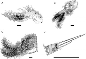
First pair of cirri (Cirrus I) is placed a little apart from the second pair and lies on the prosoma, close to the side of the mouth (Figure 2A). The pedicel is stout and somewhat curved anteriorly. The second joint is very short and irregular in outline as it forms the base for insertion of the two rami. There is a band of long plumose setae on the convex posterior margin of the two segments of the pedicel. The two rami are unequal in length; the longer anterior ramus has an average of 19 segments (Table 1). The first few segments are very indistinctly differentiated. The lower segments are broader than long, the higher ones become slightly longer than broad. There is a transverse row of denticles on the distal border of the inner margin of each segment. The terminal segment bears several moderately long setae. The posterior ramus is shorter and stouter with about 14 segments, formed by protuberant articles. The proximal segments are much broader than long. There is a line of long setae along the posterior border of ramus. Shorter setae occur on the anterior margin and in a row along the distal margin of segments on the adjacent face adjacent to the anterior ramus. There is presence of pectinate and foliate setae. The number of segments in two rami in adult specimens varies from 13:17 to 16:19 in the anterior and posterior rami.
Thoracic appendages: (A) Cirrus I; (B) Cirrus II; (C) Cirrus III; (D) intermediate segment of outer ramus of Cirrus IV. Scale bars: 10 μm.

Number of articles for posterior (Post.) and anterior (Ant.) rami of Cirri I–VI.

The pedicel of Cirrus II is stouter and bigger than that of Cirrus I (Figure 2B). The rami are unequal in length and taper sharply to the distal segment. The proximal segments in both rami are about five times as wide as long. The anterior faces of all segments except the terminal one in each ramus are strongly protuberant, bearing a tuft of long foliate and simple setae.
Cirrus III (Figure 2C) is characterized by the greater degree of development of the pedicel, which is larger than in any of the other cirri. The rami of this cirrus are short, approximately equal in length and resemble Cirrus II. The segments are broader than long, with a group of foliate long setae on the anterior surface. On the posterior surface, each segment bears a tuft of setae which, in some proximal segments of posterior ramus, can extend to the lateral margins of the segments.
The remaining three pairs of cirri are very similar and differ only in the length of the rami. The pedicel is more slender than those in the first three cirri and the second segment is more sharply defined than the first and narrower in anteroposterior direction. As in the preceding cirri the pedicel is slightly flattened laterally (Figure 2D).
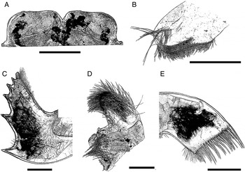
The anterior mouth field is defined by the presence of the labrum (Figure 3A). Megabalanus azoricus presents a simple labrum with median notch. Each side terminates in one big tooth.
Mouth appendages: (A) labrum; (B) mandibular palps; (C) mandibles; (D) maxillula; (E) maxilla. Scale bars: 10 μm.

The palps are attached in the anterior sides of the labrum (Figure 3B). They are paddle-like and large with the margins almost parallel and as thick as wide. At the free end there is a tuft of long flexible setae extending in a line some way down the posterior margin of the appendage. The anterior margin, which projects over the edge of the labrum, bears a long row of setae and there is a tuft of shorter setae on the outer surface of the palp, towards the distal extremity.
Mandibles (Figure 3C) with 5 acute teeth and one last (6th) tooth strongly cuspidate. Lower angle denticulate. There is a long gap between the first and the second teeth of about 1.7 times the distance between the second and the third teeth. There is a row of short setae along the dorsal margin of the mandible above the tooth and a row of rather stouter ones on the ventral margin. A few short setae are scattered over the surface of the appendage, near the free margin.
The maxillula (Figure 3D) is flat and somewhat rectangular in outline with straight anterior border and there is no trace of a notch. The lower proportion does not present projections but has one large spine at apical angle followed by nine moderate spines.
The maxilla (Figure 3E) is bilobed, consisting of an oval main lobe with a protuberant lower lobe roughly semi-circular in outline. The margin towards the mouth is invested with setae, longer at the distal extremity and extending a short way down the margin away from mouth. This appendage presents simple setae in both lobes and pinnate setae on lower margin of distal lobe and upper margin of proximal lobe.
ANATOMY
Megabalanus azoricus is hermaphroditic with simultaneous development of both testis and ovary. The ovaries are composed of multiple spherical lobules consisting of branched ovarian tubules interspersed in a connective tissue stroma. The testes consist of numerous lobed seminiferous tubules lying around the alimentary tube. The basidorsal tubular penis is annulated, longer than Cirrus VI, and covered with setae.
MORPHOMETRY
The morphometric feature with the largest dimension is the rostrum 36.05 ± 0.61 mm high while that with the smallest is the opercular plate with a width of 12.95 ± 0.17 mm. The plates show great similarity in dimensions with a strong relation between length and width (e.g. right tergum width = 0.72*length + 0.84; R2 = 0.89). The relations between the two opercular plates scutum and tergum are also very similar (R2 = 0.87); therefore, we decided to use the right operculum length as a reference measure.
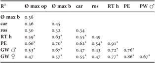
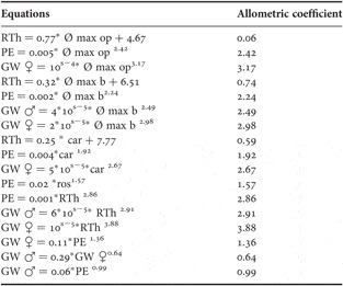
The functions describing the variation between the morphometric features are presented in Table 2. All the relations marked with an asterisk had an allometric coefficient greater than 0.5 and are analysed in Table 3 where allometric coefficients (b) are also presented.
Coefficient of determination (r2) of the calculated functions for the analysed morphometric parameters and weight of Megabalanus azoricus parts: Ø max op, operculum maximum diameter; Ø max b, basal maximum diameter; car, carinal height; ros, rostral height; RTh, right tergum height; PE, edible weight; GW ♂, male gonad weight; GW ♀, female gonad weight.

*, functions with R2 superior to 0.5, relations explored in Table 3.
Relations between the morphometric parameters of Megabalanus azoricus: Ø max op, operculum maximum diameter; Ø max b, basal maximum diameter; car, carinal height; ros, rostral height; RTh, right tergum height; PE, edible weight; GW ♂, male gonad weight; GW ♀, female gonad weight.
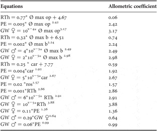
The edible weight shows a strong relationship with all analysed parameters (r2 ≥ 0.50) and the gonad relates strongly to all linear features measured. The strongest correlation obtained was 0.91 between, right tergum height versus edible weight (Table 3), a metric parameter and the biomass.
The allometric coefficients show a positive relationship with all the features measured and analysed.
DISCUSSION
This study confirmed all the previous descriptions performed based on the wall plates and tergum and scutum features. In addition, we add a completed description of the thoracic appendages and internal anatomy of the ovaries and tests. This complete description of M. azoricus for the Azores will allow future comparisons with other exemplars from Madeira, Canarias, and St Helena.
The standard morphometric parameters analysed for M. azoricus displayed positive relations (r 2 > 0.50) between edible mass and gonad weight. Consequently, these measurements can be used in non-destructive studies (e.g. photography) in the future, similar to those carried out by Schmidt-Nielsen (Reference Schmidt-Nielsen1984) and Klingenberg & Monteiro (Reference Klingenberg and Monteiro2005).
The allometric study revealed the existence of an isometric growth of the opercular plates. The female gonad, contrary to expectation, presents a higher ratio (4), which indicates that the growth of female gonad is very large. This fact can be justified by the presence of individuals with mature gonads throughout the year (Dionísio et al., Reference Dionísio, Rodrigues and Costa2007).
Over the years it has become evident that in descriptions more attention needs to be given to barnacle anatomy, particularly the cirral appendages and trophi, collectively called the ‘arthropodal' characters (Southward et al., Reference Southward, Burton, Coles, Dando, Defelice, Hoover, Parnell, Yamaguchi and Newman1998; Southward & Newman, Reference Southward and Newman2003) due to their taxonomic value. These characters, poorly described for Megabalanus azoricus in earlier literature, together with other valuable diagnostic characters such as the wall, opercular opening and opercular plates (Walker, Reference Walker, Harrison and Humes1992; Anderson, Reference Anderson1994) were here included in this ‘re-description’ of M. azoricus. Thus, this work constitutes an important contribution to a more accurate and better definition regarding the taxonomic entity of M. azoricus, essential for future studies of its biology, conservation and management, mainly when it represents an important food resource with great impact on human populations.
ACKNOWLEDGEMENTS
We thank João Brum, Dr Rui Costa, Dr André Amaral, Paulo Melo and Dr Paulo Torres for technical assistance. This research was funded by research project POCI/MAR/58185/2004 and FRCT/2007/M 3.1.2.