


Neurally mediated syncope and epilepsy are common causes of transient loss of consciousness (TLOC) in children and can present with overlapping clinical features, including tonic or clonic movements. Misdiagnosis is therefore frequent and may lead to unnecessary long-term anti-seizure therapy. We describe a 9-year-old girl treated for drug-resistant epilepsy (DRE) for seven years before multidisciplinary reassessment revealed cardio-inhibitory vasovagal syncope with prolonged asystole. This vignette highlights the importance of careful semiological evaluation and reanalysis in children and the value of combined electroencephalographic (EEG) and cardiac monitoring in distinguishing convulsive syncope from epilepsy.
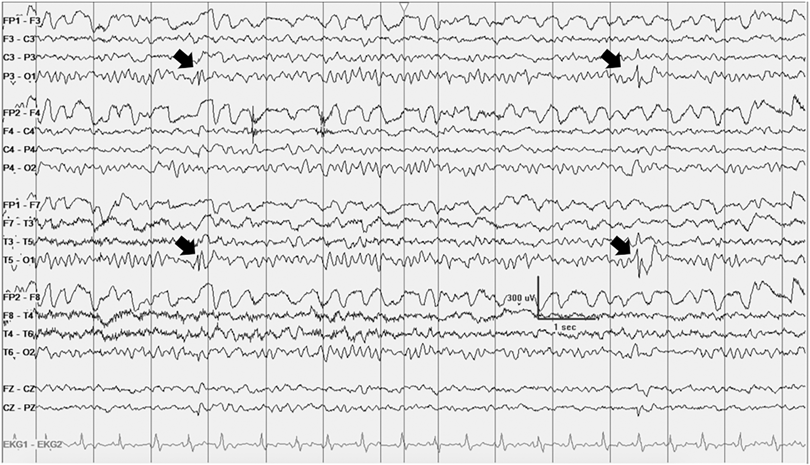
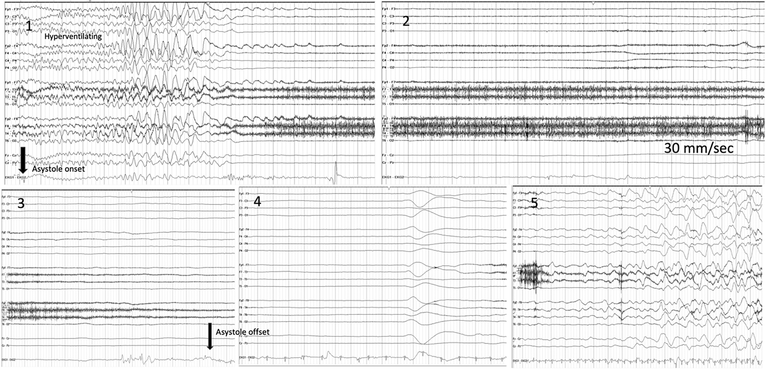
A 9-year-old girl experienced stereotyped episodes from age two: loss of tone, tonic posturing, upward eye deviation, perioral cyanosis, bilateral clonic-like jerking and occasional urinary incontinence, lasting approximately three minutes and followed by postictal confusion, drooling and vomiting. Events occurred on a monthly to bimonthly basis. EEG at age five showed left occipital epileptiform discharges (Figure 1), prompting a diagnosis of focal onset epilepsy and initiation of anti-seizure medication (ASM). By age seven, despite dual anti-seizure therapy (levetiracetam and carbamazepine), seizure frequency remained at approximately one per month. Initial cardiac evaluation, including electrocardiogram (ECG), Holter monitoring and echocardiography, did not identify structural abnormality or arrhythmogenic disorders. During her first Epilepsy Monitoring Unit (EMU) admission, a 20-second asystole was captured during an episode in the washroom and while the EEG findings were obscured by muscle artifact and unsatisfactory, a 20-second asystole was recorded. Ictal asystole (IA) from a probable insular focus was suspected and she was referred for epilepsy surgery evaluation. Insular localization was considered based on the absence of clear interictal findings, the autonomic semiology and the brief duration of events, as well as the often subtle or obscured scalp EEG abnormalities seen in insular epilepsy. MRI Brain and PET were normal. Reevaluation of her semiology, now at an older age, revealed a compelling pattern: most events occurred predominantly while standing and never during sleep or recumbency. Additional triggers included sudden temperature change and micturition. During her second EMU admission, at age 9, simultaneous video-EEG and 12-lead ECG monitoring was performed during IV insertion, with both cardiology and epilepsy teams present. The patient experienced a habitual event immediately upon needle insertion, reporting nausea and dizziness before losing consciousness, followed by generalized stiffening and rhythmic leg jerking. This corresponded to a 50-second cardiac asystole on ECG that self-terminated. Post-asystole EEG showed generalized diffuse slowing seen with cerebral hypoperfusion; the remainder was normal (Figure 2).
EEG recording, bipolar montage demonstrating focal left occipital spikes when patient was at 3 years of age. EEG = electroencephalograph.

Figure 1. Long description
Figure 1 is an EEG recording displayed in bipolar montage from a patient at 3 years of age. The recording demonstrates focal epileptiform discharges, specifically spike-and-wave activity localised to the left occipital region. These findings were interpreted at the time as supporting a diagnosis of focal onset epilepsy and led to initiation of antiseizure medication.
EEG recording in bipolar montage demonstrating the event (peripheral line insertion at the beginning of Epoch 1) followed by hyperventilation and cardiac asystole. Approximately 10 seconds after asystole onset, flattening of electrical brain activity was observed (Epoch 1). Asystole persisted for 50 seconds (Epochs 2–3) until heart rate spontaneously returned to baseline (Epochs 2–4). Return of EEG activity was noted 20 seconds after resolution of asystole (Epoch 5). Patient had normal ECG recording before and after the event. EEG = electroencephalograph.

Figure 2. Long description
Figure 2 is an EEG recording in bipolar montage displayed across five labelled epochs, captured during the patient’s habitual event triggered by peripheral IV insertion at the beginning of Epoch 1. Epoch 1: IV insertion triggers the event; the patient experiences hyperventilation, followed by onset of cardiac asystole. Approximately 10 seconds after asystole onset, flattening of electrical brain activity is observed on EEG. Epochs 2 and 3: Asystole persists for a total of 50 seconds. Epoch 2-4: Heart rate spontaneously returns to baseline. Epoch 5: Return of EEG activity is noted approximately 20 seconds after resolution of asystole, with generalized diffuse slowing seen with cerebral hypoperfusion. The patient’s ECG recording was normal both before and after the event. The overall pattern of asystole preceding EEG slowing is consistent with cardio-inhibitory vasovagal syncope.
After seven years of treatment for presumed DRE, the diagnosis was revised to cardio-inhibitory vasovagal syncope and ASM was discontinued. Pacemaker implantation was discussed but deferred given the self-limiting nature of neurocardiogenic syncope. Education and lifestyle modifications, trigger avoidance and counterpressure maneuvers led to a marked reduction in episode frequency and significant improvement in quality of life. While prolonged reflex asystole may carry significance, management decisions are individualized based on clinical context and event burden.
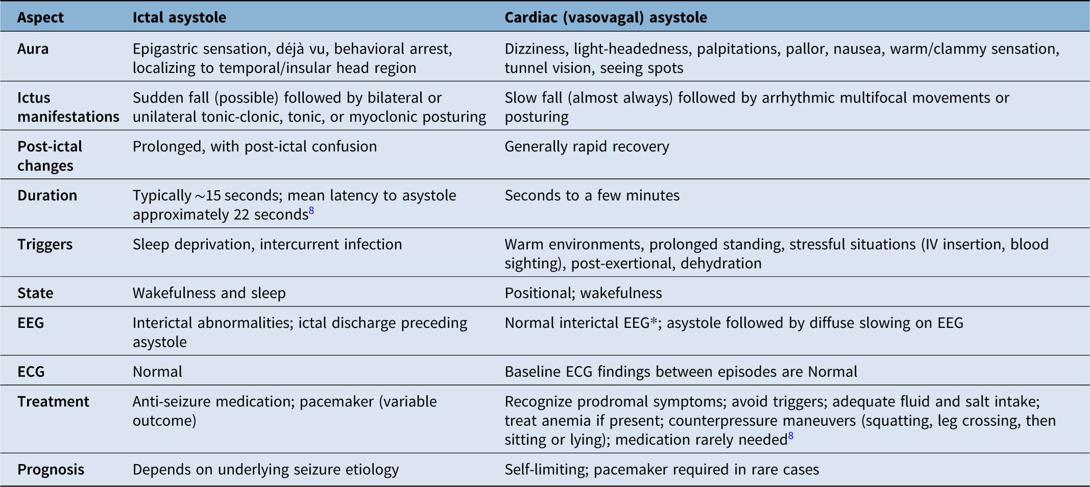
Vasovagal syncope, the most common neurally mediated syncope in children, results from cardiovascular overactivation producing hypotension and/or bradycardia.Reference Brignole, Moya and de Lange1 Up to 30% of patients diagnosed with epilepsy may have an alternative condition, with syncope and cardiac arrhythmias among the most common mimics.Reference Zaidi, Clough, Cooper, Scheepers and Fitzpatrick2 Convulsive syncope, syncope accompanied by motor phenomena due to cerebral hypoperfusion, can closely resemble a focal or generalized seizure. This distinction is particularly challenging when IA is suspected (Table 1). IA occurs in approximately 0.3%–0.4% of patients with epilepsy, reflects ictal autonomic dysregulation and is most often associated with temporal, frontal, or insular foci.Reference van der Lende, Surges, Sander and Thijs3 Critically, in IA the electrographic ictal discharge precedes the cardiac arrest, whereas in cardio-inhibitory syncope, asystole drives secondary EEG slowing.Reference Monté, Monté, Boon and Arends4 In our patient, incidental left occipital epileptiform discharges, benign findings reported in up to 6.5% of healthy children,Reference Borusiak, Zilbauer and Jenke5 may have provided misleading electrographic support for an epilepsy diagnosis.
Key features distinguishing ictal asystole from cardiac (vasovagal) asystole

Table 1. Long description
Table 1 is a two-column comparison table. The left column describes features of ictal asystole; the right column describes cardiac (vasovagal) asystole. Ten row categories are compared: (1) Aura: Ictal asystole presents with epigastric sensation, déjà vu, behavioural arrest, and symptoms localising to the temporal or insular head region. Cardiac asystole presents with dizziness, light-headedness, palpitations, pallor, nausea, warm or clammy sensation, tunnel vision, and seeing spots. (2) Ictus manifestations: Ictal asystole involves a sudden fall (possible), followed by bilateral or unilateral tonic-clonic, tonic, or myoclonic posturing. Cardiac asystole involves a slow fall (almost always), followed by arrhythmic multifocal movements or posturing. (3) Post-ictal changes: Ictal asystole involves prolonged recovery with post-ictal confusion. Cardiac asystole involves generally rapid recovery. (4) Duration: Ictal asystole typically lasts approximately 15 seconds, with a mean latency to asystole of approximately 22 seconds. Cardiac asystole lasts seconds to a few minutes. (5) Triggers: Ictal asystole is triggered by sleep deprivation and intercurrent infection. Cardiac asystole is triggered by warm environments, prolonged standing, stressful situations such as IV insertion or blood sighting, post-exertional states, and dehydration. (6) State: Ictal asystole occurs during wakefulness and sleep. Cardiac asystole is positional and occurs during wakefulness only. (7) EEG: Ictal asystole shows interictal abnormalities with an ictal discharge preceding asystole. Cardiac asystole shows a normal interictal EEG (with a footnote noting this does not exclude incidental electrographic findings), asystole followed by diffuse slowing on EEG. (8) ECG: Both types show a normal ECG. (9) Treatment: Ictal asystole is treated with antiseizure medication and pacemaker, with variable outcomes. Cardiac asystole is managed through recognising prodromal symptoms, avoiding triggers, adequate fluid and salt intake, treating anaemia if present, counterpressure manoeuvres including squatting, leg crossing, then sitting or lying, with medication rarely needed. (10) Prognosis: Ictal asystole prognosis depends on the underlying seizure aetiology. Cardiac asystole is generally self-limiting, with a pacemaker required only in rare cases.
* Does not exclude incidental electrographic findings such as those observed in the present patient.
EEG = electroencephalograph.
In the largest pediatric series of 120 children and adolescents with TLOC, vasovagal syncope accounted for 63.8% of diagnoses.Reference Choi, Han and Lee6 The modified Calgary Score, a validated clinical tool for differentiating neurally mediated syncope from epilepsy in children, demonstrates high sensitivity and specificity.Reference Zou, Wang and Zhu7 While key distinguishing features of ictal vs. cardiac asystole are summarized in Table 1, an important limitation in young children is the difficulty in identifying prodromal symptoms. In pre-verbal children, or those not yet toilet-trained, situational triggers such as micturition syncope may therefore go entirely unrecognized. When both diagnoses remain plausible, simultaneous video-EEG and ECG monitoring is essential in capturing the electroclinical correlate of spontaneous events.Reference Khalil, Shukralla and Kilbride8
This vignette reinforces that diagnostic reconsideration must be an ongoing process, particularly in children with atypical semiology. Careful history reevaluations, multidisciplinary collaboration and simultaneous EEG–ECG were the decisive elements in revisiting this patient’s diagnosis and transforming her care trajectory. Early recognition of syncope masquerading as epilepsy can be difficult in young patients and may lead to years of inappropriate therapy.
Author contributions
MCC: Case identification, literature review, and drafting of the manuscript. MK: Clinical care of the patient, critical revision of the manuscript. LA: Cardiology input and interpretation of cardiac data, critical revision of the manuscript. MNN: Conceptualization, supervision, critical revision and final approval of the manuscript. All authors approved the final version for submission.
Funding statement
MNN has received honorarium and consultation fees from PeriPharm and Jazz Pharmaceuticals. MK has received honoraria for lectures and has served advisory board from Jazz Pharmaceuticals. MCC has received honoraria from UCB Pharma, Livanova, Exeltis Pharma Mexico and Stendhal Pharma and travel support from UCB Pharma. LAD has nothing to declare.
Competing interests
None to report.

 Open access

