Introduction
Vocal fold paralysis can occur at any age, due to a variety of causes. It occurs most commonly following iatrogenic surgical injury, but can also result from: endotracheal intubation; blunt chest or neck trauma; tumours involving the skull base, neck or chest; and viral infection.
The consequences of vocal fold paralysis include: voice changes (ranging from hoarseness, breathy voice, effortful phonation and air wasting to diplophonia); airway problems (including shortness of breath with exertion, stridor, and ineffective or poor cough); and difficulty swallowing (including aspiration).
Vocal fold medialisation procedures have been carried out for many years; different techniques and materials have been used, with variable success, limitations and complications. The main aim of treatment for vocal fold paralysis is to improve voice quality and prevent aspiration. Commonly used injectable materials include fat, collagen, polytetrafluoroethylene (Teflon®), calcium hydroxyapatite, gelation sponge (Gelfoam®) and, more recently, polymethylsiloxane gel (Bioplastique™; Uroplasty BV, Geleen, Netherlands).
Bioplastique is a biphasic material and consists of solid silicone particles (ranging in size from 100 to 400 μm) suspended in a polyvinylpyrrolidone carrier. The material elicits a low-grade inflammatory reaction once injected. Collagen subsequently encapsulates and localises the silicone and, as deposition progresses, it replaces the organic component of the material in a ratio slightly higher than 1:1. The carrier itself is removed by the body and excreted by the kidneys in under four weeks. It has been observed that particles of less than 60 μm can be engulfed by macrophages and transported to regional lymph nodes. The inability of Bioplastique to migrate or to be carried by the lymph to the lymph nodes is based on this theory.
Bioplastique has been used successfully in aesthetic plastic surgical procedures,Reference Ersek, Gregory and Salisbury1 to seal leakage around speech valvesReference Rokade, Mathews and Reddy2 and to treat faecal incontinence (injected into the anal sphincter),Reference Tjandra, Lim, Hiscock and Rajendra3 all with good long term results.
Bioplastique is commonly used in Europe for vocal fold augmentation procedures, and its safety is well documented.Reference Alves, Loughran, MacGregor, Dey and Bowie4, Reference Bihari, Meszaros, Remenyi and Lichtenberger5 A recent study by Hamilton et al. Reference Hamilton, Sachidananda, Carding and Wilson6 has shown that results for Bioplastique, with regards to voice performance, are comparable to those for Isshiki thyroplasty, and that Bioplastique is possibly more suitable for patients with limited life expectancy, as it was quicker to use and had few complications.
Animal studies have shown no evidence of migration or malignant change, and minimal local tissue reaction,Reference Dewan and Byard7, Reference Nijhuis, van den Bogaard, Daemen and Baeten8 thus enhancing the evidence for Bioplastique's efficacy and clinical use.
We report a case of foreign body reaction and presumed migration of injected Bioplastique after vocal fold augmentation, requiring removal. To our knowledge, this is only the second9 reported case both of a clinical foreign body reaction to Bioplastique and of removal through a conventional microflap approach.
Case report
A 66-year-old man with a six-year history of left vocal fold paralysis thought to be secondary to a viral aetiology was referred to our centre. He had previously been treated with four medialisation procedures (three Bioplastique vocal fold injections and a thyroplasty with expanded polytetrafluoroethylene (Gore-Tex®; WL Gore & Associates, Phoenix, Arizona)) at his referring centre, with only transient improvement in his voice.
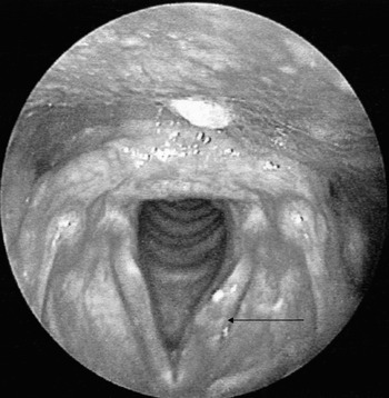
On the patient's first visit, he was observed to have a breathy voice. Stroboscopy revealed fullness in the mid-membranous region of the left vocal fold, which was impeding the mucosal wave (Figure 1). His right vocal fold was mobile and there was also some degree of abduction and adduction of his left vocal fold, and it was thought that the viral process may have resolved to some extent. The patient's voice quality was assessed pre-operatively using the grade–roughness–breathiness–asthenia–strain score, as follows: grade = three, roughness = three, breathiness = two, asthenia = one and strain = two.
Pre-operative appearance showing fullness (arrow) in mid-membranous portion of left vocal fold.

Our clinical suspicion was of misplaced Gore-Tex material. At suspension microlaryngoscopy, however, the fullness was felt more likely to be due to Bioplastique. A biopsy using the microflap technique confirmed foreign material.
Post-operatively, the patient underwent speech and language therapy, with only minimal improvement in his voice.
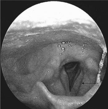
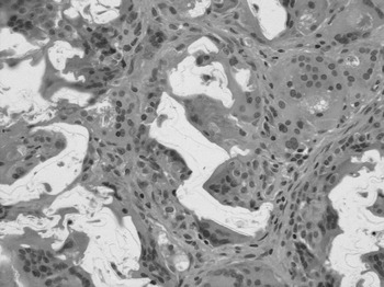
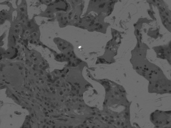
On review after a few months, the fullness was still present in the mid-membranous portion of the patient's left vocal fold, albeit less obvious. He underwent a further suspension microlaryngoscopy, with removal of further foreign body from his left vocal fold via microflap (Figure 2). Histological analysis showed multiple irregular areas of translucent, acellular material surrounded by a prominent foreign-body reaction composed of multinucleated giant cells, histiocytes and fibroblasts (Figure 3). The acellular material was non-birefringent under polarised light (Figure 4), in contrast to the intense birefringence seen with Teflon;Reference Baijens, Speyer, Linssen, Ceulen and Manni10 thus, the histological picture was that of a granulomatous reaction to Bioplastique.Reference Rajkumar, Khalil, Elloy, Sheffield and Baldwin11
Post-operative appearance showing improved left vocal fold appearance (arrow).

Medium power photomicrograph of the left vocal fold biopsy, showing the Bioplastique™ as irregularly shaped, translucent masses surrounded by a multinucleate giant cell reaction (H & E; original magnification ×250).

Medium power photomicrograph of the left vocal fold biopsy taken under polarised light, showing the absence of birefringence, characterising the material as Bioplastique™ (H & E; original magnification ×250).

Post-operative voice assessment showed the following scores: grade = two; roughness = one; breathiness = two; asthenia = one and strain = zero. This represented an overall improvement in the patient's voice quality, and, clinically, he felt that his voice quality had improved.
At the time of writing, the patient was undergoing further speech therapy.
Discussion
The main aim of treatment of vocal fold paralysis is to improve vocal fold closure with concomitant improvement in voice quality and efficacy of cough production. Factors that need to be taken into account when deciding which material to use include the general health and probable lifespan of the patient, degree of compensation, and pulmonary status.
Foreign body reactions and migration caused by injected materials have been described in the literature. Tissue foreign body reaction to Teflon has been well documented;Reference Lewy and Millet12–Reference Boedts, Roels and Kluyskens14 more recently, such reactions have been reported to Radiesse (Bioform, San Mateo, California, USA).Reference Tanna, Zalkind, Glade and Bielamowicz15 There have been reports of migration of Bioplastique after treatment for ureteric refluxReference Dewan, Hoebeke, Ehall, Chow, Edwards and Terlet16 and granuloma formationReference Lackgren, Wahlin and Stenberg17 following aesthetic surgery. However, to our knowledge, the current report represents the second published case of Bioplastique material, used for vocal fold augmentation, migrating and causing a foreign body tissue reaction.
Bioplastique granulomas can be confirmed histologically, as the material exhibits irregularly shaped, cystic spaces of varying size containing jagged, translucent foreign bodies that are non-birefringent under polarised light.Reference Rudolph, Soyer, Schuller-Petrovic and Kerl11 These specific histopathological findings allow the correct diagnosis to be made, despite sparse clinical information.
• Bioplastique™ is commonly used in Europe as an injectable material for medialisation treatment of vocal fold paralysis. Its safety and efficacy as a permanent material are well documented
• This case highlights the fact that foreign body reaction and migration are still possible with this material, albeit rare
• Although this material can cause foreign body reactions and may possibly migrate, it is removable by microlaryngoscopy via the microflap technique, with potential vocal improvement
Clinicians need to be aware of the possibility of Bioplastique migrating and causing foreign body reactions. In our case, it was difficult to be absolutely certain that the Bioplastique material had actually migrated, rather than been injected into the wrong site or undergone inadvertent medialisation due to the Gore-Tex, as the initial surgical procedures were all performed in another centre. However, as there has been evidence of migration at other sites,Reference Dewan, Hoebeke, Ehall, Chow, Edwards and Terlet16 this possibility must be considered.
We also feel that clinicians need to be aware that removal of portions of this material is achievable via microlaryngoscopy and microflap technique, as shown in this case, with reasonable improvement in voice. This differs from the problems encountered while attempting to remove Teflon granulomas endoscopically.Reference Ossoff, Koriwchak, Netterville and Duncavage18
Conclusion
Bioplastique is an injectable material which is commonly used in Europe for medialisation in cases of vocal fold paralysis. Its safety and efficacy as a permanent material are well documented. The current case highlights the fact that foreign body reaction and migration may still be possible for this material, albeit rare. We also highlight the fact that, although this material can cause foreign body reactions and may possibly migrate, it is removable by microlaryngoscopy via the microflap technique, with potential vocal improvement.