Introduction
Vestibular schwannomas, or acoustic neuromas, are benign, slow-growing tumours of the vestibular portion of the vestibulocochlear (VIIIth) cranial nerve.Reference Newton, Shakeel, Flatman, Beattie and Ram 1 Presentation is variable, with patients most commonly presenting with asymmetrical sensorineural hearing loss (SNHL).Reference Obholzer, Rea and Harcourt 2 Tinnitus, vertigo, disequilibrium and facial palsy can also be diagnostic indicators, making vestibular schwannoma a challenge to accurately diagnose clinically.Reference Obholzer, Rea and Harcourt 2 – Reference Selesnick, Jackler and Pitts 4 Radiological assessment is therefore crucial, with gadolinium-enhanced magnetic resonance imaging (MRI) of the internal auditory meatus recognised as the ‘gold standard’ for investigating asymmetrical SNHL.Reference Sidman, Carrasco, Whaley and Pillsbury 5
With such wide-ranging symptomatology for vestibular schwannoma, increasing numbers of MRI scans of the internal auditory meatus are being performed.Reference Vandervelde and Connor 6 This has led to a rise both in the number of scans reported as normal and those with incidental clinical findings.Reference Papanikolaou, Khan and Keogh 7 There is a growing body of literature addressing the occurrence of incidental findings on MRI scans performed both in healthy individuals and in those presenting with suspected vestibular schwannoma.Reference Katzman, Dagher and Patronas 8 , Reference Mirza, Malik, Ahmed, Willatt and Hughes 9
This study aimed to determine the frequency of incidental findings on MRI scans of the internal auditory meatus within our department, and to suggest how to best manage these findings when identified.
Materials and methods
We performed a retrospective review of all acoustic protocol MRI scans performed over a three-month period between May and July 2015 at the Radiology Department of Blackpool Victoria Hospital. All scans, authorised by one of four consultant otorhinolaryngologists, were performed for: asymmetrical SNHL, persistent unilateral tinnitus or other unexplained audiovestibular symptoms.
The standard protocol for the MRI scans was T2-weighted, three-dimensional constructive interference in steady state imaging, with 0.7 mm sections through the internal auditory meatus and 5 mm sections through the brain.
Reports on all scans were reviewed. Exclusion criteria included surveillance scans for known vestibular schwannoma (n = 8) and cases in which a full MRI could not be obtained because of patient factors (n = 1). A total of 109 MRI scans were assessed and included in the study.
Results
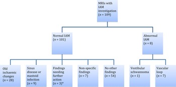
Of the 109 scans, 8 were reported to show an abnormal internal auditory meatus: there was 1 small vestibular schwannoma (0.9 per cent) and 7 vascular loops (6.4 per cent) (Figure 1). The indication for the MRI scan that revealed a vestibular schwannoma was unilateral SNHL. The indications for the MRI scans that revealed vascular loops were: unilateral SNHL (n = 2), tinnitus (n = 3) and vertigo (n = 2).
Various magnetic resonance imaging (MRI) findings in patients with unilateral audiovestibular symptoms. *These were: an empty sella with benign intracranial hypertension, a deep lobe parotid tumour and demyelinating changes. IAM = internal acoustic meatus

A significant proportion (92.7 per cent; n = 101) of the 109 scans were found to have a ‘normal’ internal auditory meatus; no other findings were reported for 54 of them (49.5 per cent). However, 28 scans (25.7 per cent) demonstrated old ischaemic changes, small vessel disease or degenerative changes that did not warrant any further investigations, except for informing these patients’ general practitioners. The age range of patients with these findings was 46–87 years, with a median age of 62 years.
Nine patients (8.3 per cent) were found to have paranasal sinus disease and evidence of fluid in the mastoid air cells. Seven patients (6.4 per cent) had non-specific findings that were of little clinical importance, such as cava septum pellucidum and dilated Virchow–Robin space.
The remaining three patients (2.8 per cent) required further action. One patient was found to have a deep lobe parotid tumour that required surgical intervention, another had a possible empty sella with benign intracranial hypertension that warranted referral to neurosurgeons, and the last patient had demyelinating changes and needed to be reviewed by neurologists.
Discussion
As stated above, MRI scanning is regarded as the gold standard investigation for ruling out internal auditory meatus tumours such as vestibular schwannoma, because of its cost-effectiveness, accuracy and superiority over other audiovestibular investigations such as auditory brainstem response (ABR).Reference Robson, Leighton, Anslow and Milford 10 , Reference Raber, Dort, Sevick and Winkelaar 11 Our study has demonstrated, however, that only a small proportion (0.9 per cent) of scans requested actually demonstrated retrocochlear pathology. This is in line with other studies, which have shown that none of the various clinical criteria that exist to try to increase the pick-up rate of vestibular schwannoma are particularly effective, with none improving beyond a 4 per cent pick-up rate,Reference Newton, Shakeel, Flatman, Beattie and Ram 1 – Reference Dawes and Jeannon 3 , Reference Vandervelde and Connor 6 and an incidental finding is more likely to be reported.
Vascular loops
Vascular loops were detected in 6.4 per cent of patients in our cohort. This conforms with previous literature, with studies by Papanikolaou et al.Reference Papanikolaou, Khan and Keogh 7 and Gupta et al.Reference Gupta, Gupta, Gupta, Gupta, Kalsotra and Sharma 12 showing incidence rates of 1 per cent and 8.66 per cent respectively. A systematic review by Chadha et al. showed an association between vascular loops in contact with the VIIIth cranial nerve, and asymmetrical SNHL and pulsatile tinnitus; however, in clinical practice, these are regarded as normal variants and can be present in up to 33 per cent of the population.Reference Chadha and Weiner 13
If a vascular loop is found to be in contact with the affected VIIIth cranial nerve, with no other causative pathology identified, neurosurgical referral could be considered. Techniques such as decompression of the internal auditory meatus, or insertion of a Silicon or Teflon cushion between the compressing artery and VIIIth cranial nerve, have been described in the literature.Reference Møller, Møller, Jannetta and Jho 14 , Reference Tash, Kier and Chyatte 15 Microvascular decompression is not widely undertaken for audiovestibular symptoms because of its invasive nature. None of the patients in our study were disturbed enough by their symptoms to consider neurosurgical intervention.
Ischaemic white matter changes
The largest proportion of incidental findings in our cohort concerned white matter changes, small vessel disease and previous ischaemic damage (25.7 per cent). Myelin and axonal loss due to small vessel disease or ischaemia is detected as white matter hyperintensities on T2-weighted MRI scans. This is secondary to chronic hypoperfusion of the white matter and disruption of the blood–brain barrier, which leads to chronic leakage of plasma into the white matter.Reference O'Sullivan, Lythgoe, Pereira, Summers, Jarosz and Williams 16
In the Rotterdam Scan Study, a population-based MRI study, 1077 members of the public underwent MRI.Reference De Leeuw, de Groot, Achten, Oudkerk, Ramos and Heijboer 17 The findings showed that the prevalence and degree of white matter change increased significantly with age. The age range of patients with these findings in our study was 46–87 years, with a median age of 62 years. Debette and Markus demonstrated an increased risk of stroke and dementia in patients with white matter hyperintensities on MRI scans.Reference Debette and Markus 18 Otorhinolaryngologists should therefore be prepared to screen these patients for stroke and dementia, or, at least, inform the patients’ general practitioners so that preventative or disease-modifying measures can be implemented.
Paranasal sinus and mastoid disease
Of our patients, 8.3 per cent had paranasal sinus or mastoid disease found on MRI scans. The rate of incidental paranasal sinus disease on T2-weighted MRI scans in the literature ranges from 24.7 per centReference Moser, Panush, Rubin, Honigsberg, Sprayregen and Eisig 19 to 49.2 per cent.Reference Patel, Chavda, Violaris and Pahor 20 These variable rates can be influenced by environmental factors such as scanning in the winter, and patient risk factors such as sex, age, smoking and previous sinus disease, which all increase the incidence of incidental sinus disease.Reference Tarp, Fiirgaard, Christensen, Jensen and Black 21
Demyelinating disease
One of our patients was found to have demyelinating changes and was subsequently referred to the neurology department. In the absence of clinical symptoms and signs suggestive of multiple sclerosis, no further investigations were required.
The term ‘radiologically isolated syndrome’ was first used by Okuda et al.,Reference Okuda, Mowry, Beheshtian, Waubant, Baranzini and Goodin 22 in 2009, to describe patients who fulfilled the Barkhof criteria for the diagnosis of multiple sclerosis based on MRI findings, in the absence of any symptoms or clinical findings suggestive of multiple sclerosis.Reference Barkhof, Filippi, Miller, Scheltens, Campi and Polman 23 These authors found that more than 50 per cent of patients with radiologically isolated syndrome showed radiological progression of demyelination, and 30 per cent went on to be diagnosed with multiple sclerosis.Reference Okuda, Mowry, Beheshtian, Waubant, Baranzini and Goodin 22 Radiologically isolated syndrome can therefore be considered as a precursor for the development of multiple sclerosis. Onward referral to neurologists is highly recommended for these patients, even though they may be free of clinical signs and symptoms at the time of diagnosis.
Anatomical variants
We encountered anatomical variants such as dilated Virchow–Robin spaces and cavum septum pellucidum, which are of no clinical relevance. However, to the untrained otorhinolaryngological observer, these findings could cause concern if not flagged by the reporting radiologist as being of no clinical relevance. Other anatomical variants that have little health relevance are mega cisterna magna, callosal lipoma and asymmetrical ventricles. 24
Cost-effectiveness and alternatives
The cost-effectiveness of performing MRI scans to diagnose vestibular schwannoma has long been the subject of debate. Limitations of both contrast-enhanced computed tomography (CT) and ABR have been discussed, with studies such as that by Raber et al. showing that contrast-enhanced CT is no more accurate than ABR yet three times more costly.Reference Raber, Dort, Sevick and Winkelaar 11 Furthermore, ABR has low sensitivity and specificity compared to MRI in detecting lesions causing asymmetrical SNHL.Reference Cueva 25 However, cost-analysis concerns should not dictate or discourage the clinical investigation of vestibular schwannoma, as delayed diagnosis can impact on the preservation of the facial nerve and auditory function during any subsequent surgical intervention. It is important, however, to consider the proportion of these scans which are returned as normal. Despite this, clinical diagnosis remains difficult, and, at present, more accurate diagnostic or investigative criteria have not been suggested.
Magnetic resonance imaging could not be tolerated by one of our patients because of claustrophobia. In such cases, contrast-enhanced CT of the temporal bones can be used, and ABR may be considered as it can be sensitive in identifying vestibular schwannomas greater than 1 cm in size.Reference Stachler, Chandrasekhar, Archer, Rosenfeld, Schwartz and Barrs 26 , Reference Fortnum, O'Neill, Taylor, Lenthall, Nikolopoulos and Lightfoot 27
An increasing number of MRI scans are being requested for patients presenting with unilateral audiovestibular symptoms, in order to rule out internal auditory meatus lesions. The majority of these scans report a normal internal auditory meatus, with an incidental finding other than a vestibular schwannoma much more likely to be present. If these findings require no further investigation or management, patients should be reassured, and tinnitus therapy, balance clinic or hearing aids issued if appropriate.Reference Reeves, Mason, Prosser and Kiernan 28
Otorhinolaryngologists should also be mindful that hearing loss could stem from dysfunction anywhere along the auditory pathway (inner ear, vestibulocochlear nerve or higher processing centres of the brain). Therefore, pathology concerning the higher auditory pathways in addition to abnormalities within the internal auditory meatus should not be overlooked, and should be imaged accordingly in patients with unilateral audiovestibular symptoms. Fishman et al. described a few case studies in which the observed pathology (meningioma, demyelinating disease and widespread vasculitis) seemed to have caused the hearing loss, either as a direct effect on the vestibulocochlear nerve or via higher auditory pathways.Reference Fishman, Shoeb and Abramovich 29
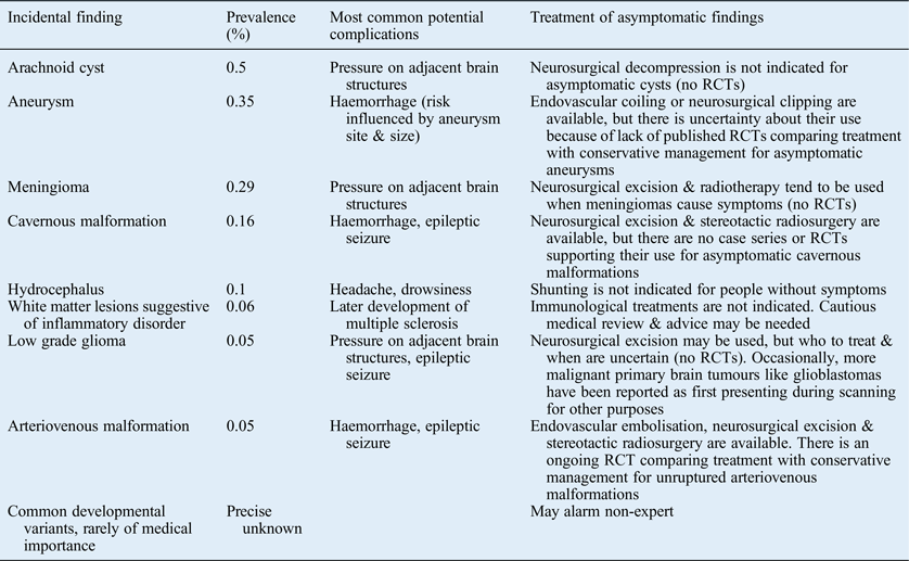
In the case of incidental findings requiring intervention, otorhinolaryngologists should have a basic level of understanding of the underlying pathology so that patients can be informed and counselled in clinic, prior to onward referral to the appropriate specialty. Further guidance can be sought by using the ‘Management of Incidental Findings Detected during Research Imaging’ document produced by the Royal College of Radiologists (Table I). 24
-
• Unilateral audiovestibular symptoms are common; internal auditory meatus magnetic resonance imaging (MRI) is used to rule out vestibular schwannomas
-
• Most internal auditory meatus MRI scans are normal, but incidental findings are not uncommon
-
• The vestibular schwannoma pick-up rate remains quite low despite various screening criteria
-
• This cohort study reports on the frequency of incidental findings and their management with reference to literature
-
• The study aimed to highlight the importance of imaging higher auditory pathways
-
• It aimed to improve otorhinolaryngologists’ knowledge in managing patients with incidental findings and in performing any onward referral

We suggest formulating a local algorithm in conjunction with radiology colleagues for incidental findings, particularly for use in units without easily available specialist neuroradiological, neurological or neurosurgical advice, in order to avoid unnecessary concern over the reporting of normal anatomical variants and to facilitate a more efficient referral on to allied specialties.