INTRODUCTION
The recognition of lameness as a major welfare disease of dairy cattle has increasingly focused attention on means of improving knowledge of hoof (claw) biology (Vermunt Reference Vermunt and Zemljic2004). Lameness is well recognized to have a complex aetiology with risk factors related to ‘intrinsic’ animal characteristics interacting with those derived from the ‘extrinsic’ environment. It is frequently caused by lesions of individual claws arising from failure of function of the outer horn and inner basement membrane and dermis which constitute the external tissues of its integumental cover. Such lesions are frequently associated with disruption of horn production by processes which are inadequately understood but which may be related to inflammatory laminitis (Lischer & Ossent Reference Lischer, Ossent and Shearer2002; Thoefner et al. Reference Thoefner, Pollitt, van Epps, Milinovich, Trott, Wattle and Andersen2004) and/or enzyme-induced changes in dermis and basement membrane affecting suspension of body weight by the laminar region (Tarlton et al. Reference Tarlton, Holah, Evans, Jones, Pearson and Webster2002; Webster et al. Reference Webster, Knott and Tarlton2005), or in sole (Hendry et al. Reference Hendry, Knight, Galbraith and Wilde2003). Such changes may increase body weight-bearing on soft tissues of the sole and in the presence of poorly-formed (Kempson & Logue Reference Kempson and Logue1993) or thin horn (van Amstel et al. Reference van Amstel, Shearer and Palin2004), or adverse flooring conditions (Hinterhofer et al. Reference Hinterhofer, Ferguson, Apprich, Haider and Stanek2005) reduce resistance to tissue damage and so increase susceptibility to lesion formation. Means of reducing susceptibility to the effects of such changes in weight-bearing or to other events causing claw horn lameness will be assisted by better knowledge of claw biology and in particular claw horn production. Among the factors which influence growth and development of cellular and extracellular claw tissues of the claw are nutrients which include sulphur-containing amino acids (Tomlinson et al. Reference Tomlinson, Muelling and Fakler2004).
Claw horn is produced by proliferation and differentiation of specialized epidermal cells (Budras et al. Reference Budras, Geyer, Maieri, Muelling, Lischer and Ossent1998; Galbraith et al. Reference Galbraith, Rae, Omand, Hendry, Knight and Wilde2006a) located on the basement membrane adjacent to the underlying dermis. The epidermis is avascular and receives nutrients, regulatory mitogens and morphogens from the vascularized dermis. Proliferation of basal epidermal cells occurs by mitosis with one daughter cell normally remaining to divide further and the other committing to differentiation and suprabasal migration. The process of differentiation involves the progressive expression of a range of genes including those encoding for keratins as major components of intermediate filaments (IFs), and intermediate filament associated proteins (IFAPs) which form an important part of the bovine solear tissue proteome (Galbraith et al. Reference Galbraith, Flannigan, Swan and Cash2006b). These proteins contain variable quantities of cysteine which is essential for the formation of inter- and intra-molecular disulphide bond formation in the synthesis of the cytoskeleton. The cytoskeleton contributes to physical characteristics and functional properties of terminally differentiated cells typical of the epidermal integument (Gillespie Reference Gillespie and Goldsmith1991; Budras et al. Reference Budras, Geyer, Maieri, Muelling, Lischer and Ossent1998; Galbraith et al. Reference Galbraith, Rae, Omand, Hendry, Knight and Wilde2006a). Such terminal differentiation also involves a form of apoptosis resulting in enucleation and partial dehydration of the cells to produce the ‘cornified’ horn end product.
One nutritional approach to enhancing the supply of L-cysteine in vivo has been to feed supplementary protein or intestinally-available L-methionine which increased yield and rates of growth of wool in sheep (Hynd Reference Hynd1989) and hair fibre in goats (Galbraith et al. Reference Galbraith, Mengal, Scaife, Lischer and Ossent1998; Souri et al. Reference Souri, Galbraith and Scaife1998). There is also some evidence of responses to L-methionine supplementation in rates of growth of claw wall horn in dairy cows (Metcalf et al. Reference Metcalf, Marsh, Johnston, May and Livesey1998) and Angora goats (Galbraith et al. Reference Galbraith, Mengal, Scaife, Lischer and Ossent1998). The importance of synthesis of keratins and associated proteins in the cellular structures of good quality horn is well established (Kempson & Logue Reference Kempson and Logue1993). However, the optimal concentrations of sulphur-containing amino acids required for the proliferation and differentiation of constituent horn-forming epidermal cells and the precise mechanisms by which such processes are controlled are not known. L-cysteine contributes to the composition of ‘low’ and ‘high’ sulphur amino acid-containing integumental protein (Gillespie Reference Gillespie and Goldsmith1991) in proportions considerably in excess of those provided by rumen microbial proteins and dietary protein in normal diets of ruminants (Galbraith Reference Galbraith2000). L-methionine is well recognized as an essential amino acid in the nutrition of many animal species. It has a range of roles in supporting molecular and biochemical function in cells and in contributing to synthesis of L-cysteine by transulphuration (Reis Reference Reis, Rogers, Reis, Ward and Marshall1989). However, little is known about the quantitative relationship between methionine supply and its uptake and utilization by cells of the integument and in particular the bovine claw. Although not previously studied in claw tissue, mechanisms of uptake of methionine may be considered in the context of general knowledge of its transmembrane transport in animal cells. Particular reference is made to catalysis of uptake (1) by, for example, Systems A and L involving sodium-dependent and sodium-independent neutral amino acid transporters respectively (Hyde et al. Reference Hyde, Taylor and Hundal2003) and (2) by gap junctions involving connection proteins such as Connexin 43 which has been identified immunohistochemically in claw epidermis by Frohberg-Wang et al. (Reference Frohberg-Wang, Muelling, Budras and Zemljic2004). Gap junctions are well recognized to mediate transport of solutes and signalling molecules in epidermal keratinocytes in skin (Gerido & White Reference Gerido and White2004). A third component involves transport across the basement membrane which separates dermis and epidermis by mechanisms which include diffusion (Budras et al. Reference Budras, Geyer, Maieri, Muelling, Lischer and Ossent1998). The work reported herein was part of a programme that measured the responses of bovine claw tissue to methionine supply in vivo and in vitro. It utilized a well-established bovine in vitro claw explant system (Hendry et al. Reference Hendry, Lancelott, Knight, Kempson and Wilde1995, Reference Hendry, MacCallum, Knight and Wilde1999, Reference Hendry, MacCallum, Knight and Wilde2001) which previously demonstrated synthesis of proteins, under a variety of conditions, by incorporation of 35S-label from supplied L-[35S]-methionine or L-[35S]-methionine and L-[35S]-cysteine mixtures, and incorporation of 3H-methyl thymidine into DNA as an indicator of cellular proliferation. The present study was designed to extend this work by quantifying the relationships between L-methionine and L-cysteine supply in the culture medium, uptake into intracellular pools and incorporation into protein fractions using L-[35S]-methionine. The work tested the hypothesis that such incorporation of 35S into cellular proteins is not limited by uptake of L-methionine, under concentrations typically found to be physiological in vivo, but is rather determined by mechanisms including those limiting its utilization for protein biosynthesis within the cell. Effects of differences of L-methionine supply on behaviour of epidermal cells in the basal and immediately supra-basal regions were studied by measurements of synthesis of DNA and effect on markers for proliferation and apoptosis.
The methodological approach of Shennan and colleagues (Hurley et al. Reference Hurley, Wang, Bryson and Shennan2000) used to determine transport characteristics of amino acids in mammary tissue explants was applied in the present study to the measurement of L-methionine uptake and incorporation in explants of the bovine claw.
MATERIALS AND METHODS
Claw tissue, preparation of explants, culture conditions and radiochemicals
Methods for the collection of claws, preparation of explants and culture conditions were as described previously (Hendry et al. Reference Hendry, Lancelott, Knight, Kempson and Wilde1995, Reference Hendry, MacCallum, Knight and Wilde1999, Reference Hendry, MacCallum, Knight and Wilde2001). Briefly, right hind lateral claws of crossbred female beef cattle (18–20 months old) and Holstein-type dairy cattle of varying ages, but all in excess of 30 months, were collected immediately post mortem at a commercial abattoir and rapidly transported in ice to the laboratory, typically within 1 h of slaughter. Claws without apparent lesions and subsequently shown to have healthy underlying soft tissue were selected. The central region of the sole of each claw was opened dorso-ventrally from horn to pedal bone by two parallel incisions using an autopsy saw. The outer horn was removed and sole material, intact and containing all tissue types including epidermis and underlying dermis, was cut into columnar explants weighing 30–50 mg each and measuring approximately 0·5 (thickness)×2 (width)×3 (length) mm, respectively. Explants were cultured for various lengths of time as described below in multi-well plates in an atmosphere of air: CO2 (0·95: 0·05) at 37°C in basal culture medium (Dulbecco Modified Eagle Medium/F-12 Nutrient Mix (DMEM/F12)) (1:1, Invitrogen) containing the normal concentrations of other amino acids and prepared initially without L-methionine, L-cysteine or L-cystine. The basal culture medium was variously supplemented to provide the required concentrations of these sulphur-containing amino acids. It should be noted that DMEM/F12 normally contains L-methionine, L-cysteine and L-cystine at 114, 145 and 130 μmol/litre, respectively. Radiochemicals were purchased from GE Healthcare (Little Chalfont, UK) as follows: L-[35S]-methionine (>1000 Ci/mmol); [methyl-3H]-thymidine (70–86 Ci/mmol); [U-14C]-sucrose (460–740 mCi/mmol).
Time course of methionine uptake by explants
The time course of methionine uptake was measured by incubating freshly prepared explants in DMEM/F12 medium customized to contain 1·0 mmol unlabelled L-methionine/litre and radioactively-labelled trace L-[35S]-methionine (1·0 μCi/ml) for up to 60 min. Explants were removed at pre-determined times, rinsed in radioisotope-free fresh culture medium, gently blotted, weighed and placed in vials containing 10% trichloroacetic acid (TCA) for 16 h to precipitate TCA-insoluble macromolecules. Following centrifugation (13 000 g for 3 min) to remove particulate and precipitated material, radioactivity in the TCA-soluble supernatant was counted. This fraction represents free amino acid taken up by the whole tissue in intracellular and extracellular space and subsequently released. The water content of representative explants was determined by drying to constant weight at 110°C. The extracellular space of the tissue was estimated from parallel studies which measured the distribution of sucrose (as [U-14C]-sucrose (0·5 μCi/ml)) between tissue and incubation medium according to Hurley et al. (Reference Hurley, Wang, Bryson and Shennan2000). The method is based on the impermeability of sucrose to cells and its equilibration with extracellular fluid only. Concentrations of L-methionine in intracellular pools were thus quantified (Shennan & McNeillie Reference Shennan and McNeillie1994) according to the equation:
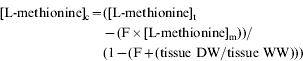
where: c=intracellular L-methionine concentration (μmol/kg intracellular water), t=tissue L-methionine concentration (μmol/kg wet weight), m=incubation medium L-methionine concentration (μmol/litre), F=tissue extracellular space (expressed as a fraction); DW=dry weight; WW=wet weight.
Effect of L-methionine and L-cysteine concentrations on L-methionine uptake
Additional studies on uptake of L-methionine were conducted under conditions typically applying to longer term incubations in culture medium and which determined kinetics of uptake as affected by concentrations of unlabelled amino acid in culture medium. Effects on uptake of concentrations of unlabelled L-methionine ranging from 0 to 20 mmol/litre and in normal DMEM/F12 culture medium at concentrations of L-methionine (114 μmol/litre) and of L-cysteine (145 μmol/litre) and L-cystine (130 μmol/litre) and at various proportions of these were measured after 21 h pre-incubation. Explants were transferred to fresh media of similar composition and supplemented with L-[35S]-methionine. Intracellular concentrations, as an index of uptake, were then measured following removal of explants from media after 30 min as described above. The specific activity of L-[35S]-methionine in each incubation culture medium was determined by counting the radioactivity in 100 μl samples and relating to quantity of unlabelled L-methionine present.
L-methionine incorporation, protein and DNA synthesis, autoradiography and immunoblotting
A second objective of the current study was to quantify the incorporation of 35S label supplied by L-[35S]-methionine into macromolecular protein fractions as a function of unlabelled L-methionine, L-cysteine and L-cystine concentrations in culture medium. Explants were prepared as described above and incubated for 21 h in a range of concentrations and proportions of L-methionine, L-cysteine and L-cystine in DMEM/F12 followed by 3 h in similarly composed medium containing L-[35S]-methionine (6 μCi/ml) and [methyl-3H]-thymidine (2·5 μCi/ml) to provide measures of (a) L-methionine incorporation and protein synthesis and (b) DNA synthesis, respectively (Hendry et al. Reference Hendry, Lancelott, Knight, Kempson and Wilde1995; Goulet et al. Reference Goulet, Poitras, Rouabhia, Cusson, Germain and Auger1996). These concentrations were guided by information on concentrations in blood of sulphur-containing amino acids from studies on dairy cattle (Galbraith et al. unpublished) that, for L-methionine, ranged from approximately 16 to 25 μmol/litre and 50 μmol/litre (Shennan et al. Reference Shennan, Miller and Calvert1997). Following incubation, explants were rinsed in radioisotope-free fresh culture medium, gently blotted and snap-frozen in liquid nitrogen prior to grinding (Freezer Mill 6750, Glen Creston Ltd). Powdered tissue was suspended in 0·1 mol/litre sodium phosphate pH 7·4, 2·0 mol/litre sodium chloride containing 0·2 mmol/litre phenylmethylsulphonyl fluoride and 16·2 mmol/litre 3-[(3-cholamidopropyl)dimethyl-ammonio]-1-propane sulphonate (CHAPS) and protein concentrations of the tissue homogenates measured (Bradford Reference Bradford1976). Protein and DNA synthesis were determined in explant homogenates by precipitation and washing with 1·22 mol/litre TCA with a final dissolution in 19·4 mol/litre formic acid for 15 h at 37°C, then scintillation counting with corrections for isotopic dilution calculated from the ratio of L-[35S]-methionine (radioactively labelled) to unlabelled L-methionine in the original incubation medium.
The pattern of L-methionine uptake into cells and extracellular matrix was determined in explants pulsed with L-[35S]-methionine (1·0 μCi/ml) for 30 min in culture medium containing 1·0 μmol/litre methionine and then incubated in radiolabel-free fresh media for 24 h. Explants were sectioned by cryostat (10 μm thick) then emulsion-dipped (K5, Ilford) and developed on day 14. The pattern of distribution of L-[35S]-methionine in protein fractions was determined in explants incubated for 24 h in media containing 1·0 or 30 μmol/litre methionine and L-[35S]-methionine at the same specific activity of 1·0 μCi/μmol/litre unlabelled L-methionine. These explants were extracted by TCA (1·22 mol/litre) precipitation, re-solubilized in 8 mol/litre urea and subjected to conventional sodium dodecyl sulphate-polyacrylamide gel electrophoresis (SDS-PAGE) (120 g/litre of acrylamide: bis-acrylamide mix; 30: 0·8) at a loading of 10 μg protein per lane. Protein bands in gels were detected with zinc stain (Bio-Rad) and then subjected to either autoradiography or transfer to polyvinylidene difluoride (PVDF) membrane (NEN Life Sciences). Membranes were tested for detection of cytokeratins (CK) including CK5/6 (mouse anti-human, 1:1000, Chemicon) and CK10 (mouse anti-human, 1:1000, Novacastra) with electrogenerated chemiluminescence (ECL) methodology (New England Biolabs). Negative controls were prepared under similar conditions by omitting the primary antibody. Gels and immunoblots were examined in duplicate and comparisons made between major bands by scanning densitometry (Gel Doc 2000, Bio-Rad).
Histology and immunohistochemistry
Conventional histological and immunohistochemical studies were conducted to determine cell cycle activity of epidermal and dermal cells of explants which had been incubated in the presence of either 1·0 or 30·0 μmol L-methionine/litre. Markers of DNA synthesis during the S phase, as a prerequisite to cell division, were as follows: the synthetic thymidine analogue, bromodeoxyuridine (BrdU), was added to culture medium for measurement of uptake by cells; expression of proliferating cell nuclear antigen (PCNA) which associates with DNA polymerase δ; the Feulgen stain which provides a semi-quantitative measure of the increases of DNA in each cell. Markers for programmed cell death activity were: Bcl-2 which is an anti-apoptotic member of a gene family which has additional members with anti- and pro-apoptotic activity; the TUNEL (Terminal transferase deoxyuridine triphosphate (dUTP) nick end labelling) detection system for the presence of fragmentation of DNA during apoptosis.
Explants were removed from culture medium after 48 or 72 h, rinsed, gently blotted and snap frozen in liquid nitrogen. Cryosections (10 μm thick) of explant tissue were then prepared and fixed in 40 g paraformaldehyde/litre for 10 min followed by washing in PBS and treatment with trypsin (10·0 g/litre) for 10 min. Endogenous peroxidase was blocked by incubation in 30 g/litre hydrogen peroxide followed by incubation with 10 ml/litre horse serum (Vector Laboratories Inc, Burlingame, CA) in PBS for 20 min to block non-specific binding. Sections were exposed to primary antibodies (monoclonal mouse anti-rat PCNA, Sigma) 1:3000 in PBS or polyclonal mouse anti-human Bcl-2 (Oncogene) 1:50 in PBS and non-immune mouse IgG (Sigma) or PBS as controls, for 18 h at 4°C. Sections were washed and blocked as before and then incubated with biotinylated universal secondary antibody (Vector) for 30 min followed by washing and incubation with ABC reagent (Vector) for 30 min. Sections were mounted in styrolite and visualized by light microscopy. Quantitative image analysis (Image Pro-Plus Version, 4.50.19, Media Cybernetics Inc, USA) was applied to count positively-stained cells in the basal and immediately suprabasal epidermis (Cruchley et al. Reference Cruchley, DeSouza, Williams, Greenspan and Greenspan1998) as a function of basement membrane length (one unit=1·2 mm) for 18 papillae.
For measurement of BrdU incorporation, the explants were pulsed with 0·05 mmol BrdU/litre for 6 h followed by incubation in fresh media and removal at 24, 48 or 72 h. Other explants were incubated for 24 or 48 h prior to a 6 h pulse of 0·05 mmol BrdU/litre BrdU, and then removed at 48 or 72 h, respectively. Cryostat sections of the explants were then treated with 2 M HCl (1 h at 37°C) to denature DNA before exposure to monoclonal mouse anti-BrdU (1:1000, Sigma) with visualization using the secondary antibody system described above.
DNA in the basal cells lining the basement membrane and immediately superbasally, was localized by a modified Feulgen method. Cryostat sections were fixed in 4% paraformaldehyde and then immersed in glacial acetic acid: ethanol (1:3) (Clarks Fixative) for 2 h. The sections were then hydrated through an ethanol series before immersion in 1 M HCl at 60°C for 6 min followed by Schiff's Reagent (Sigma) for 20 min. Sections were mounted conventionally and examined by light microscopy for quantitative image analysis. Apoptosis of basal epidermal cells was measured using TUNEL methodology following the manufacturer's recommendations using digoxigenin-dUTP antibodies (Apoptag kit; Oncor, Inc., Gaithersburg, MD). Positive cells were counted as described above.
Data analysis
Data were examined statistically by conventional one-way ANOVA (Minitab v 13.3, General Linear Model) followed by Tukey's method for pairwise comparisons of means. The data were obtained from independent samples collected from individual explants which were harvested and tested for measured parameters in response to differences in tissue culture conditions and following different lengths of incubation. Differences in means were considered significant at P<0·05 for all tests used. In studies which investigated kinetic parameters of L-methionine uptake in explants, rates of uptake, V, against concentrations [S] of L-methionine at 30 min of incubation, were linearized by conventional Lineweaver-Burke plot to determine values for V max and Michaelis constant (K M). These values were used to generate curves relating V and [S] according to the Michaelis–Menten equation, V=V max [S]/[S]+K M using Microsoft Excel. This software was also used to test, by regression analysis, the linearity of certain relationships for the data obtained.
RESULTS
Parameters of uptake
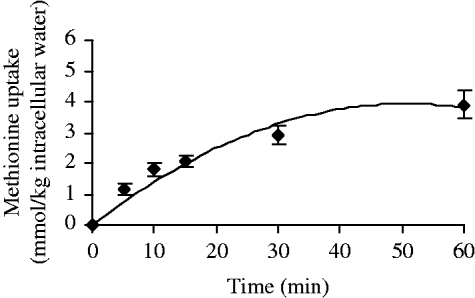
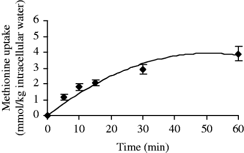
The time course of uptake of L-methionine into intracellular pools of freshly prepared bovine (beef) sole tissue explants in culture medium containing tracer L-[35S]-methionine and 1·0 mmol unlabelled L-methionine/litre is shown in Fig. 1. The pattern of uptake is time-dependent and essentially linear for the first 10 min (y=0·1893x, R 2=0·97, P<0·01) and then becomes curvilinear. The data suggested that maximum intracellular concentration was achieved at approximately 30 min with steady state concentration being maintained to the 60 min measurement. Concentrative uptake, in which the intracellular concentration exceeded that of external culture media, occurred between 5 and 8 min after exposure of explants to the culture medium containing unlabelled L-methionine and tracer L-[35S]-methionine.
Time course of L-methionine uptake by beef cattle solear tissue. Freshly prepared explants were incubated in DMEM/F12, L-methionine-deficient medium supplemented with 1·0 mmol/litre unlabelled L-methionine and 1·0 μCi/ml L-[35S]-methionine. Values are shown as means±s.e.m. for n=9 replicate cultures.
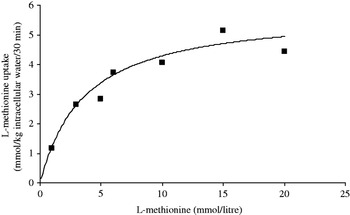
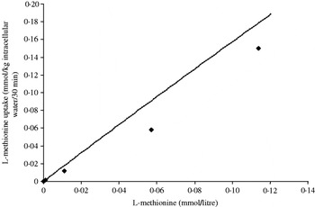
The uptake of L-methionine at concentrations ranging from 1·0 to 20 mmol/litre was measured, after 21 h pre-incubation, in fresh culture medium containing tracer L-[35S]-methionine (Fig. 2). Kinetic parameters for uptake after 30 min indicate the presence of a saturable curve and when subjected to a Lineweaver–Burke analysis (y=0·6178x+0·1712, R 2 =0·99, P<0·01, not shown) gave K M and V max values of 3·61 mmol/litre and 5·84 mmol/kg intracellular water/30 min, respectively. In Fig. 2 the data points are presented together with the line obtained by substituting the values calculated for K M and V max into the Michaelis–Menten equation, V=V max [S]/[S]+K M. Measurements were also made on uptake by beef cattle solear explants exposed to physiological concentrations of up to 114 μmol/litre, which is the normal concentration for L-methionine in DMEM/F12 culture medium (Fig. 3). The data points are presented together with the line obtained by substituting the same values calculated for K M and V max (see above) into the Michaelis–Menten equation. The rate of L-methionine uptake so calculated presents as relating approximately linearly to L-methionine in the culture media over the concentration range studied.
Effect of L-methionine concentration on its uptake by beef cattle solear tissue. Explants were incubated for 21 h in DMEM/F12, L-methionine-deficient medium supplemented with 0–20 mmol/litre methionine, followed by 30 min incubation in similarly supplemented fresh media containing 1·0 μCi/ml L-[35S]-methionine and uptake measured with corrections made for isotope dilution. Values for V max and KM, calculated by Lineweaver–Burke plot, were used to calculate the curve line according to the Michaelis–Menten equation (see Material and Methods section). Individual points are mean values for n=9 replicate cultures.

Effect of L-methionine concentration on its uptake by beef cattle solear tissue under normal culture conditions. Explants were incubated for 21 h in DMEM/F12, L-methionine-deficient medium supplemented with up to 114 μmol/litre L-methionine, followed by 30 min incubation in similarly supplemented fresh media containing 1·0 μCi/ml L-[35S]-methionine. Uptake of [35S] was measured with corrections made for isotope dilution. Values for V max and K M, calculated by Lineweaver–Burke plot were used to calculate the curve line according to the Michaelis–Menten equation (see Materials and Methods section). Individual points are mean values for n=12 replicate cultures.

Parameters of incorporation: (a) synthesis of proteins, (b) synthesis of DNA and (c) effects of L-cysteine and L-cystine
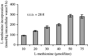
(a) The effect of L-methionine concentrations in tissue culture medium, on synthesis of proteins in explants, was measured in TCA-precipitated fractions solubilized in formic acid. Results from studies measuring incorporation of L-methionine, expressed as a function of weight of intracellular water, in explants from dairy cows (Fig. 4) showed a concentration-dependent (μmol unlabelled L-methionine/litre (P<0·05): 10<30, 40, 50, 75; 20<40, 50, 75; 40<50, 75) stimulation in protein synthesis with a peak occurring at 50 μmol/litre concentration in culture medium. Other results (not shown) on solear explants from beef heifers based on incorporation in relation to tissue wet weight indicated maximal incorporation at 30 μmol/litre L-methionine.
Effects of unlabelled L-methionine concentration on protein synthesis in dairy cattle solear tissue. Following 21 h incubation, explants were incubated with 6·0 μCi/ml L-[35S]-methionine and its incorporation was measured after 3 h. Values are shown as means±s.e.m. for n=12 replicate cultures with s.e.d. (df=66).

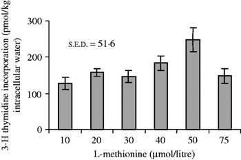
(b) Similarly, the magnitude of incorporation of [methyl-3H]-thymidine in the TCA insoluble macromolecular fraction, as a marker for DNA synthesis, was dependent on concentration (μmol unlabelled L-methionine/litre (P<0·05): 10, 20, 30<50) in culture medium (Fig. 5). The highest value occurred at 50 μmol/litre for dairy cow solear explants. Data obtained for beef heifers indicated maximum incorporation per mg tissue wet weight at 40 μmol L-methionine/litre (not shown).
Effects of L-methionine concentration on DNA synthesis in dairy cattle solear tissue. Following 21 h incubation, explants were incubated with 2·5 μCi/ml [3H methyl]-thymidine and its incorporation was measured after 3 h. Values are shown as means±s.e.m. for n=12 replicate cultures with s.e.d. (df=66).

(c) In addition, inclusion of L-cysteine and L-cystine in the medium (at proportions of 0·1, 0·5 and 1·0 of normal medium where normal values in DMEM/F12 medium were 145 and 130 μmol/litre, respectively) had no effect on uptake or incorporation of L-methionine or incorporation of [methyl-3H]-thymidine by beef heifer explants. Means values (±s.e.), for example, for incorporation (per kg intracellular water/3 h) of L-methionine (nmol) were 106±10·9, 107±12·9 and 79·0±13·4 and for methyl-thymidine (pmol), 94·5±13·6, 113±16·8 and 106±16·3, respectively.
Cell cycle behaviour
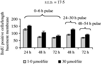
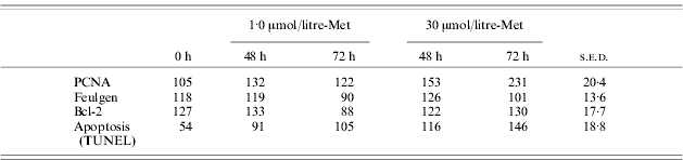
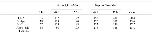
Numbers of basal and immediately suprabasal epidermal cells positive for PCNA expression were greater at the higher (30 μmol/litre) than lower (1·0 μmol/litre) concentration of L-methionine tested at 72 h (Table 1). Reductions in expression of genomic DNA by Feulgen were evident for both concentrations of L-methionine at 72 h compared with measurements made at 0 and 48 h. A significantly greater number of cells were positive for BrdU at 48 h when pulsed from time 0 or 24 h at 30, compared with 1·0 μmol/litre L-methionine concentration in culture medium (Fig. 6). Similar trends were apparent at 24 (P=0·052) and 72 h (P=0·085). A significantly more frequent cellular expression of the apoptosis inhibitor Bcl-2 was evident at 72 h, in response to 30 μmol/litre L-methionine on comparison with 1·0 μmol/litre. Tests for apoptosis in basal and immediately suprabasal epidermal cells by TUNEL gave time-associated increases (P<0·05) in positive signal compared with time 0 and a greater apoptotic response to 30 μmol/litre, compared with 1·0 μmol L-methionine/litre, at 72 h (Table 1).
Effects of 1·0 or 30 μmol/litre L-methionine concentration on BrdU incorporation in dairy cattle solear tissue. Explants were pulsed with 0·05 mmol/litre BrdU for 6 h at various stages through the culture and then removed at 24, 48 and/or 72 h, cryosectioned and immunostained for detection of BrdU in individual cells. Cell counts are presented per unit length (1·2 mm) of basement membrane Values are shown as means±s.e.m. for n=6 replicate cultures with s.e.d. (df=50).

The effect of L-methionine concentration on proliferation (PCNA and Feulgen) and apoptosis (Bcl-2 and apoptosis by TUNEL assay) markers in bovine sole explants after 48 or 72 h. Epidermal cell counts (means with s.e.d. (df=25)) in histological sections are presented per unit length (1·2 mm) of basement membrane for n=6 replicate cultures.
Localization of 35S from L-[35S]-methionine supplementation
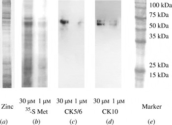
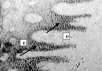
Conventional zinc staining demonstrated a typical pattern for integumental tissue extracts separated by SDS-PAGE (Fig. 7). Protein banding was evident in the molecular weight range for keratins (44–69 kDa) and intermediate filament-associated proteins (12–25 kDa). A similar pattern was evident in autoradiographs showing incorporation of label in a similar range of proteins. Results for immunoblots for separated proteins showed positive signals for the detection systems for CK5/6 and CK10 which co-localized with bands shown to be positive by autoradiography. L-methionine concentrations in culture media of 30 μmol/litre compared with 1·0 μmol/litre, produced stronger signals in both autoradiographic and immunoblotting systems (results were consistent between duplicates) indicating a greater support of protein synthesis in extracts of explants. Comparisons of major bands by scanning densitometry gave differences in signal which ranged between 2·6–3·5 fold for autoradiography and 1·7 and 2·4 for CK5/6 and CK 10 immunoblotting, respectively. Comparisons of the location of 35S label in cryosections of explants exposed to a 30 min pulse of L-[35S]-methionine and incubated for 24 h showed its relative absence in the dermis with clear evidence of retention in basal and suprabasal epidermal cells (Fig. 8).
Single dimension SDS-PAGE of extracts of beef cattle solear explants following 24 h incubation in DMEM/F12, L-methionine-deficient medium supplemented with 1·0 or 30 μmol/litre L-methionine. From left, (a) zinc stain, (b) autoradiographs for L-[35S]-methionine with similar isotope dilution (similar ratio of labelled to unlabelled L-methionine), (c) immunoblots for CK5/6, (d) immunoblots for CK10 and (e) molecular mass marker.

Autoradiograph of cryosection of beef cattle solear explant incubated in DMEM/F12, L-methionine-deficient medium supplemented with 1·0 μmol/litre unlabelled L-methionine. Explants were pulsed at time 0 with 1·0 μCi/ml L-[35S]-methionine for 30 min with further incubation in radiolabel-free fresh medium, removal at 24 h and processing. Arrows show autoradiographic signal in epidermis. D, dermis; E, epidermis. Magnification×128.

DISCUSSION
The epidermis of the mammalian integument is the specialized outermost tissue which produces end products such as skin, hair and claw horn. Such end products arise from proliferation and differentiation of epidermal cells under the influence of the underlying dermis. Such differentiation typically includes expression of genes encoding for (a) keratins which form IFs of the cytoskeleton and (b) high sulphur (cysteine)-containing IFAPs. The combination of IFs and IFAPs in formation of intermolecular disulphide bonds is an essential process in the formation of functionally effective horn in the bovine claw. Such bond formation has been described histochemically (Budras et al. Reference Budras, Geyer, Maieri, Muelling, Lischer and Ossent1998). The supply of sulphur amino acids and the relationship between L-methionine and L-cysteine have been well recognized as important in the growth and properties of mammalian hair and other integumental tissues (Gillespie Reference Gillespie and Goldsmith1991) but are inadequately quantified. For example, it is clear that hair growth in sheep may be increased by provision of dietary protein (Hynd Reference Hynd1989) or in Angora goats by intestinally absorbable L-methionine supplements (Souri et al. Reference Souri, Galbraith and Scaife1998). However, similar supplementation of methionine to goats (Galbraith et al. Reference Galbraith, Mengal, Scaife, Lischer and Ossent1998) or dairy cattle (Metcalf et al. Reference Metcalf, Marsh, Johnston, May and Livesey1998) produced initial responses which were not sustained beyond the initial 4 or 6 week periods of study. In addition, Laven & Livesey (Reference Laven and Livesey2004) observed the absence of significant effect on development of haemorrhages in cattle hoof horn. These differences in response probably reflect differences in the quantities of sulphur-containing amino acids required to support synthesis of sheep wool or mohair fibre (kg quantities per year) compared with requirements for claw horn deposition (c. 700 g/year).
Major similarities in the morphology of the dermis and epidermis of the hair follicle and horn-producing structures of the bovine claw are well recognized (Hendry et al. Reference Hendry, Lancelott, Knight, Kempson and Wilde1995; Galbraith Reference Galbraith1998).
Applying regression analysis to the present results for kinetics of L-methionine uptake using 1·0 mmol/litre extracellular concentration, suggested initial linearity for up to 10 min and concentrative uptake (when intracellular concentration exceeded extracellular concentration) occurring within 8 min. This was followed by a curvilinear pattern with maximum accumulation being attained by approximately 30 min with this steady state maintained at least until 60 min. Measurements of the relationship between uptake and concentration of methionine in culture medium were made following pre-incubation of 21 h. Measurements were taken after 30 min which was shown to produce approximate maximal uptake and steady state conditions required to support protein and DNA synthesis in subsequent studies. This approach produced a saturable curve which obeyed Michaelis–Menten kinetics and gave values for K M and V max of 3·61 mmol/litre and 5·84 mmol/kg intracellular water/30 min, respectively. It is recognized that taking measurements after the initial linear uptake and following exposure to in vitro culture conditions may have altered sensitivity of uptake mechanisms compared with those present in vivo or in freshly prepared tissues. However, applying the in vitro model to measurement of uptake under lower (0·114 mmol/litre approximates to 0·03 of K M) and more physiological L-methionine concentrations in normal culture media, provided no evidence of saturation of uptake mechanisms. The results for explants both freshly prepared and after 21 h incubation both indicate a capacity for substantial uptake of L-methionine. The concentrations in the mmol/litre range suggest the presence of low affinity transport mechanisms, the characteristics of which for L-methionine (e.g. Systems A and L) have been summarized previously (Hyde et al. Reference Hyde, Taylor and Hundal2003). While there appear to be no reports in the literature describing kinetics of L-methionine transport in other tissues of the mammalian integument there is evidence in other tissues for low affinity mechanisms determining uptake of other amino acids. For example, low affinity transport with a K M in excess of 1·0 mmol/litre has similarly been described for lysine in porcine mammary tissue (Hurley et al. Reference Hurley, Wang, Bryson and Shennan2000). These authors also indicated the possibility of a more complex analysis of kinetic data to that of the single saturable curve used in their work. This observation is consistent with the likelihood that compositionally complex tissue models comprise of more than one uptake system. These may, in addition to transporters, include gap junction-facilitated transport between epidermal cells and trans-basement membrane systems and so are potentially more complex than in vitro models based on cell culture. However, the results from the present in vitro model do suggest that net uptake of claw soft tissues has the capacity to be increased in response to increases in extracellular concentrations considerably in excess of those typically observed in vivo (i.e. up to 50 μmol/litre (Shennan et al. Reference Shennan, Miller and Calvert1997)). Similarly, the absence of effects on uptake of L-methionine of inclusion of L-cysteine and L-cystine at concentrations up to those present in normal DMEM/F12 media suggests that uptake was non-competitive at the concentrations tested. Issues concerning the possible effects of alterations in L-methionine concentration on uptake of other amino acids which share common transport mechanisms can be considered in the context of the apparently high capacity of these systems (K M in mmol/litre) and the lower (μmol/litre) concentrations which optimize responses in incorporation to methionine supply. However, obtaining data to describe such effects will require further study.
In terms of the localization in explants, autoradiographic studies showed major concentration in basal and suprabasal epidermis with very limited retention of L-[35S]-methionine in the dermis. This result is consistent with systems of nutrient transportation in vivo which include routes from vascular dermis into avascular epidermis and involving gap junction-mediated intercellular epidermal transport (Gerido & White Reference Gerido and White2004). Further analysis by PAGE indicated that supplied L-methionine supported synthesis of proteins of a range of molecular weights including those associated with keratins (44–69 kDa) and lower molecular weight intermediate-filament associated proteins (IFAPs) (12–25 kDa). Differences in the 35S signal indicated a concentration-dependent response to supply (30>1·0 μmol/litre). The latter IFAP fraction is known to be rich in cysteine which is particularly important in the formation of disulphide trans-molecular bonds (Gillespie Reference Gillespie and Goldsmith1991). Co-localization of CK5/6 and CK10 by immunoblots with bands of autoradiographic signal gives further evidence that supplied methionine supported protein synthesis at both basal and suprabasal levels, respectively. It was also evident that the immunochemical signal for these CK was less at the lower concentration of L-methionine supply in culture medium of explants. Such results also suggest the maintenance of synthetically competent epidermal cells with patterns of protein expression typically similar to those of cell culture systems from other regions of the integument (Goulet et al. Reference Goulet, Poitras, Rouabhia, Cusson, Germain and Auger1996).
It may be concluded from the present work that apparent uptake capacity for L-methionine is (a) well above concentrations observed in normal blood concentrations in vivo of the order of 50 μmol/litre (Shennan et al. Reference Shennan, Miller and Calvert1997) and (b) did not limit incorporation into the TCA-precipitated macromolecular fraction of the dermal and epidermal tissue in vitro. In contrast, the incorporation of L-methionine as an indicator of protein synthesis was shown to be concentration-sensitive within the normal haematological range. Initial studies on solear explant tissues of beef heifers indicated maximal intracellular incorporation between concentrations in media of 11·7 and 57 μmol/litre with evidence, on the basis of a less sensitive measurement of expression as a proportion of wet tissue weight (includes the weight of largely extracellular dermis), of a peak around 30 μmol/litre. Values based on intracellular water and derived from subsequent studies in dairy cattle peaked at approximately 50 μmol/litre. Similar investigation of the effect of extracellular L-methionine supply on apparent DNA synthesis also indicated a concentration-dependent response with maximal synthesis and apparent proliferative activity again obtained at 50 μmol/litre. It is interesting to note that these concentrations in the range of 30–50 μmol/litre correspond reasonably to the value (50 μmol/litre) for L-methionine given for the systemic blood of dairy cows (Shennan et al. Reference Shennan, Miller and Calvert1997), although concentrations in the local environment of internal hoof tissue appear not to have been described. It is also recognized that variation in the values obtained may relate to factors such as differences in age and physiological stage of animals from which explants were derived.
Further evidence of concentration-dependent effects of L-methionine on proliferative activity in the epidermal basal and immediately suprabasal region was obtained for the proliferation marker PCNA and for uptake of BrdU. Signals for both were greater in media containing 30 μmol L-methionine/litre compared with 1·0 μmol/litre with significant differences or non-significant trends recorded after 48 or 72 h incubation. Similar patterns of expression have been demonstrated in tissues such as human tongue epithelia (Cruchley et al. Reference Cruchley, DeSouza, Williams, Greenspan and Greenspan1998).
Mechanisms of cellular response to increasing L-methionine supply may be expected to include (i) reductions in limitations in L-methionine availability per se, for protein synthesis (ii) contributions to production, by transulphuration, of cysteine for which there is particular demand for synthesis of IFAPs and proteins in IFs and (iii) provision of methyl groups for synthesis of polyamines which are essential for synthesis of DNA and protein in cells undergoing mitosis (e.g. Reis Reference Reis, Rogers, Reis, Ward and Marshall1989). More recent considerations, although not defined in bovine claw tissue, have included a more direct role of amino acids and their availability in the modulation of gene expression. Among suggested mechanisms are sensing by amino acid transporters and signalling by the kinase mTOR (mammalian target of rapamycin) pathway (e.g. Hyde et al. Reference Hyde, Taylor and Hundal2003).
Results from histological studies on basal and immediately suprabasal cellular activity were particularly interesting in comparing responses to 1·0 μmol/litre or 30 μmol L-methionine/litre after 72 h incubation. There was evidence for a greater expression of the apoptosis inhibitor Bcl-2 at the higher L-methionine concentration for which numbers of positive cells were similar to those recorded at the beginning of the culture. Such numbers were significantly decreased for the lower L-methionine concentration consistent with increases in the apoptotic (TUNEL) signal. However, the anti-apoptotic result for 30 μmol/litre methionine was confounded by an apparently greater apoptotic (TUNEL) response under similar conditions. It is not clear whether this latter result for TUNEL derives from greater relative expression of pro-apotopic compounds such as Bcl-2-associated X protein (BAX), which was not measured, or represents a problem with methodology such as discussed for other studies (e.g. Labat-Moleur et al. Reference Labat-Moleur, Guillermet, Lorimier, Robert, Lantuejoul, Brambilla and Negoescu1998). It is also possible that basal cells were undergoing faster turnover (i.e. greater rates of proliferation and apoptosis under conditions of the study) with the balance in favour of proliferation. The data for PCNA, Feulgen and BrdU for basal and immediately suprabasal cells support this possibility. While enucleation occurs normally in later differentiation of epidermal horn-forming cells, the effects of such early apoptosis, without increased proliferation, may be expected to reduce production of suprabasal cells and subsequent protein gene expression. Such a reduction was not obtained and the increases in protein and apparent DNA synthesis also observed in incorporation studies and in synthesis of individual keratin proteins would consequently be expected to contribute to increased deposition of claw horn in vivo. It is evident that the use of TUNEL methodology in hoof tissue requires further investigation.
In terms of responses in vivo, results from one study (Galbraith et al. unpublished) showed that supplementation of dairy heifers throughout pregnancy with 5–6 g/day rumen-protected L-methionine which produced small although significant (P<0·05) increases in mean systemic blood concentrations (23·2 v. 19·2 μmol/litre), had no effect on growth characteristics of, or concentrations of L-methionine or L-cysteine in, claw wall horn collected 2–3 days post partum (Galbraith et al. Reference Galbraith, Rae, Omand, Hendry, Knight and Wilde2006a). These results contrast with those of Clark & Rakes (Reference Clark and Rakes1982) who reported increases in rates of growth but reductions in the incorporation of L-cysteine into upper claw wall horn of dairy cattle supplemented for 70–90 days with the quantitatively greater 30 g/day of methionine hydroxy analogue. It is apparent that supplementation of the diet in vivo to produce a range of systemic concentrations of L-methionine to beyond 50 μmol/litre will be required to test the validity of the above in vitro data.
In conclusion, the results from the present study provide new information on the effect of variation in extracellular L-methionine supply on the physicochemical, protein synthetic and proliferative behaviour of bovine solear epidermis and dermis in vitro. Extrapolation to nutritional supply in vivo suggests the appropriateness of delivery of systemic concentrations of 50 μmol/litre L-methionine to maximize proliferative and protein depositional activity in solear epidermis. Future work could usefully investigate (a) the value and means of delivering such optimal concentrations in vivo and (b) the mechanisms of sensing and signal transduction in the dermal-epidermal unit and horn-forming cells in response to such nutrient supply.
The authors acknowledge the funding of the work by the Biotechnology and Biological Sciences Research Council, and the Scottish Executive Environment and Rural Affairs Department. We thank Dr Trevor Stuchbury for advice on analysis of data for kinetics of amino acid transport. Technical assistance provided by Ms M Wallace, Mr J Struthers and Mr M Birnie is gratefully acknowledged.