Invasive aspergillosis (IA) affects immunocompromised patients, notably those undergoing allogeneic hematopoietic stem-cell transplantation (HSCT) or patients developing neutropenia following cytotoxic therapy for hematologic malignancies.Reference Gerson, Talbot, Hurwitz, Strom, Lusk and Cassileth 1 – Reference Segal 3 Hospital-acquired IA in neutropenic patients has been described in both epidemic and endemic settings.Reference Arnow, Sadigh, Costas, Weil and Chudy 4 However, the nosocomial origin of IA in patients who are not overexposed and especially in patients outside the setting of allogeneic HSCT is still a matter of debate.Reference Berthelot, Loulergue and Raberin 5 A prospective 4-year study monitoring fungal contamination of patients treated in a bone marrow transplant unit and 2 hematology wards supported a causal relationship between environmental fungal contamination and IA.Reference Alberti, Bouakline and Ribaud 6 Numerous studies have been published supporting the impact of air filtration systems such as high-efficiency particulate air (HEPA) filtration or laminar air flow (LAF) in reducing contamination with airborne fungi and the frequency of IA.Reference Berthelot, Loulergue and Raberin 5 , Reference Cornet, Levy and Fleury 7 , Reference Hahn, Cummings, Michalek, Lipman, Segal and McCarthy 8 In other studies, implementation of mobile air decontamination units (eg, Plasmair, AirInSpace, Montigny-le-Bretonneux, France) during hospital construction work or renovations resulted in a significant reduction in overall fungal contamination.Reference Poirot, Gangneux and Fischer 9 , Reference Sixt, Dalle and Lafon 10
In this study, we evaluated the impact of a mobile air decontamination system (Plasmair) on the level of exposure to airborne fungal contamination and on the risk that neutropenic patients develop IA outside the setting of allogeneic HSCT. We took advantage of a 2-phase equipment installation of Plasmair systems in the hematology intensive care unit (hICU), with partial equipment during the first 2 years, to compare frequencies of IA in Plasmair-equipped and non-equipped rooms during this first period.
PATIENTS AND METHODS
Patients
Data were retrospectively collected for patients hospitalized in the hICU of Hôpital de Versailles for treatment of hematologic malignancies or for autologous HSCT and who developed chemotherapy-induced neutropenia (polymorphonuclear count <0.5 g/L lasting ≥7 days). Patients diagnosed with acute leukemia received the standard combination of daunorubicin and the conventional dose of cytarabine as induction therapy (3+7 protocol) followed by intermediate doses (for patients >60 years) or high doses (for patients<60 years) of cytarabine for 2 or 3 consolidation cycles. BEAM chemotherapy with carmustine, etoposide, cytarabine, and melphalan was used as conditioning regimen for autologous stem cell transplantation. Patients with non-Hodgkin’s lymphoma were included in the study after the administration of high-dose cyclosphosphamide. Patients with aplastic anemia were treated with thymoglobulin and steroids. Prophylaxis with oral posaconazole (300 mg/day) was stratified according to local guidelines, particularly in patients with acute myeloid leukemia during induction. Regarding antifungal pre-emptive therapy, patients who were suspected to have acquired IA received intravenous caspofungin (70 mg/day on day 1, and 50 mg/day beginning on day 2) until the diagnosis of IA was confirmed or rejected. Antimicrobial prophylaxis was not applied at a specified level. If IA was confirmed, patients were treated intravenously with voriconazole (6 mg/kg twice on day 1 and 4 mg/kg beginning on day 2). No other antifungal prophylaxis was used. In case of febrile episodes, patients received piperacillin-tazobactam (4 g every 8 hours by intravenous infusion over 30 minutes) as first-line treatment, which was replaced by ceftazidime if the fever persisted at day 4 combined with vancomycin if a methicillin-resistant Staphylococcus aureus was suspected. With respect to antibiotics and antifungal agents, patient care did not differ in the absence or presence of Plasmair.
Patients were included from the beginning of the hospitalization during which they developed neutropenia. No patients undergoing allogeneic HSCT are managed in the hICU ward. A mobile air decontamination system (Plasmair, AirInSpace, Montigny-le-Bretonneux, France) was installed in the hICU in a 2-phase program during which half of the rooms were equipped during the first phase (2009–2010). This unit comprises 15 beds; all rooms were included in the analysis; and Plasmair-equipped and non-equipped rooms were located in the same area. No construction work was undertaken during the study period. Patients were assigned to Plasmair-equipped (Plasmair group) or non-equipped rooms (no Plasmair group) depending on their availability. As each hospital room encounter was coded using the 10th revision of the International Classification of Diseases (ICD-10) for billing purposes. We identified patients to be included as those with an encounter in the first Plasmair installation phase (2009–2010) with an ICD-10 D70.1 code (agranulocytosis secondary to cancer chemotherapy) as well as a code between C81 and C96 (malignant neoplasms of lymphoid, hematopoietic, and related tissue). Duration of neutropenia per encounter was determined from medical records. The following data were collected for each patient for the period under study (2009–2010): (1) gender, (2) age, (3) initial diagnosis, (4) hospital room air filtration equipment (ie, Plasmair or no Plasmair), (5) total number of days spent at hospital while neutropenic during the 2-year study period (for most patients, several distinct hospital room encounters were combined), and (6) prophylaxis with posaconazole. In the second phase (from 2011 onward) all rooms were equipped with the Plasmair; therefore, no data were collected during this period. No other HEPA filters equipped hICU rooms. Included patients were followed until October 2011. This study was approved by the hospital ethics committee.
Mobile Air Decontamination System: Plasmair
Plasmair is a European Community (EC)–labeled mobile air decontamination unit. Airborne organisms are destroyed in a 3-step process comprising exposure to high electric fields, subsequent ionization, and electrostatic nano filtration. The basic air handling level during filtration in Plasmair-equipped wards meets the ISO 7 requirements.Reference Gangneux, Robert-Gangneux and Gicquel 11
Environmental Monitoring
Environmental samples were collected routinely 4 times per year according to local procedures. Surface samples were collected at 5 locations in each room by wiping a surface of ~25 cm2 with a moist cotton-tipped swab. A threshold of 5 colony-forming units (CFU)/25 cm2 was set to define a positive sample. In each room, 1 air sample was collected and 1 particle count was performed, each during a 5-minute period. A microbial air sampler loaded with Sabouraud agar Petri dishes was used, allowing the analysis of 0.5 m3. A threshold of 5 CFU/m3 was set to define positivity. In the absence of recommended thresholds for rooms without HEPA filters or LAF devices, surface and air thresholds were defined arbitrarily as previously described.Reference Bergeron, Reboux, Poirot and Laudinet 12 – Reference Sautour, Sixt and Dalle 14 A Biotest ErgoTouch Pro 2 handheld airborne particle counter (EMD Millipore, Molsheim, France) was used for particle counts. We evaluated particles >0.5 μm (commensurate with the size of Aspergillus spores). A threshold of 350,000 particles/m3 was set to define positivity to be in agreement with ISO 7 classification.Reference Gangneux, Robert-Gangneux and Gicquel 11 No outdoor data were collected during the study.
Diagnosis of Invasive Aspergillosis
A galactomannan antigen (GM) assay was performed twice per week systematically for all hICU patients. The test has a threshold of serum GM detection of an optical density index (ODI) of 0.5.Reference Pinel, Fricker-Hidalgo and Lebeau 15 Computed tomography (CT) scans were performed only for patients with persistent fever or respiratory symptoms.
All cases of suspected IA were recorded throughout the study period and were evaluated by the local committee for IA comprising 2 hematologists, 2 mycologists, an infectious diseases specialist, and a radiologist. The revised EORTC/MSG criteria were used to classify IA cases as definite or probable according to clinical, microbiological, and imaging criteria.Reference De Pauw, Walsh and Donnelly 16 Bronchoalveolar lavage (BAL) was performed whenever possible. Cases falling into the category of possible IA were not taken into account.
Statistical Methods
The proportion of hospitalization days in rooms equipped with a Plasmair device was computed for each patient. As this proportion was strongly bivariate with most patients having a probability close to 0 (mostly hospitalized in non-equipped room) or close to 1 (mostly hospitalized in Plasmair-equipped rooms), this proportion was subsequently dichotomized into 2 groups (<50% or >50%). We first compared baseline characteristics between patients in Plasmair-equipped rooms versus non-equipped rooms using the Wilcoxon signed-rank test for quantitative potential confounding factors and a Fisher’s exact test for the qualitative factors. We then tested the association between Plasmair and IA using Fisher’s exact test. We also tested the association between IA and patient baseline characteristics (age, sex, number of neutropenia days, prophylaxis with posaconazole and initial diagnosis) to assess potential confounding factors for the association between Plasmair and IA using either the Wilcoxon signed-rank test or Fisher’s exact test. We then performed a multivariate logistic regression analysis with IA as the dependent variable and Plasmair and all potential confounding factors as explaining variables. Regarding environmental monitoring, we compared the proportion of positive samples in Plasmair-equipped rooms with that in non-equipped rooms using Fisher’s exact test. Statistical analyses were performed using R version 3.0.2 software.
RESULTS
Patients
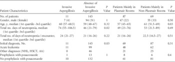
A total of 167 patients with chemotherapy-induced neutropenia hospitalized in the hICU between January 2009 and December 2010 were initially identified. Among them, 156 patients were included and 11 patients were excluded (8 patients with neutropenia lasting <7 days and 3 patients due to the lack of data about the room’s air filtration equipment). Baseline characteristics of included patients are shown in Table 1. Most patients (70.5%) were treated for acute leukemia; 20.5% had autologous HSCT; 6.5% received chemotherapy for non-Hodgkin’s lymphoma; and 2.5% had aplastic anemia.
Patients and Clinical Characteristics

NOTE. HSCT, hematopoietic stem cell transplant; NHL, non-Hodgkin’s lymphoma; AA, aplastic anemia.
A total of 14 patients treated with high-dose cytarabine as consolidation therapy received prophylactic antifungal therapy with pozaconazole. A total of 82 patients received caspofungin in pre-emptive therapy.
Environmental Contamination
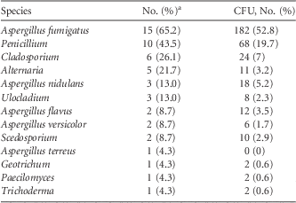
During the study period, 136 surface samples and 149 air samples were collected and analyzed. In non-equipped rooms, 5.9% of the surface samples were positive above the threshold and none of the surface samples were positive in Plasmair-equipped rooms (no significant difference, P=.10) (Table 2). On the other hand, the rate of air contamination in Plasmair-equipped rooms was significantly lower than in non-equipped rooms (7.3% and 19.4%, respectively; P=.04). The species isolated from positive air and surface samples are shown in Table 3. Aspergillus fumigatus was the most commonly identified species, with 28.8% of positive samples. Penicillium was found in 19.2% of positive samples. Particle counts were below the threshold in 43 of 45 samples in Plasmair-equipped rooms, whereas the threshold was exceeded in 28 of 30 samples in non-equipped rooms (P<.001) (Table 2).
Environmental Contamination: Fungal Load of Air Samples Collected in Rooms With or Without Plasmair, Particle Counting and Surface Contamination

NOTE. CFU, colony-forming unit.
a Samples are noted as positive because CFU or particle count exceeded the threshold.
b Fisher’s exact test.
c Surface contamination threshold, 5 CFU/25 cm2.
d Air contamination threshold, 5 CFU/m3.
e Particle count threshold, 350,000/m3.
Isolated Species From Positive Air and Surface Samples Collected During the Study Period

NOTE. CFU, colony-forming units.
a The no. of positive samples from air and surfaces was 23.
Incidence of Invasive Aspergillosis
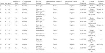
During the study period, 11 cases of probable IA were diagnosed. Notably, no IA case had signs of IA at admission. Patient characteristics are shown in Table 4. The serum GM assay was positive in all cases. We diagnosed 2 cases using BAL, and these cultures were positive with A. fumigatus. CT scans showed a halo sign in 8 patients and a cavitation in 4 patients associated with dense lesions and macronodules. Of these patients, 9 received casponfungin, and 2 received voriconazole the diagnosis was confirmed. Subsequently, all patients received voriconazole therapy. Of all patients in our study cohort, 6 died due to their hematological malignancies and 1 died due to IA (no autopsy was performed).
Characteristics of the 11 Patients With Acute Leukemia who Developed Probable Invasive Aspergillosis

NOTE. IA, invasive aspergillosis; EORTC, European Organization for Research and Treatment of Cancer; BAL, bronchoalveolar lavage; M, male; F, female; AL, acute leukemia; LND, last news date.
a Threshold: optical density index (ODI) =0.5.
Relationship Between IA Incidence and Equipment With Plasmair Units
We estimated the proportion of days the patients (who were all neutropenic) spent in Plasmair-equipped rooms. This proportion was strongly bivariate, with values <0.1 or >0.9 for 100 of 156 patients (among these, 54 patients had only 1 stay). Therefore, we dichotomized the patients into 2 groups (proportion of days in Plasmair-equipped rooms <50% or >50%). We detected a significant association between IA and these 2 groups, with 1 of 87 IA cases in the Plasmair group versus 10 of 69 cases in the no-Plasmair group (P=.02; OR=0.11; 95% CI, 0.00– 0.84) (Table 1). Considering the group of 100 patients described above who stayed almost exclusively in Plasmair-equipped rooms (45 patients) or in non-equipped rooms (55 patients), the association between IA and Plasmair equipment remains significant, with 6 cases of IA in non-equipped rooms versus no case of IA in Plasmair-equipped rooms (P=.03). When analyzing on potential confounding factors, we found an association between age and the Plasmair group (P=.03), duration of neutropenia and IA (P=.05), and initial diagnosis and IA (P=.03) (Table 1). When performing a multivariate logistic analysis with IA as the outcome and the Plasmair group, age, duration of neutropenia, and initial diagnosis as explanatory variables to take into account all potential confounding factors, the association between IA and the Plasmair group remained significant (P=.03; OR=0.05; 95% CI, 0.01–0.22).
DISCUSSION
Previous studies have suggested a relationship between environmental fungal contamination and the incidence of IA in patients with hematologic malignancies. A positive link was demonstrated between IA incidence and the degree of fungal air or surface contamination.Reference Alberti, Bouakline and Ribaud 6 This link was more pronounced in wards not equipped with HEPA filters. Similar conclusions were reported from a large 7-year surveillance study in a university hospital including patients with hematologic malignancies, chronic immunologic disorders, and chronic pulmonary diseases.Reference Garnaud, Brenier-Pinchart and Thiebaut-Bertrand 17
Nonetheless, formal demonstration of a direct effect of air filtration systems on the incidence of IA is lacking. Most published studies emphasize the impact of fungal contamination during unusual exposure, such as that encountered during construction work. These studies were conducted in the setting of allogeneic HSCT, where patients are known to be at higher risk of IA.Reference Benet, Nicolle and Thiebaut 18 Abdul Salam et alReference Abdul Salam, Karlin, Ling and Yang 19 published data supporting the effectiveness of portable HEPA filters in a retrospective study that, in contrast to the present study, included possible IA and that compared incidence rates of IA during pre-installation and a post-installation periods. The air samples were comparable to those analyzed in previous studies in wards equipped with Plasmair.Reference Bergeron, Reboux, Poirot and Laudinet 12 , Reference Sautour, Sixt and Dalle 14 The absence of an apparent significant association between the equipment of rooms with Plasmair and low CFU counts in surface samples in the present study could be due to the small number of samples analyzed. Surface cleaning procedures were similar in Plasmair-equipped and non-equipped rooms.
In addition, Aspergillus threshold concentrations for preventing IA have yet to be defined for high-risk patients. Concentrations of 0.1–1 CFU/m3 of air in the absence of construction work have been considered.Reference Munoz, Burillo and Bouza 20 However, concentrations as low as 1 CFU/m3 of air may be responsible for IA in high-risk patients.Reference Arnow, Sadigh, Costas, Weil and Chudy 4 For this reason, the absence of any CFU in the air of LAF-equipped rooms should be required.Reference Gangneux, Adjide and Bernard 13 So far, no threshold values for wards not equipped with HEPA filters or LAF have been established. Cutoff values concerning air and surface contamination have been set arbitrarily by the local Aspergillosis committee at Hôpital de Versailles.
In this study, we demonstrated the impact of mobile air decontamination systems on fungal airborne contamination outside the setting of allogeneic HSCT. We exploited the opportunity of a 2-phase equipment installation program of mobile air decontamination systems in an hICU, resulting in concomitantly Plasmair-equipped and non-equipped rooms. This unique situation allowed us to study patients assigned to each set of these rooms, with baseline characteristics of the 2 patient populations being comparable.
A limitation of our study lies in the relatively small number of probable cases of IA recorded during the study period, while the incidence of IA in French hematology units was comparable.Reference Bocquet, Patris, Dumartin, Gottot, Rykner and Brucker 21 Moreover, although patients were allocated to Plasmair-equipped and non-equipped rooms only depending on their availability, the design of this study does not reach the level of evidence of a prospective double-blinded randomized trial.
In this retrospective study, we did not take into account the date of IA diagnosis because it could not be determined with certainty in all cases. Furthermore, when IA was diagnosed, patients were not moved from a non-equipped room to a Plasmair-equipped room, which avoided additional bias.
Another matter of debate concerns the level of air filtration needed for patients with acute leukemia or patients undergoing autologous as opposed allogeneic HSCT. It has been suggested that HEPA filters combined with LAF systems may be the most efficient means of controlling environmental fungal contamination, at least in the setting of allogeneic HSCT.Reference Alberti, Bouakline and Ribaud 6 Using mobile air decontamination systems without LAF systems in the context of pre-emptive antifungal therapy, we observed a very low incidence of IA. This low incidence of IA was also associated with a very low rate of environmental fungal contamination, suggesting that mobile air decontamination systems, such as Plasmair, may be efficient and convenient alternatives to HEPA filters and LAF devices for patients not undergoing allogeneic HSCT.
In summary, we have demonstrated that a mobile air decontamination system (Plasmair) significantly reduced the incidence of IA in neutropenic patients with hematologic malignancies. Such systems may represent an efficient and convenient alternative to HEPA filtration combined with LAF.
ACKNOWLEDGMENTS
The authors would like to thank Le Bihan Marie Renée and Breton Nelly for technical assistance and the Centre Hospitalier de Versailles for editorial assistance. The authors also thank Ekkehard Collatz for reviewing the English usage.
Financial support: No financial support was provided relevant to this article.
Potential conflicts of interest: All authors report no conflicts of interest relevant to this article.