Congenitally corrected transposition of the great arteries is a rare form of CHD. Congenitally corrected transposition of the great arteries is reported to occur in both situs solitus and in situs inversus. Approximately 90–95% of cases occur with situs solitus, and 5–10% occur with situs inversus.Reference Feingold, O’Sullivan and del Nido 1 In rare cases, transposition events may be accompanied by single coronary artery anomalies.Reference Ismat, Baldwin and Karl 2 Furthermore, atrioventricular block associated with isolated dextrocardia or situs inversus or atrioventricular discordance cases have been reported in the literature.Reference Shenthar, Rai and Walia 3 , Reference Huhta, Maloney and Ritter 4 In this case report, for the first time in the literature, a young situs inversus patient with congenitally corrected transposition, single coronary artery anomaly, and atrioventricular block together is presented.
Case report
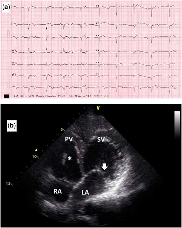
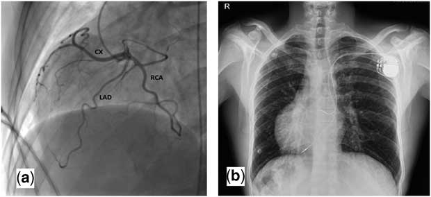
A 37-year-old man was admitted to the outpatient department with the complaint of chest pain and dizziness for the past several days. The symptoms worsened with exertion and were not related with the body position. There was no history of syncope or neurological involvement. He took no medications and had no known diseases. On initial examination, cardiac sounds were heard on the right side of the chest, the blood pressure was 130/70 mmHg, the pulse was 52 per minute, the respiratory rate was 16 per minute, and the oxygen saturation was 98% while he was breathing ambient air. The remainder of the examination was normal. Electrocardiogram on admission revealed atrioventricular dissociation and complete block (Fig 1a). Echocardiography demonstrated dextrocardia with congenitally corrected transposition of great arteries and mild systemic – morphologically tricuspid – valve insufficiency (Fig 1b). Abdominal ultrasound demonstrated reversal of abdominal viscera. Serum biochemical and haematologic parameters were within normal limits. There was no obvious cause of the heart block. The coronary angiography showed a single coronary artery arising from a single ostium, which originated from the left coronary sinus and supplying the whole heart (Fig 2a). CT coronary angiography confirmed dextrocardia, besides single coronary artery anomaly without interarterial course and atherosclerosis. The patient was subsequently referred for implantation of a DDD-R pacemaker. By means of the left subclavian vein, atrial and ventricular leads were advanced through and fixed in the right auricle and venous ventricular apex, morphologically left, respectively. The leads were connected to a double-chamber pacemaker (Fig 2b). The patient recovered uneventfully and was discharged 3 days after the procedure without complication.
Standard 12-lead ECG showing complete atrioventricular block obtained on admission (a). Apical four-chamber view of the heart illustrating reversal insertion of ventricles. The left sided atrioventricular valve is more apically positioned (arrow), thus the right ventricle (systemic ventricle) is left sided and connected to the left atrium (b). LA=left atrium; PV=pulmonic ventricle; RA=right atrium; SV=systemic ventricle; (*) Pacemaker lead

Left anterior oblique cranial angulation view shows single coronary artery anomaly (a). Chest X-ray demonstrating the correct placement of the DDD-R pacemaker (b).

Discussion
We present a case of congenitally corrected transposition of the great arteries in situs inversus with a single coronary artery and complete atrioventricular block. To the best of our knowledge, this combination of abnormalities has not been reported previously.
The risk of complete atrioventricular block in patients with atrioventricular discordance has been demonstrated.Reference Huhta, Maloney and Ritter 4 A reviewed data analysis on 107 patients showed that with increasing follow-up the risk of natural onset atrioventricular block continued at a rate of ~2% per year after diagnosis.Reference Huhta, Maloney and Ritter 4 A long course of the atrioventricular bundle and fibrosis in the bundle was demonstrated by Walmsley report.Reference Walmsley 5 It might be helpful to explain the mechanism of complete atrioventricular block in patients with atrioventricular discordance.
The majority of patients with situs inversus have no other associated pathology and enjoy a normal life period. On the other hand, congenital heart anomalies are associated with up to 3% of situs inversus patients and can affect the life span.Reference Torgensen 6 Atrioventricular block associated with isolated dextrocardia or situs inversus cases has been reported in the literature.Reference Shenthar, Rai and Walia 3 Similarly, the risk of complete atrioventricular block in patients with atrioventricular discordance has been demonstrated.Reference Huhta, Maloney and Ritter 4 The presence of both clinical conditions in our case suggests that this combination can often cause atrioventricular block. Therefore, a closer follow-up of congenitally corrected transposition patients with situs inversus may be considered. Also, our case was accompanied by a single coronary artery anomaly.
Single coronary is an anomalous condition in which the single coronary artery originates from either the left or right aortic sinus and supplies the entire heart. These cases are rare, occurring in ~0.024% of the population according to Lipton’s study.Reference Lipton, Barry and Obrez 7 According to the literature, situs inversus and single coronary artery occurring together is even more rare and there were no accompanying conduction system defects.Reference Wester, Ernst and Mast 8 Accordingly, it can be assumed that single coronary artery anomaly does not present an additional risk for atrioventricular block. However, the presence of interarterial course is important for sudden cardiac death in patients with single coronary artery anomaly. In our case, there was no interarterial course in the tomography.
Venous return anomalies might be seen in patients with situs inversus who have associated congenital cardiac defect.Reference Pott, Brar and Valderrabano 9 Pacemaker implantation may be difficult in these patients owing to the complex anatomical arrangement. Thus, information about anatomy and/or coexisting congenital abnormalities, which may preclude percutaneous approach, should be gained before the procedure. There was no venous return anomaly in our case. We successfully implanted DDD-R pacemaker by using the left subclavian vein.
Coexistence of single coronary artery and congenitally corrected transposition in a situs inversus patient was reported only in one case in the literature.Reference Kuwata, Yoneyama and Izumo 10 Atrial septal defect was also presented in this case; however, atrioventricular block was not accompanied. In our case, however, atrioventricular block was accompanied. This is the first case in the literature in which situs inversus with congenitally corrected transposition of great arteries, single coronary artery anomaly, and complete atrioventricular block co-exist in a patient.
Acknowledgements
None.
Financial Support
This research received no specific grant from any funding agency or commercial or not-for-profit sectors.
Conflicts of Interest
None.
Ethical Standards
Informed consent was obtained from all individual participants included in this case.