Introduction
Abdominal angiostrongyliasis (AA) is a disease caused by the nematode Angiostrongylus costaricensis, which inhabits the mesenteric arteries of its hosts (Morera & Céspedes, Reference Morera and Céspedes1971). Wild rodents serve as definitive hosts, while terrestrial molluscs are the intermediate hosts (Morera & Céspedes, Reference Morera and Céspedes1971).
In humans, the disease often becomes apparent through intestinal lesions characterized by infarction, pseudotumour or acute appendicitis (Graeff-Teixeira et al., Reference Graeff-Teixeira, Camillo-Coura and Lenzi1991). In the most severe cases, with intestinal obstruction or perforation, AA requires surgical treatment. Finding the mechanism whereby the parasite causes ischaemic lesions is crucial for developing strategies for the prevention and treatment of associated complications (Mousa, Reference Mousa and Mousa2004).
The use of anthelmintics has not been deemed efficient against AA, as intestinal lesions could be aggravated by the death of the parasite inside the arteries (Morera & Bontempo, Reference Morera and Bontempo1985; Mentz & Graeff-Teixeira, Reference Mentz and Graeff-Teixeira2003). Anti-inflammatory drugs, such as betamethasone, are not efficacious either, causing deterioration of the lesions in some cases (Fante et al., Reference Fante, Dieterish and Rodriguez2008).
Anticoagulants and thrombolytics could help in the prevention of ischaemic intestinal lesions (Mousa, Reference Mousa and Mousa2004). There are several commercially available anticoagulants that interfere with different stages of blood coagulation (Harter et al., Reference Harter, Levine and Henderson2015). Heparins are widely used, including enoxaparin, a low-molecular-weight heparin, for the treatment of ischaemia and infarction (Silvain et al., Reference Silvain, Beygui, Barthélémy, Pollack, Cohen, Zeymer, Huber, Goldstein, Cayla and Collet2012). Enoxaparin inhibits the conversion of prothrombin to thrombin and reduces the conversion of fibrinogen to fibrin, preventing clot formation. It also reduces coagulation factors and inactivates factor X (Harter et al., Reference Harter, Levine and Henderson2015).
A previous study utilized prophylactic doses of enoxaparin for the treatment of mice infected with larvae (L3 stage) of A. costaricensis. The enoxaparin-treated animals did not show any differences in the development and severity of intestinal lesions and in mortality rate when compared to untreated ones. It is hypothesized that the low dose used in this study was not efficient in preventing thrombogenesis (Rodriguez et al., Reference Rodriguez, Porto, dos Santos Ferrari, Marcolan, da Silva, Graeff-Teixeira and Fornari2011).
Therefore, due to the lack of treatments against AA, the aim of the present study was to assess the effect of high doses of enoxaparin on the prevention of ischaemic intestinal lesions and, consequently, on the survival of mice infected experimentally with A. costaricensis.
Materials and methods
Animals
Twenty-four male Swiss mice aged from 6 to 8 weeks and weighing 25–38 g were used. The animals were placed in appropriate cages with water and food ad libitum.
Isolation of A. costaricensis larvae
The larvae (L3) were isolated from infected Phyllocaulis variegatus, Limax maximus and Meghimatium pictum slugs, in Marau, State of Rio Grande do Sul, Brazil, after detection of a positive case of AA in a local resident. The slugs were stored in moist plastic jars and fed chayote (Sechium edule); after 20 days they were euthanized and the tissues were digested with 0.03% pepsin and 0.7% hydrochloric acid at 37°C for 12 h. Thereafter, the modified Baermann technique was used for isolation of L3 larvae.
Drugs
Enoxaparin (Clexane®, Aventis, Maisons-Alfort, França; vials containing 20 mg) was injected subcutaneously. The drug was diluted in water and prepared in insulin syringes at doses of 2.5 mg/kg, according to the weight of each animal. The volume of placebo in the control group was identical to the volume of enoxaparin used in the treated group. For both sedation and euthanasia, isoflurane (Isoforine®, Cristália, São Paulo, Brazil) was administered by inhalation using an induction chamber.
Experimental design
The mice were infected with ten L3 larvae of A. costaricensis, then were split into two groups of 12 animals as follows: Group 1, control, given placebo; Group 2, treated with enoxaparin.
Experimental protocol
On day 1, the mice (n = 24) were sedated with isoflurane and infected by oral administration (gavage) of ten L3 larvae of A. costaricensis. After infection, the mice were kept in appropriate cages and monitored daily for food acceptance, defecation, activity and weight. From day 15, the animals in Group 1 were given daily doses of sterile water by injection (placebo); at the same time, the animals in Group 2 received daily doses of enoxaparin (2.5 mg/kg) injected subcutaneously. Both groups received 1.6 mg/ml of paracetamol (Tylenol®, Janssen, São José dos Campos, São Paulo, Brazil), on a daily basis, for pain relief. The experimental period covered 50 days, and the animals were sacrificed on the last day by inhaled isoflurane and then submitted to necropsy.
Necropsy and histological analysis
The gross aspect of the gastrointestinal tract was examined for signs of ischaemic lesions, peritonitis (fibrin deposition) and pseudotumours. The specimens were fixed in 10% formalin for 24 h. For the microscopic analysis, the specimens were embedded in paraffin and sectioned in a microtome (at 5 μm thickness). The slides were stained with haematoxylin and eosin and examined by two independent pathologists, who were unaware of the treatment given to each animal. The following microscopic aspects were investigated: (1) presence of infarction; (2) identification of adult parasites, larvae and eggs; (3) eosinophilic infiltrate (quality and quantity); (4) granuloma formation; and (5) characterization of vasculitis and thrombosis.
Statistical analysis
The differences in proportions were analysed by the chi-square test. However, Fisher's exact test, as a substitute for the chi-square test, was used in the tables in which the estimated values of counts were smaller than 5. Survival data were analysed by the Kaplan–Meier method, and comparison between groups was performed by the log-rank test. The data were considered to be significantly different when P < 0.05.
Results
Mortality
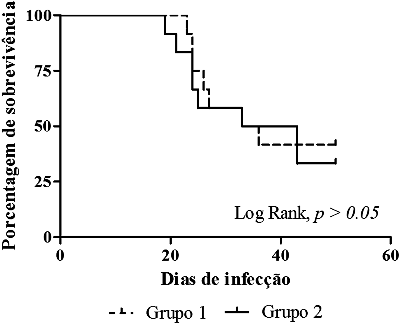
Fifteen animals (62.5%) died between 19 and 43 days after infection. Of these, seven (46.7%) participated in the control group and eight (53.3%) were in the group treated with enoxaparin. At the end of the experiment, nine animals survived (37.5%) (fig. 1).
Survival curve stratified by the treatments used in mice infected with Angiostrongylus costaricensis (Log Rank test, P < 0.05).

Gross findings
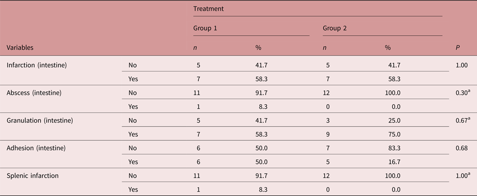
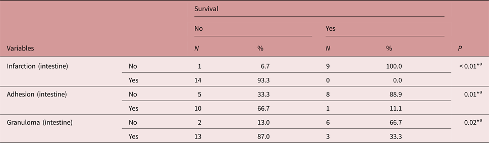
The results of the gross analyses did not indicate significant differences according to Fisher's exact test, as shown in table 1. Intestinal lesions included infarction, granulation in the serosa and adhesion was performed in both groups.
Gross findings in mice infected with Angiostrongylus costaricensis and treated with placebo (Group 1) or enoxaparin (Group 2).

* Probability lower than 5% (P < 0.05) indicates significant difference (chi-square test).
a Fisher's exact test replaced chi-square test when the estimated counts were smaller than 5 (2 × 2 table).
Microscopic findings
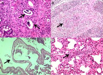
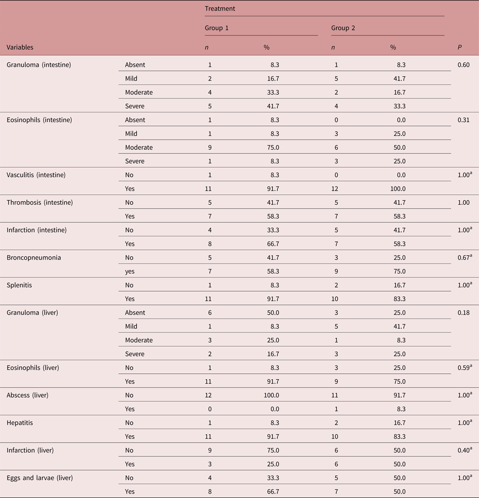
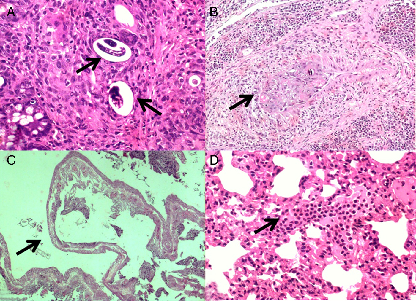
In the intestine, where granulomas (fig. 2A) were present, no significant difference was observed in their levels of severity between Group 1 (91.7%) and Group 2 (91.7%). On the other hand, Group 2 had a higher proportion of mild granuloma (41.7%) than Group 1 (16.7%) (table 2). The presence of thrombosis (fig. 2B) was the same for both groups, i.e. 58.3%; infarction (fig. 2C) was detected in 66.7% of Group 1 animals, compared to 58.3% of the animals in Group 2 (table 2).
Microscopic findings: (A) granulomas in the submucosal layer with eggs and larvae (magnification 200×); (B) arterial thrombosis (magnification 100×); (C) intestinal infarction (magnification 50×); (D) bronchopneumonia with neutrophilic infiltrate (200×).
Microscopic findings in mice infected with Angiostrongylus costaricensis and treated with placebo (Group 1) or enoxaparin (Group 2).

*Probability lower than 5% (P < 0.05) indicates significant difference (chi-square test).
a Fisher's exact test replaced chi-square test when the estimated counts were smaller than 5 (2×2 table).
In the lungs, there was no difference between treatments in terms of granuloma severity, presence of thrombi and presence of eggs and larvae, which were absent in both groups. Bronchopneumonia (fig. 2D) was detected in 58.3% of animals from Group 1 and in 75% of animals from Group 2 (table 2).
Likewise, in the spleen, there were no significant differences between the groups regarding infarction (G1: 16.7%; G2: 25%), granuloma (G1: 25%; G2: 16.7%), thrombosis (G1: 16.7%; G2: 0%), eosinophilic infiltrate (G1: 75%; G2: 58.3%), eggs and larvae (G1: 16.7%; G2: 25%), nor in the liver regarding infarction (G1: 25%; G2: 50%), granuloma (G1: 50%; G2: 75%), abscesses (G1: 0%; G2: 8.3%), hepatitis (G1: 91.7%; G2: 83.3%), eosinophilic infiltrate (G1: 91.7%; G2: 75%), and eggs and larvae (G1: 66.7%; G2: 50%), as shown in table 2. There were no findings in the brain and kidneys of animals from either group.
As to the survival of animals in both groups, 93% (14/15) of those that died had intestinal infarction, with granulation in 87% of the cases (13/15) and adhesions in 66.7% (10/15) (table 3).
Gross findings in mice infected with Angiostrongylus costaricensis associated with survival.

* Probability lower than 5% (P < 0.05) indicates significant difference (chi-square test).
a Fisher's exact test replaced chi-square test when the estimated counts were smaller than 5 (2 × 2 table).
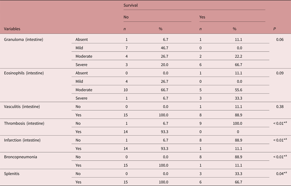
Thrombi and intestinal infarction were present in 93.3% (14/15) and bronchopneumonia and splenitis in 100% of the animals that died (table 4).
Microscopic findings in mice infected with Angiostrongylus costaricensis associated with survival.

* Probability lower than 5% (P < 0.05) indicates significant difference (chi-square test).
a Fisher's exact test replaced chi-square test when the estimated counts were smaller than 5 (2×2 table).
Increased proportions of mild to moderate granuloma and eosinophilic infiltrate were observed in the animals that had an early death compared to those that survived for up to 50 days.
Discussion
In this study, we found that high doses of enoxaparin (2.5 mg/kg) (Andújar et al., Reference Andújar, Matoses, Rodríguez and Navarro2016) do not have a protective effect against AA, since the survival rate of mice did not vary between the treated and control groups, and both gross and microscopic findings were similar between them.
There is no effective drug treatment against AA, although another study demonstrated that levamisole is efficient in the eradication of A. costaricensis larvae when given orally at the onset of infection in mice (Ishii et al., Reference Ishii, Terada, Fujiu and Sano1989) and in the elimination of the adult parasite in vitro (Terada et al., Reference Terada, Rodriguez, Dharejo, Ishii and Sano1986).
In severe cases, surgical intervention is the only treatment choice available for AA, with removal of the affected intestinal segment (Graeff-Teixeira et al., Reference Graeff-Teixeira, Camillo-Coura and Lenzi1991). Due to the lack of an efficacious drug treatment, and as one of the main complications of the disease consists of thrombogenesis, the use of anticoagulants could be a way to prevent thromboembolic events and to reduce morbidity and mortality (Rodriguez et al., Reference Rodriguez, Porto, dos Santos Ferrari, Marcolan, da Silva, Graeff-Teixeira and Fornari2011).
Therefore, a previous study was carried out using daily preventive doses of enoxaparin (0.57 mg/kg) in mice infected with A. costaricensis. The treatment was not effective in preventing intestinal lesions and in reducing the mortality rate (Rodriguez et al., Reference Rodriguez, Porto, dos Santos Ferrari, Marcolan, da Silva, Graeff-Teixeira and Fornari2011). The low dose of enoxaparin used in the study could explain its inefficacy in preventing the lesions caused by the parasite.
So, in the present study, we used the same experimental design as the previous study, but we administered a higher dose of enoxaparin (2.5 mg/kg daily), which, nevertheless, was not efficient in the treatment of AA.
Gross findings were very similar in the control and treated groups. The mice showed granulation in the serosa, adhesion of the intestine to the abdominal wall and intestinal infarction. In a previous study, mice infected experimentally with A. costaricensis also developed these lesions. However, in the present study, there were a smaller number of intestinal abscesses and a higher prevalence of intestinal infarction, which affected 14 animals, compared to four in the previous study. Intestinal infarction is the main complication of AA, eventually leading to death (Graeff-Teixeira et al., Reference Graeff-Teixeira, Camillo-Coura and Lenzi1991; Waisberg et al., Reference Waisberg, Corsi, Rebelo, Vieira, Bromberg, dos Santos and Monteiro1999), and its higher prevalence is due probably to the larger virulence of the parasites, which were isolated from slugs near where a resident diagnosed with AA lived (Rafaluk et al., Reference Rafaluk, Gildenhard, Mitschke, Telschow, Schulenburg and Joop2015).
In the microscopic analysis, the lesions were similar in both groups. Vascular granuloma, eosinophilic infiltrate, thrombosis and intestinal necrosis were observed. Granuloma and eosinophilic infiltrate were also detected in the spleen and in the liver. The microscopic findings are quite similar to those described by Rodriguez et al. (Reference Rodriguez, Porto, dos Santos Ferrari, Marcolan, da Silva, Graeff-Teixeira and Fornari2011), showing that high-dose enoxaparin was not efficient in reducing the prevalence of lesions.
Statistical analyses were also performed, with classification of animals into survivors and non-survivors, regardless of the treatment used. Of the 15 mice that died during the experiment, 14 had infarction and intestinal necrosis, which probably led to their deaths. Other frequent findings in non-survivors were adhesion of the intestine to the abdominal wall, granulation of the serosa and thrombosis, demonstrating worse outcomes for these animals (de Azevedo et al., Reference de Azevedo, Rodriguez, Porto, Graeff-Teixeira and Fornari2011).
The therapeutic doses of enoxaparin consist of 1.5 mg/kg given subcutaneously every 24 h (Andújar et al., Reference Andújar, Matoses, Rodríguez and Navarro2016). Drug uptake through this route of administration is approximately 100%, with an average half-life of 4.5 h (Andújar et al., Reference Andújar, Matoses, Rodríguez and Navarro2016). In this study, we used 2.5 mg/kg, whereas in a previous study the mice given 5 mg/kg showed excellent anticoagulant activity and a half-life of 5 h (Li et al., Reference Li, Lee, Sim, Toida, Linhardt and Kim2004). Hence, the inefficacy of enoxaparin in the prevention of intestinal lesions caused by A. costaricensis was not because of insufficient anticoagulation. Thus, thrombogenesis might not be the major cause of vascular occlusion and intestinal infarction; instead, this could be attributed to vascular granulomas and to the parasite itself (Rodriguez et al., Reference Rodriguez, Porto, dos Santos Ferrari, Marcolan, da Silva, Graeff-Teixeira and Fornari2011), which may explain the inefficacy of the anticoagulant used.
We conclude that high doses of enoxaparin neither prevent intestinal lesions nor increase the survival of mice infected experimentally with A. costaricensis. Finally, we also emphasize the need for further studies to check the importance of the inflammatory reaction in AA pathogenesis.
Conflict of interest
None.
Ethical standards
The experiment was performed at the laboratory animal facility of the Institute of Biological Sciences of Universidade de Passo Fundo, State of Rio Grande do Sul, Brazil. The study protocol (no. 030/2014) was approved by the Animal Research Ethics Committee of Universidade de Passo Fundo.