Introduction
The foliose lichen genus Phaeophyscia, first described by Moberg, was originally included in the genus Physcia (Schreb.) Michx. and comprises c. 50 species worldwide. This group occurs on diverse substrata, including bark, wood, bryophytes, rock and soil, in a wide range of habitats (Moberg Reference Moberg1977; Aptroot & Sipman Reference Aptroot and Sipman1991). Phaeophyscia is characterized by a foliose thallus, white or orange-red medulla, paraplectenchymatous upper and lower cortex, lecanorine apothecia with Physcia or Pachysporaria-type ascospores, ellipsoid conidia, and an absence of atranorin (Moberg Reference Moberg1977; Aptroot & Sipman Reference Aptroot and Sipman1991).
Numerous authors have conducted taxonomic studies of Phaeophyscia around the world, from North America (Esslinger Reference Esslinger1978a, Reference Esslingerb; Brodo & Sharnoff Reference Brodo, Sharnoff, Brodo, Sharnoff and Sharnoff2001), East Africa (Moberg Reference Moberg1983), China (Moberg Reference Moberg1995; Hu & Chen Reference Hu and Chen2003; Li & Zhao Reference Li and Zhao2006), Japan (Kashiwadani Reference Kashiwadani1975, Reference Kashiwadani1984a, Reference Kashiwadanib; Harada Reference Harada2016) and Europe (Moberg Reference Moberg1974, Reference Moberg1977, Reference Moberg1978, Reference Moberg1994; Poelt Reference Poelt1974; Hale Reference Hale1983) to Russia (Moberg Reference Moberg1995). Little attention has been paid to the phylogenetics of this group, although some molecular studies of several species have been conducted as part of studies on Physconia Poelt, Physcia (Schreb.) Michx., Anaptychia Körb., Heterodermia Trevis. and other related groups (Helms et al. Reference Helms, Friedl and Rambold2003; Cubero et al. Reference Cubero, Crespo, Esslinger and Lumbsch2004; Lücking et al. Reference Lücking, Del Prado, Lumbsch, Will-Wolf, Aptroot, Sipman, Umaña and Chaves2007; Lohtander et al. Reference Lohtander, Ahti, Stenroos and Urbanavichus2008; Gaya et al. Reference Gaya, Högnabba, Holguin, Molnar, Fernández-Brime, Stenroos, Arup, Søchting, van den Boom and Lücking2012).
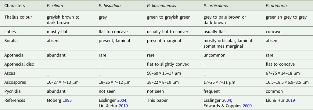
Previously, eight species of Phaeophyscia have been reported from Pakistan (for instance, see Table 1), of which half were added by Ahmad (Reference Ahmad1965), viz. Phaeophyscia ciliata (Hoffm.) Moberg, P. endococcina (Körb.) Moberg, P. hispidula (Ach.) Essl. and P. orbicularis (Neck.) Moberg. Bhatty & Iqbal (Reference Bhatty and Iqbal1978) added P. sciastra (Ach.) Moberg and the remaining three species were reported by Aptroot & Iqbal (Reference Aptroot and Iqbal2012), viz. P. exornatula (Zahlbr.) Kashiw., P. melanchra (Hue) Hale and P. primaria (Poelt) Trass. In this study, we combined morphology, chemistry and phylogenetic analysis with the aim of clarifying the relationships among Physciaceae and investigating support for a potential new species.
Comparison of morphological characters of selected Phaeophyscia species recorded in Pakistan and which are similar to P. kashmirensis.

Materials and Methods
Study area and collection
A survey of the Jhelum Valley in Azad Jammu and Kashmir, undertaken in 2021, focused on increasing the biotic knowledge of the lichens of Pakistan. Azad Jammu and Kashmir, which lies in the north-eastern part of Pakistan, comprises an area of 13 297 km2, with mountains ranging from 360 to 6325 m in altitude, and is characterized by siliceous rocks and undulating terrain (Dar et al. Reference Dar, Cochard, Shreshta and Ahmad2012). The collected samples were deposited in the herbarium of the Institute of Botany, University of the Punjab, Lahore (LAH).
Morphological characterization
Specimens were examined macro and micromorphologically under a stereomicroscope (Meiji Techno, EMZ-5TR, Japan) and a compound microscope (SWIFT M4000-D), with a 9MP camera system, respectively. For anatomical investigation, hand-cut sections of thallus and apothecia were mounted in water and potassium hydroxide (K). A minimum of 20 measurements in water were made for each diagnostic feature from two samples; length × width of ascus and dimensions of ascospores were measured. The dimensions of ascospores and ascus are presented as smallest value recorded–largest value recorded.
Chemical characterization
Chemistry was analyzed using standard spot tests. These were performed with 10% K, 5% para-phenylenediamine in ethanol (P) and calcium hypochlorite solution (C). Secondary chemistry was analyzed using thin-layer chromatography (TLC) using solvent system C, following standard methods (Orange et al. Reference Orange, James and White2010).
DNA extraction, PCR amplification and sequencing
Genomic DNA was extracted directly from a portion of thallus with apothecia from each specimen using a modified 2% CTAB method (Gardes & Bruns Reference Gardes and Bruns1993). The ITS-nrDNA region (internal transcribed spacer of the nrDNA) was amplified using the primer pair ITS1F (forward primer) (Gardes & Bruns Reference Gardes and Bruns1993) and ITS4 (reverse primer) (White et al. Reference White, Bruns, Lee, Taylor, Innis, Gelfand, Sninsky and White1990), following the amplification protocol of Khan et al. (Reference Khan, Khalid and Lumbsch2018). PCR products were visualized on a 1% agarose gel with ethidium bromide (Sambrook & Russel Reference Sambrook, Russel, Sambrook and Russel2001). PCR products were sent to Tsingke, China for sequencing. Sequences were assembled using BioEdit (Hall Reference Hall1999). BLAST analysis was used to retrieve highly similar sequences of the nrITS region. The maximum query coverage and percent identity of the sequences, along with related taxa, were noted. Sequences retrieved from GenBank and sourced from relevant literature were used in an initial alignment, which was trimmed and then realigned using web-PRANK with default settings (Löytynoja & Goldman Reference Löytynoja and Goldman2010). On the CIPRES Portal (Miller et al. Reference Miller, Pfeiffer and Schwartz2010), the HYK + G + I model was selected using jModelTest (Posada Reference Posada2008). Using RAxML-HPC2 v.8.1.11 on CIPRES, a maximum likelihood (ML) analysis was also implemented (Stamatakis Reference Stamatakis2014), with 1000 bootstraps used for rapid bootstrapping. FigTree v. 1.4.3 (Rambaut et al. Reference Rambaut, Suchard, Xie and Drummond2014) was used for visualizing the phylogeny from the ML analysis.
Results
Molecular phylogenetic analyses
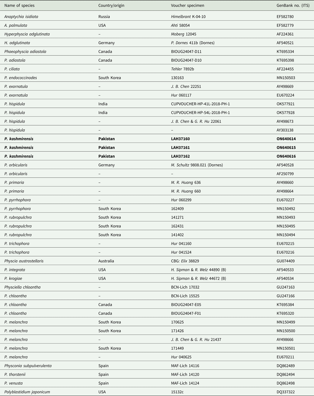
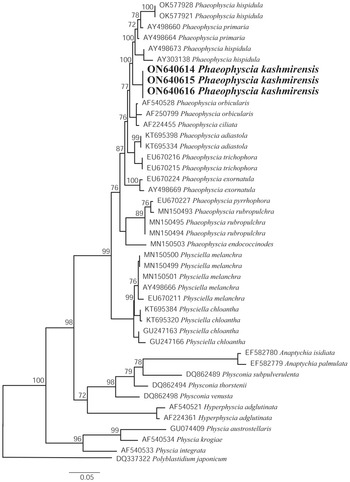
ITS sequences of the holotype of the new species and of the collections CKR-03 and CKR-4 were identical. The final ITS dataset consisted of 42 sequences, including Polyblastidium japonicum (M. Satô) Kalb (DQ337322) as outgroup (Liu & Hur Reference Liu and Hur2019) (see Table 2 for voucher details). The aligned ITS1-5.8S-ITS2 region comprised 542 sites, of which 289 were conserved and 241 variable; 186 sites were parsimony-informative. In the analysis of the ITS region (Fig. 2), three newly generated sequences formed a separate clade in a strongly supported sister relationship with the Phaeophyscia hispidula group, justifying the recognition of an undescribed species which is presented here.
Voucher information and GenBank Accession numbers of sequences used in the ITS phylogenetic analysis of Phaeophyscia and related species. New sequences are in bold.

Taxonomy
Phaeophyscia kashmirensis Fayyaz, Afshan & Khalid sp. nov.
MycoBank No.: MB 844145
The species is characterized by a green to greyish green thallus, usually narrow (0.5–2 mm), flat to convex lobes with abundant marginal soralia, black, dense small rhizines, small Physcia-type ascospores 18–22 × 8–10 μm and an absence of pycnidia.
Type: Pakistan, Azad Jammu and Kashmir, Garhi Dupatta, 34°36ʹN, 73°35ʹE, 817 m alt., on siliceous rock, 2 October 2021, I. Fayyaz & N. S. Afshan (CKR-23) (LAH37160—holotype).
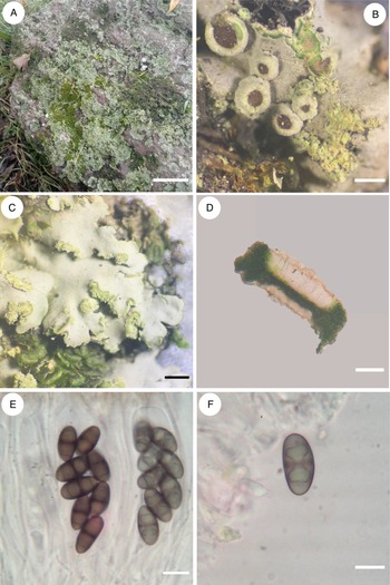
(Fig. 1)
Phaeophyscia kashmirensis. A, foliose thallus. B, apothecia. C, lobe with rhizines. D, section of an apothecium. E, ascus. F, ascospore. Scales: A = 2 cm; B = 0.5 mm; C = 2 mm; D = 75 μm; E = 16 μm; F = 8 μm. In colour online.

Thallus foliose, green when fresh but greyish green when dry, loosely attached to substratum, very variable in size, 5–10 cm diam., orbicular to irregular. Lobes irregularly or dichotomously branched, without pruina, lobe tips creamy to paler, flat to slightly convex, imbricate, usually upturned near the tips, 0.5–2 mm wide. Soralia frequently present, marginal. Lobules absent. Cortex dark brown, paraplectenchymatous, 15–20 μm thick. Algal layer 25–30 μm thick. Photobiont cells globose, 10–15 μm diam. Medulla white. Lower surface black, usually becoming pale to brown towards lobe tips, rhizinate. Rhizines dense, black, simple, 1–2 mm long. Lower cortex paraplectenchymatous, blackish brown, 22–35 μm thick.
Apothecia rather rare, up to 0.5 mm diam., sessile to very short stipitate, the margin entire or becoming irregularly crenate, lacking cortical hairs, epruinose. Disc flat to slightly convex. Epihymenium light brown, 20–24 μm. Hymenium hyaline, 70–80 μm. Hypothecium hyaline to light brown, 35–40 μm. Ascus cylindrical to clavate, 50–60 × 15–17 μm. Ascospores dark brown, ellipsoid, Physcia-type, 18–22 × 8–10 μm. Paraphyses hyaline, branched and anastomosing, 2–3 μm thick.
Pycnidia not found.
Chemistry
Thallus K−, C−, KC−, P−; no substances detected by TLC.
Etymology
The specific epithet ‘kashmirensis’ (Latin) refers to the type locality of Azad Jammu and Kashmir.
Habitat and distribution
The known collections of the new species are from Himalayan moist temperate forest in Azad Jammu and Kashmir. The specimens were found on siliceous rocks. The coniferous forest is dominated by species such as Pinus roxburghii Sarg., Pyrus pashia L., Quercus oblongata D. Don and Q. glauca Thumb. The maximum daily temperature of the region varies from 30–32 °C during the summer, with an average winter temperature of 4 °C, and there is moderate rainfall.
Additional specimens examined
Pakistan: Azad Jammu and Kashmir: Chikar (34 $^\circ $36ʹN, 73
 $^\circ $35ʹE), 1607 m alt., on rock, 2021, I. Fayyaz & N. S. Afshan (CKR-03); ibid., 2021, I. Fayyaz & N. S. Afshan (CKR-04).
Discussion
Three collections of this taxon were made from Garhi Dupatta and Chikar (Muzaffarabad District, Azad Jammu and Kashmir). The two sites vary significantly in terms of elevation, vegetation and climatic conditions. However, phylogenetic analysis confirmed that the three collections of Phaeophyscia kashmirensis form a monophyletic clade with good support (Fig. 2).
Phylogeny of Phaeophyscia and related species based on a maximum likelihood (ML) analysis of the ITS region. The tree was rooted using Polyblastidium japonicum. The new species, Phaeophyscia kashmirensis, is shown in bold.

In the phylogeny, Phaeophyscia kashmirensis appeared as sister species to the P. hispidula group, comprising the sorediate P. hispidula and the non-sorediate P. primaria. The new taxon has a green to greyish green thallus and usually narrow, flat to convex lobes (0.5–2 mm wide). Phaeophyscia hispidula differs in having a grey thallus and broad, flat to concave lobes 1–3 mm wide, while P. primaria differs in the greenish grey to grey thallus, broad, concave lobes 2–8 mm wide, absence of soralia, and smaller ascospores (16.5–18.5 × 6.9–8.5 μm) (Liu & Hur Reference Liu and Hur2019).
Phaeophyscia orbicularis differs in the mostly laminal (to marginal) soralia and the often presence of skyrin (a yellow-orange pigment) in the uppermost part of medulla (Edwards & Coppins Reference Edwards, Coppins, Smith, Aptroot, Coppins, Fletcher, Gilbert, James and Wolseley2009). Phaeophyscia ciliata differs in the darker (grey-brown to dark brown) thallus and absence of soralia (Moberg Reference Moberg1995). These differences, together with the phylogenetic analysis, confirm the novelty of this species.
Acknowledgements
We are highly grateful to the University of the Punjab, Lahore, Pakistan for providing project no. D/72/EST-I (dated 14/01/2022) to conduct this research work. We also wish to thank the anonymous reviewers for their valuable comments that greatly improved the manuscript.
Author ORCIDs
Iram Fayyaz, 0000-0001-6193-1069; Najam-ul-Sehar Afshan, 0000-0003-4538-3626; Abdul Nasir Khalid, 0000-0002-5635-8031.