INTRODUCTION
The construct of mild cognitive impairment (MCI) encapsulates the intermediate stage between normal ageing and dementia in which cognitive decline is present yet activities of daily living are preserved (Albert et al., Reference Albert, DeKosky, Dickson, Dubois, Feldman, Fox, Gamst, Holtzman, Jagust, Petersen, Snyder, Carrillo, Thies and Phelps2011; Flicker, Ferris, & Reisberg, Reference Flicker, Ferris and Reisberg1991; Petersen et al., Reference Petersen, Smith, Waring, Ivnik, Tangalos and Kokmen1999). Development of the concept of MCI has supported a proliferation of studies of the prodromal stages of dementia, but most of the focus has been in MCI later diagnosed as Alzheimer’s disease (MCI-AD). Less work has been devoted to prodromal dementia with Lewy bodies (DLB), the second most common form of neurodegenerative dementia (Donaghy & McKeith, Reference Donaghy and McKeith2014). MCI-LB refers to MCI in individuals who will eventually be characterised as DLB, although DLB may have other prodromal presentations, such as rapid eye movement sleep behaviour disorder (RBD). While DLB is diagnosed when dementia precedes or accompanies parkinsonism (with the latter not necessarily occurring), another Lewy body dementia (LBD), Parkinson’s disease dementia (PDD), is defined as dementia occurring in the context of existing PD. The differentiation between DLB and PDD is operationalised by a “one year rule”, so that PDD is diagnosed when PD occurs a year or more before dementia. LBDs share neuropathological hallmarks (Lewy bodies and Lewy neurites; Aarsland, Reference Aarsland2016; Boeve, Reference Boeve2012; Braak et al., Reference Braak, Del Tredici, Rüb, de Vos, Steur and Braak2003) and appreciable overlap in clinical presentation, including neuropsychiatric symptoms, autonomic features such as constipation and orthostatic dizziness, sleep disturbances, and response to cholinergic therapy (Burn & McKeith, Reference Burn and McKeith2003; Donaghy, O’Brien, & Thomas, Reference Donaghy, O’Brien and Thomas2015; Thomas et al., Reference Thomas, Hendriksen, Piggott, Ferrier, Perry, Ince and O’Brien2006). Indeed, the DLB/PDD Working Group has suggested that “there is no clinical symptom that absolutely distinguishes DLB and PDD” (Lippa et al., Reference Lippa, Duda, Grossman, Hurtig, Aarsland, Boeve, Brooks, Dickson, Dubois, Emre, Fahn, Farmer, Galasko, Galvin, Goetz, Growdon, Gwinn-Hardy, Hardy, Heutink, Iwatsubo, Kosaka, Lee, Leverenz, Masliah, McKeith, Nussbaum, Olanow, Ravina, Singleton, Tanner, Trojanowski and Wszolek2007, p. 817).
Cohort studies suggest neuropsychological impairment in PD is not solely a late-stage issue, with subtle deficits detectable up to 7 years before diagnosis (Darweesh et al., Reference Darweesh, Verlinden, Stricker, Hofman, Koudstaal and Ikram2017). Because cognitive impairment may not always be detected or reported at presentation (Aarsland et al., Reference Aarsland, Kurz, Beyer, Bronnick, Piepenstock Nore and Ballard2008; reviewed in Goldman, Williams-Gray, Barker, Duda and Galvin, Reference Goldman, Williams-Gray, Barker, Duda and Galvin2014), many patients with an early alpha-synucleinopathy may be labelled as PD-MCI once parkinsonism advances, and PDD if loss of autonomy occurs, despite longer-standing cognitive deficits. Indeed, updated PD diagnostic criteria state that dementia at the time of parkinsonism onset is not an excluding factor for diagnosis (Postuma et al., Reference Postuma, Berg, Stern, Poewe, Olanow, Oertel, Obeso, Marek, Litvan, Lang, Halliday, Goetz, Gasser, Dubois, Chan, Bloem, Adler and Deuschl2015). Therefore, a PD-MCI diagnosis may also capture prodromal DLB and much existing work in PD-MCI may include prodromal DLB patients (Fujishiro, Nakamura, Sato, & Iseki, Reference Fujishiro, Nakamura, Sato and Iseki2015; Massa et al., Reference Massa, Arnaldi, De Cesari, Girtler, Brugnolo, Grazzini, Bauckneht, Meli, Morbelli, Pardini, Sambuceti, De Carli, Tiraboschi and Nobili2019). Massa et al. (Reference Massa, Arnaldi, De Cesari, Girtler, Brugnolo, Grazzini, Bauckneht, Meli, Morbelli, Pardini, Sambuceti, De Carli, Tiraboschi and Nobili2019), in recognition of the relevance of this diagnostic issue, recently reported on a small case series of patients that presented first with cognitive impairment and later developed motor symptoms consistent with PD (mean time to PD diagnosis = 2.9 years, range 0.5–6.1), with nearly 50% having converted to dementia at follow-up. Massa et al. (Reference Massa, Arnaldi, De Cesari, Girtler, Brugnolo, Grazzini, Bauckneht, Meli, Morbelli, Pardini, Sambuceti, De Carli, Tiraboschi and Nobili2019) note that these patients meet both prodromal DLB (McKeith et al., Reference McKeith, Boeve, Dickson, Halliday, Taylor, Weintraub, Aarsland, Galvin, Attems, Ballard, Bayston, Beach, Blanc, Bohnen, Bonanni, Bras, Brundin, Burn, Chen-Plotkin, Duda, El-Agnaf, Feldman, Ferman, Ffytche, Fujishiro, Galasko, Goldman, Gomperts, Graff-Radford, Honig, Iranzo, Kantarci, Kaufer, Kukull, Lee, Leverenz, Lewis, Lippa, Lunde, Masellis, Masliah, McLean, Mollenhauer, Montine, Moreno, Mori, Murray, O’Brien, Orimo, Postuma, Ramaswamy, Ross, Salmon, Singleton, Taylor, Thomas, Tiraboschi, Toledo, Trojanowski, Tsuang, Walker, Yamada and Kosaka2017) and PD with cognitive presentation (Postuma et al., Reference Postuma, Berg, Stern, Poewe, Olanow, Oertel, Obeso, Marek, Litvan, Lang, Halliday, Goetz, Gasser, Dubois, Chan, Bloem, Adler and Deuschl2015) criteria. Moreover, utilising neuroimaging data, they conclude that, “the distinction between prodromal DLB and PD with a cognitive presentation seems blurry at the pathophysiological level, and these patients might be labelled as affected with prodromal LBD, as suggested by some authors (Donaghy & McKeith, Reference Donaghy and McKeith2014; McKeith, Reference McKeith, Taylor, Thomas, Donaghy and Kane2016)” (Massa et al., Reference Massa, Arnaldi, De Cesari, Girtler, Brugnolo, Grazzini, Bauckneht, Meli, Morbelli, Pardini, Sambuceti, De Carli, Tiraboschi and Nobili2019, p. 14).
As such, a review of the neuropsychological profile of MCI-LB should be informed by current research in cognition in PD-MCI. In terms of neuropsychological presentation, PDD and DLB may be indistinguishable when advanced, with cognitive deficits typically observed in the executive function (EF), visuospatial and memory domains (Aarsland, Reference Aarsland2016). However, some differences between LBD groups have been reported, with DLB performing more poorly than PDD in tasks requiring EF and attentional ability (Downes et al., Reference Downes, Priestley, Doran, Ferran, Ghadiali and Cooper1998; Gnanalingham, Byrne, Thornton, Sambrook, & Bannister, Reference Gnanalingham, Byrne, Thornton, Sambrook and Bannister1997; Mondon et al., Reference Mondon, Gochard, Marqué, Armand, Beauchamp, Prunier, Jacobi, de Toffol, Autret, Camus and Hommet2007). The current systematic review aims to evaluate existing research on cognition in MCI-LB and PD-MCI.
METHODS
Search Strategy and Scope
The present review examined studies that used standardised tests of domain-level cognitive impairment in MCI associated with Lewy body disease. Key terms of the search (updated January 2019) of seven databases (Medline, Embase, Psychinfo, PubMed, ProQuest, Scopus, ScienceDirect) were chosen to target the three defining aspects of the studies of interest: (1) patients with clinical symptoms consistent with Lewy body neuropathology (“Lewy bodies,”), (2) patients in a predementia stage (“MCI”), and (3) the use of neuropsychological methods (“neuropsychology”). Synonyms were created for each term and exploded with medical subject headings. Results were limited to human studies in peer-reviewed English language journals excluding reviews, meta-analyses, abstracts, case studies, commentaries, discussion papers, editorials, and conference proceedings. Articles that were primarily imaging or eye-tracking studies were removed. Titles and abstracts were reviewed independently by two authors (JC, BL) and discrepancies were discussed to achieve consensus.
Inclusion and Exclusion Criteria
Included studies measured at least one cognitive domain with an established neuropsychological task (previously published in ≥10 peer-reviewed journal articles). Composite scores of standardised tasks were accepted. Exclusion criteria were: unclear, insufficient, or non-clinical PD diagnostic criteria; unspecified MCI classification; n < 10 per group; use of global measures [e.g. Mini-Mental State Exam (MMSE)]; domain-level composite data only; or no healthy controls (HCs). Where studies included a third comparison group, patient and HC data were extracted. Baseline data were extracted from longitudinal studies.
Data Review, Extraction, and Synthesis
The following variables for each cognitive outcome measure were extracted: first author, year of publication, country of publication, participant numbers, participant age means and standard deviations (SD), PD diagnostic criteria, MCI criteria, disease duration, and outcome measure means, SDs and p-values. The direction of effect sizes (ES) was reversed as appropriate to reflect deficits as negative ES. Bias-corrected ES Hedges’ g (Hedges & Olkin, Reference Hedges and Olkin1985) and 95% confidence interval (CI) based on the pooled estimate of SD were calculated and plotted by cognitive domain. For commonly reported tasks, summary ES and 95% total CIs were calculated using Cochrane Reviews’ Review Manager (RevMan; The Nordic Cochrane Centre, Reference Centre2014) fixed effect model and inverse variance. Due to different testing parameters and scoring across studies (i.e. z-scores), the summary ES were calculated as the standardised mean difference (SMD) to avoid overestimation of the overall difference (Hedges’ adjusted g; Deeks & Higgins, Reference Deeks and Higgins2010).
RESULTS
The Evidence Base
The database search resulted in >7000 articles; 6286 after removing duplicates. Initial title review yielded 713 references that were screened by abstract. Articles were typically removed for topic irrelevance, especially not targeting early Lewy body disorders, only measuring cognition globally, or being primarily an imaging study. The full text of the remaining 73 articles was reviewed by both reviewers and any ambiguity discussed for consensus. Reasons for removal at full-text screen were: absence of HCs (n = 7), unclear or insufficient PD diagnosis (n = 11), irrelevant patient (n = 10) or control (n = 9) groups, inappropriate cognitive tasks (n = 5), ≤10 participants tested (n = 2), being primarily an imaging study (n = 1), and cohort redundancy with other retained papers (n = 8; the paper providing the most comprehensive cognitive task data was chosen). Studies excluded for absences of HCs (n = 7) generally compared PD-MCI with PD with normal cognition (PD-NC) or normative data. They were reviewed informally for relevancy (see supplementary table).
MCI-LB Results
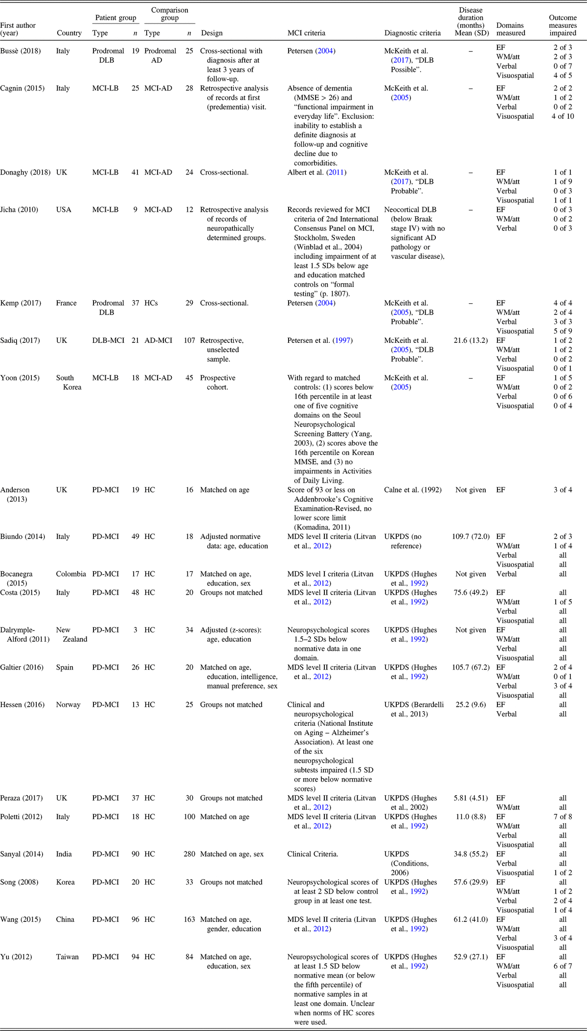
Table 1 displays details of retained papers. The search strategy yielded only one study in MCI-LB that met full inclusion criteria (Kemp et al., Reference Kemp, Philippi, Phillipps, Demuynck, Albasser, Martin-Hunyadi, Schmidt-Mutter, Cretin and Blanc2017). Six studies measured cognition in MCI-LB, but did not use a HC group. One paper had only nine patient subjects (Jicha et al., Reference Jicha, Schmitt, Abner, Nelson, Cooper, Smith and Markesbery2010). Given the small number and heterogeneity of MCI-LB results, these papers were retained for narrative review.
Details of all included studies (MCI-LB and PD-MCI)

MCI = mild cognitive impairment; DLB = dementia with Lewy bodies; AD = Alzheimer’s disease; PD = Parkinson’s disease; HC = healthy controls; MMSE = Mini-Mental State Examination; UKPDS = UK Parkinson’s Disease Society; MDS = Movement Disorder Society; SD = standard deviation; EF = executive function; WM/att = working memory/attention.
Kemp et al. (Reference Kemp, Philippi, Phillipps, Demuynck, Albasser, Martin-Hunyadi, Schmidt-Mutter, Cretin and Blanc2017), the only paper meeting full inclusion criteria, considers neuropsychological scores as well as social cognitive ability. Patients are classified as “prodromal DLB” (n = 37) for meeting MCI criteria (Petersen, Reference Petersen2004) and having at least two core symptoms of DLB (McKeith et al., Reference McKeith, Dickson, Lowe, Emre, O’Brien, Feldman, Cummings, Duda, Lippa, Perry, Aarsland, Arai, Ballard, Boeve, Burn, Costa, Del Ser, Dubois, Galasko, Gauthier, Goetz, Gomez-Tortosa, Halliday, Hansen, Hardy, Iwatsubo, Kalaria, Kaufer, Kenny, Korczyn, Kosaka, Lee, Lees, Litvan, Londos, Lopez, Minoshima, Mizuno, Molina, Mukaetova-Ladinska, Pasquier, Perry, Schulz, Trojanowski and Yamada2005). After adjusting for age, sex, and education level, prodromal DLB scored significantly lower than HCs (n = 29) on 14 of 21 extracted outcome measures, representing EF, attention and processing speed, working memory (WM), visuospatial memory, visuoconstructive ability, and verbal memory. The largest ES were reported on semantic verbal fluency (EF; g = −1.65) and digit symbol substitution (attention/processing speed, g = −1.16) tasks. The other significant ES were in the g = −0.53 to −0.93 range. Prodromal DLB showed impaired immediate and delayed free recall relative to controls. However, the authors suggested intact “real verbal memory (i.e. storage)” in the majority of their patients (29 of 37; outcome measure not specified) and emphasised retrieval deficits as related to executive impairments (Kemp et al., Reference Kemp, Philippi, Phillipps, Demuynck, Albasser, Martin-Hunyadi, Schmidt-Mutter, Cretin and Blanc2017). Kemp et al. (Reference Kemp, Philippi, Phillipps, Demuynck, Albasser, Martin-Hunyadi, Schmidt-Mutter, Cretin and Blanc2017) also concluded that verbal memory (free and cued selective reminding test total recall and recognition, g = −0.60) was better preserved than visual recognition memory [Delayed Matching to Sample-48 Items (g=−0.79)] in their sample.
All studies in MCI-LB used clinical diagnostic criteria except for Jicha et al. (Reference Jicha, Schmitt, Abner, Nelson, Cooper, Smith and Markesbery2010), where neuropathological diagnoses were utilised. No significant differences between MCI-LB and MCI-AD in global cognitive measures (e.g. MMSE) were reported. Excluding Kemp et al. (Reference Kemp, Philippi, Phillipps, Demuynck, Albasser, Martin-Hunyadi, Schmidt-Mutter, Cretin and Blanc2017), 28 of the remaining 79 (35.44%) extracted outcome variables showed differential performance between MCI-LB and MCI-AD after bias correction (see description in Table 1 and individual statistics in the supplementary materials). This tally does not include individual tasks removed that did not meet a priori exclusion criteria for publication in at least 10 peer-reviewed journals. Twenty-one variables showed poorer performance by MCI-LB relative to MCI-AD in visuospatial, attention, and psychomotor speed. Both Bussè et al. (Reference Bussè, Caffarra, Rossi, Zorzi, Fragiacomo, Camporese, Pompanin, Di Bernardo and Cagnin2018) and Cagnin et al. (Reference Cagnin, Bussè, Gardini, Jelcic, Guzzo, Gnoato, Mitolo, Ermani and Caffarra2015) reported poorer performance in MCI-LB in WM, visuospatial and visuoconstructive ability, and EF, with magnitudes ranging from medium (g = −0.56, Object Decision Visuospatial Test, Cagnin et al., Reference Cagnin, Bussè, Gardini, Jelcic, Guzzo, Gnoato, Mitolo, Ermani and Caffarra2015) to large (g = −0.95, Digit Span Backwards, Cagnin et al., Reference Cagnin, Bussè, Gardini, Jelcic, Guzzo, Gnoato, Mitolo, Ermani and Caffarra2015) and very large (g = −3.45, Pentagon Test number of angles, Busse, Hensel, Gühne, Angermeyer, & Riedel-Heller, Reference Busse, Hensel, Gühne, Angermeyer and Riedel-Heller2006). Donaghy et al. (Reference Donaghy, Taylor, O’Brien, Barnett, Olsen, Colloby, Lloyd, Petrides, McKeith and Thomas2018) showed deficits in MCI-LB relative to MCI-AD of medium ES in attention (Digit Vigilance, g = −0.56), EF (difference between Choice and Simple Reaction Time, g = −0.53), and visuospatial function (Angle Task, g = −0.55). Sadiq et al. (Reference Sadiq, Whitfield, Lee, Stevens, Costafreda and Walker2017) reported impaired scores on EF (letter fluency, g = −0.62) and attention (Trails B, g = −0.51), while Yoon, Kim, Moon, Yong and Hong (Reference Yoon, Kim, Moon, Yong and Hong2015) only showed impairment in EF (g = −0.72; Stroop Colour test) and there were no significant differences in Jicha et al. (Reference Jicha, Schmitt, Abner, Nelson, Cooper, Smith and Markesbery2010) after bias correction. MCI-LBs performed significantly better than MCI-AD on verbal memory outcome measures (immediate recall, delayed recall, and recognition) in three studies (Bussè et al., Reference Bussè, Caffarra, Rossi, Zorzi, Fragiacomo, Camporese, Pompanin, Di Bernardo and Cagnin2018; Cagnin et al., Reference Cagnin, Bussè, Gardini, Jelcic, Guzzo, Gnoato, Mitolo, Ermani and Caffarra2015; Yoon et al., Reference Yoon, Kim, Moon, Yong and Hong2015) with generally large ES (g = 0.66–3.80, mean g = 1.82).
The studies showing deficits in MCI-LB in the domains of WM and attention, EF and visuospatial function were not wholly consistent, as some outcome variables in the same domains did not reach significance. For example, Donaghy et al. (Reference Donaghy, Taylor, O’Brien, Barnett, Olsen, Colloby, Lloyd, Petrides, McKeith and Thomas2018) reported significant differences in only one of the nine outcome measures of attention and psychomotor speed (Digit Vigilance).
Additionally, both Yoon et al. (Reference Yoon, Kim, Moon, Yong and Hong2015) and Sadiq et al. (Reference Sadiq, Whitfield, Lee, Stevens, Costafreda and Walker2017) also compared MCI-LB/DLB-MCI to an MCI-stable group at follow-up. In Yoon et al. (Reference Yoon, Kim, Moon, Yong and Hong2015; 5 years of follow-up), MCI-LB was impaired in the domains of visuospatial ability and memory [Rey–Osterrieth Complex Figure (ROCF) recognition and copy], EF (Stroop Colour, verbal semantic fluencies), WM/attention (Digit Span Forwards and Backwards), and verbal learning and memory (Seoul Verbal Learning Test delayed recall) relative to MCI that did not convert to dementia. Sadiq et al. (Reference Sadiq, Whitfield, Lee, Stevens, Costafreda and Walker2017; mean follow-up 2.8 years) found that DLB-MCI patients are also impaired in EF (letter fluency) and visuoconstruction (clock drawing test) relative to stable-MCI.
PD-MCI Evidence Base
Demographic and clinical characteristics of participants
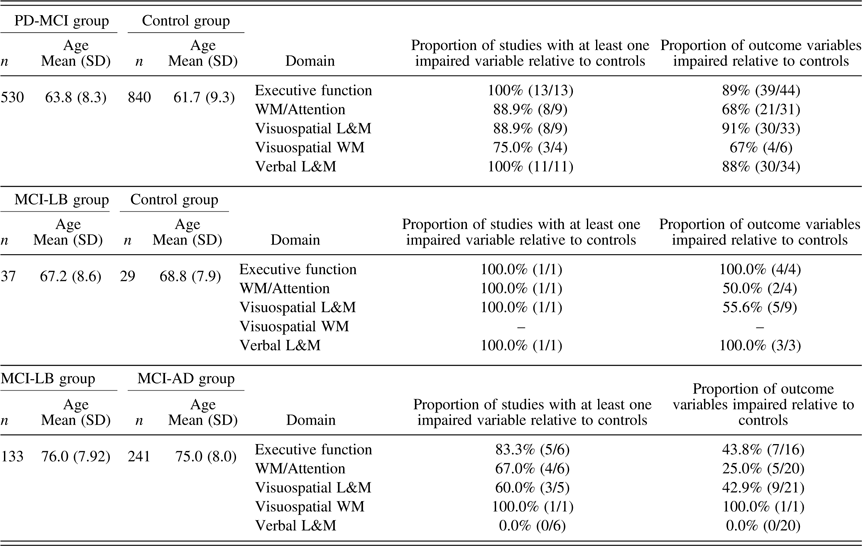
Details of quantitatively reviewed articles are summarised in Table 1 and group characteristics are summarised in Table 2. Some studies did not provide details of age (but were age-matched) or disease duration. For PD diagnosis, studies cited United Kingdom PD Society Brain Bank criteria (UKPDS; Hughes, Daniel, Kilford, & Lees, Reference Hughes, Daniel, Kilford and Lees1992) or equivalent, and National Institute of Neurological Disorders and Stroke criteria (Gelb et al., Reference Gelb, Oliver and Gilman1999). To define PD-MCI, seven studies used MDS criteria (Litvan et al., Reference Litvan, Goldman, Tröster, Schmand, Weintraub, Petersen, Mollenhauer, Adler, Marder, Williams‐Gray, Aarsland, Kulisevsky, Rodriguez-Oroz, Burn, Barker and Emre2012). The remaining studies used a mix of clinical and neuropsychological diagnostic criteria, including both normative and control data as impairment criteria.
Summary of impairments in PD-MCI from retained paper (n = 13) across neuropsychological domains

SD = standard deviation; PD = Parkinson’s Disease; MCI = mild cognitive impairment; WM = working memory, L&M = learning and memory.
Exclusion of tasks
Unclear task information or use of unestablished tasks (<10 published peer-reviewed references) necessitated the removal of variables from 8 of 13 studies. While not leading to removal from analyses, details provided were often also insufficient regarding the length of delay in memory tasks. Only the two most commonly reported measures per task and paper were retained in order to focus synthesis and avoid redundancy (e.g. for card sorting tasks, “categories achieved” and “perseverative errors” were used). Similarly, closely related variables administered for more experimental rationales were removed to avoid redundancy (e.g. from Costa et al. (Reference Costa, Zabberoni, Peppe, Serafini, Scalici, Caltagirone and Carlesimo2015), “Drawings Copy” was extracted while “Drawings with Landmarks Copy” was not). Sums were not extracted if separate scores were also provided, for example, with Digit Span Total (Yu et al., Reference Yu, Wu, Tai, Lin, Cheng and Hua2012).
Domain-Level Cognitive Assessments
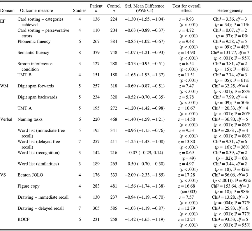
Outcome measures were organised into the domains of WM, EF, visuospatial, and verbal function, corresponding to their face validity. This conflicted with a number of papers that assigned outcome measures to domains of which they were not the primary target. For example, verbal fluency tasks were referred to as language or memory tasks in five papers when they are most commonly accepted as measures of EF (MacCarthy, Reference MacCarthy1972). Overall, every study showed worse performance in patients than HCs in at least one cognitive domain. Table 2 displays the number of outcome variables retained in each domain for each group, after removal described above, and how many were impaired. Table 3 displays summary ES of frequently reported outcome variables. See supplementary materials for individual ES and CI, and for figures displaying data not included below. Across domains, there was a pattern of smaller ES reported by studies with larger sample sizes.
Results of tests for commonly reported outcome measures for PD-MCI participants

PD = Parkinson’s disease; MCI = mild cognitive impairment; EF = executive function; WM = working memory; VS = visuospatial; TMT = Trail Making Test; JOLO = judgement of line orientation; ROCF = Rey–Osterrieth complex figure.
Executive function, working memory, and attention
A variety of EF and WM tasks were used, with ES ranging from moderate (g = −0.45; Yu et al., Reference Yu, Wu, Tai, Lin, Cheng and Hua2012, semantic fluency) to very large (g = −2.47; Sanyal, Banerjee, & Rao, Reference Sanyal, Banerjee and Rao2014; semantic fluency). EF was the most frequently targeted domain. Commonly reported outcome measures were from card sorting, verbal fluency, and Stroop tasks, with patients showing summary impairments in the medium (g = −0.63) to large (g = −1.30) range (Table 3). Verbal fluency tasks, commonly used measures of EF (Baddeley, Reference Baddeley1996), can be separated into semantic and phonemic fluency paradigms. Both showed large ES (Figure 1a and 1b), but PD-MCI was somewhat more impaired in semantic fluency tasks.
Bias-corrected effect sizes of (a) phonemic verbal fluency scores and (b) semantic verbal fluency scores in PD-MCI relative to controls in individual studies (circles) and as a summary effect size by group (diamonds).

PD-MCI showed a lower proportion of only 21 of 30 impaired WM variables, but with a similar range in summary ES (g = −0.52 to −1.65) as in EF. Six studies included WM measures that in particular require psychomotor speed, including Trails A and B. While both summary ES are large, there was greater impairment on Trails B, which requires EFs, than Trails A. All four studies that used Trails B reported significant and large deficits (above 1 SD), with a SMD of g = −1.65 (−1.93, −1.37). A similar divergence was observed between Digit Span Forwards (g = −0.52) and Backwards (g = −0.69).
Visuospatial domain
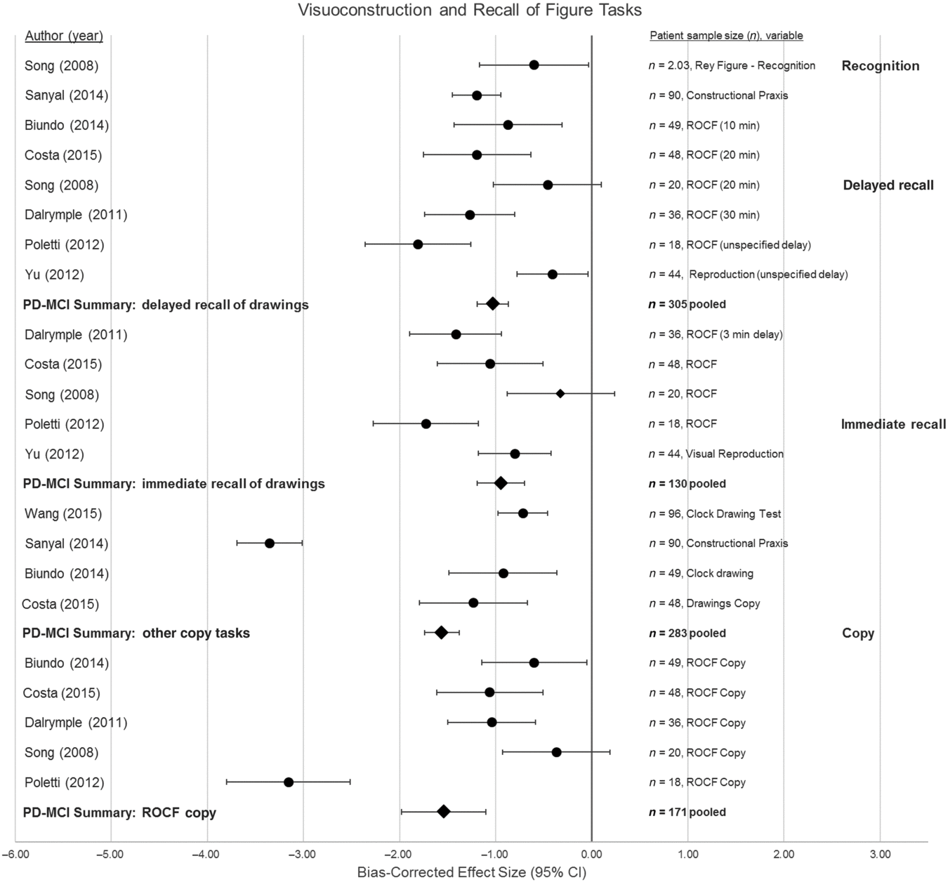
Patients performed significantly worse than controls on 30 of the 33 visuospatial memory variables. The largest SMD computed from the evidence base overall was in judgement of line orientation (JOLO) (g = −2.09; Benton, Hamsher, Varney, & Spreen, Reference Benton, Hamsher, Varney and Spreen1983), making visuospatial function the most frequently and severely impaired domain. Only Song, Kim, Jeong, Song and Lee (Reference Song, Kim, Jeong, Song and Lee2008) reported a lack of difference in visuospatial function (ROCF copy, immediate and delayed recall). Figure copying and recall was a common visuospatial paradigm, but recall conditions varied substantially in the length of delay. Summary ES for the PD-MCI group showed more pronounced deficits in figure copying than in either immediate or delayed recall. Song et al. (Reference Song, Kim, Jeong, Song and Lee2008) also tested delayed figure recognition and found a moderate deficit in PD-MCI relative to controls (g = −0.60). However, potential outliers (Poletti et al., Reference Poletti, Frosini, Pagni, Baldacci, Nicoletti, Tognoni, Lucetti, Del Dotto, Ceravolo and Bonuccelli2012; Sanyal et al., Reference Sanyal, Banerjee and Rao2014) complicate interpretation (Figure 2).
Bias-corrected effect sizes of visuospatial copying and recall tasks in PD-MCI relative to controls in both individual studies (circles) and as a summary effect size by group (diamonds). BVMT = Brief Visuospatial Memory Test; ROCF = Rey–Osterrieth Complex Figure.

Six outcomes from four studies targeted the visuospatial component of WM, per fractionated models proposed by Baddeley and Hitch (Reference Baddeley and Hitch1974), Logie and Pearson (Reference Logie and Pearson1997), and others. These include the Corsi Test (Corsi, Reference Corsi1972) and Pattern Recognition Memory (Robbins et al., Reference Robbins, James, Owen, Sahakian, Lawrence, McInnes and Rabbitt1998). A summary ES computed from Corsi Tests administered by Poletti et al. (Reference Poletti, Frosini, Pagni, Baldacci, Nicoletti, Tognoni, Lucetti, Del Dotto, Ceravolo and Bonuccelli2012), Costa et al. (Reference Costa, Zabberoni, Peppe, Serafini, Scalici, Caltagirone and Carlesimo2015), and Biundo et al. (Reference Biundo, Weis, Facchini, Formento-Dojot, Vallelunga, Pilleri and Antonini2014) suggested a moderate impairment in PD-MCI relative to controls, with Poletti et al. (Reference Poletti, Frosini, Pagni, Baldacci, Nicoletti, Tognoni, Lucetti, Del Dotto, Ceravolo and Bonuccelli2012) having the only significant finding.
Verbal domain
Patients performed significantly worse than controls in 30 out of 34 of the verbal domain variables. Each study that tested verbal learning and memory reported at least one significant difference between groups. However, three studies measuring verbal learning and memory did not find a significant difference between groups: California Verbal Learning Test in Galtier, Nieto, Lorenzo and Barroso (Reference Galtier, Nieto, Lorenzo and Barroso2016), Hopkins Verbal Learning Test (HVLT) and Boston Naming Test in Song et al. (Reference Song, Kim, Jeong, Song and Lee2008), and HVLT in Wang et al. (Reference Wang, Tang, Yan, Chen, Xu, Liu, Wang and Guo2015). Two-thirds of significant outcome variables pertained to word list recall tasks. Most immediate and delayed free recall scores indicated significant impairment, but the three studies measuring recognition did not show a significant difference between patients and controls. Very large ES were also found in naming tasks (g = −1.40).
Discussion
Summary of Findings
MCI-LB
In this structured review, only one study on the cognitive profile of MCI-LB utilised a control group. Kemp et al. (Reference Kemp, Philippi, Phillipps, Demuynck, Albasser, Martin-Hunyadi, Schmidt-Mutter, Cretin and Blanc2017) reported prodromal DLB to have a profile of diffuse impairments in the domains of EF, processing speed, attention, WM, visuospatial ability and visuospatial construction, verbal memory and language. Kemp et al. (Reference Kemp, Philippi, Phillipps, Demuynck, Albasser, Martin-Hunyadi, Schmidt-Mutter, Cretin and Blanc2017) provided evidence of immediate and delayed verbal recall deficits in prodromal DLB compared to controls, although recognition memory was intact. This study indicates that a substantial number of MCI-LB will present with verbal memory impairment at this early stage and it should not therefore be taken as a reliable indicator of prodromal AD (Kemp et al., Reference Kemp, Philippi, Phillipps, Demuynck, Albasser, Martin-Hunyadi, Schmidt-Mutter, Cretin and Blanc2017). However, ES of verbal memory impairment reported in MCI-LB (g = −058; Kemp et al., Reference Kemp, Philippi, Phillipps, Demuynck, Albasser, Martin-Hunyadi, Schmidt-Mutter, Cretin and Blanc2017) were almost half that of the pooled estimate for PD-MCI (g = −1.25).
Given that the largest deficits overall in prodromal DLB were in EF (g = −1.65) and processing speed (g = −1.16), it is possible that domain-general impairments could at least partly account for deficits across other cognitive functions (Kemp et al., Reference Kemp, Philippi, Phillipps, Demuynck, Albasser, Martin-Hunyadi, Schmidt-Mutter, Cretin and Blanc2017). Slowed speed of processing, executive dysfunction, or praxic and perceptual impairments in prodromal DLB could confound interpretation of deficits in more complex tasks such as visual construction or naming tests, as well as the greater impairments on visual versus verbal memory scores reported by Kemp et al. (Reference Kemp, Philippi, Phillipps, Demuynck, Albasser, Martin-Hunyadi, Schmidt-Mutter, Cretin and Blanc2017). Executive dysfunction, for example, has the potential to impair cognition at various stages, including encoding and retrieval in the case of memory function, thereby compounding the impact (Bryan et al., Reference Bryan, Luszcz and Pointer1999) Kemp et al.’s (Reference Kemp, Philippi, Phillipps, Demuynck, Albasser, Martin-Hunyadi, Schmidt-Mutter, Cretin and Blanc2017) emphasis on the role of EF in prodromal DLB is in line with a recent report that another measure of EF (verbal fluency) had the highest predictive accuracy for conversion to DLB in idiopathic RBD patients (Génier Marchand et al., Reference Génier Marchand, Postuma, Escudier, De Roy, Pelletier, Montplaisir and Gagnon2018). EF similarly emerges as a potential explanatory factor in PD-MCI results, as discussed further below.
Only six other studies (Bussè et al., Reference Bussè, Caffarra, Rossi, Zorzi, Fragiacomo, Camporese, Pompanin, Di Bernardo and Cagnin2018; Cagnin et al., Reference Cagnin, Bussè, Gardini, Jelcic, Guzzo, Gnoato, Mitolo, Ermani and Caffarra2015; Donaghy et al., Reference Donaghy, Taylor, O’Brien, Barnett, Olsen, Colloby, Lloyd, Petrides, McKeith and Thomas2018; Jicha et al., Reference Jicha, Schmitt, Abner, Nelson, Cooper, Smith and Markesbery2010; Sadiq et al., Reference Sadiq, Whitfield, Lee, Stevens, Costafreda and Walker2017; Yoon et al., Reference Yoon, Kim, Moon, Yong and Hong2015) resulted from the systematic review and used MCI-AD rather than a control group for comparison. This illustrates both the dearth of neuropsychological research in MCI-LB and the current focus on AD in the field. However, the small number of results in this key population may also relate to the challenge of identifying MCI-LB patients. Both DLB and PDD are under-diagnosed, and regional (geographic) differences in prevalence rates suggest varying clinical awareness of Lewy body disease (Kane et al., Reference Kane, Surendranathan, Bentley, Barker, Taylor, Thomas, Allan, McNally, James, McKeith and Burn2018). The retained studies provided limited evidence for MCI-LB and MCI-AD differentiation, as only about a third of extracted measures differed between groups and the findings within domains are sometimes inconsistent, with MCI-LB statistically differing from MCI-AD on some but not all tasks in the same domain. Verbal learning and memory appeared preserved in MCI-LB relative to MCI-AD, in line with the pronounced memory encoding deficits of AD (Lange et al., Reference Lange, Bondi, Salmon, Galasko, Delis, Thomas and Thal2002; Martin, Brouwers, Cox, & Fedio, Reference Martin, Brouwers, Cox and Fedio1985). However, without the use of HCs, it cannot be inferred that verbal impairment relative to controls should not be expected in MCI-LB cohorts.
PD-MCI
Our quantitative analyses emphasise that there are significant deficits in PD-MCI relative to HCs. Overall, the highest proportion and most severe of deficits was observed in the visuospatial domain, although the verbal and EF domains were also consistently impaired. One of the largest differences between PD-MCI and controls was in Benton’s JOLO task (Benton et al., Reference Benton, Hamsher, Varney and Spreen1983). JOLO, unlike many other visuospatial tasks, requires minimal motor skill and is free of practice effects (Montse, Pere, Carme, Francesc, & Eduardo, Reference Montse, Pere, Carme, Francesc and Eduardo2001). As such it may be particularly useful in tracking cognitive decline with PD progression. There was less consistency in the attention and WM data, with half of the papers showing deficits in some but not all reported tasks. Similarly, the magnitude of deficit on semantic verbal fluency tasks differed widely between studies, suggesting unreliability when quantifying the extent of executive dysfunction in patients. Our review found a slightly larger ES of impairment in semantic than phonemic verbal fluency tasks in PD-MCI, in line with findings from a meta-analysis in PD (Henry & Crawford, Reference Henry and Crawford2004). Semantic fluency tasks make demands on temporal functions and are typically challenging for AD patients (Henry, Crawford, & Phillips, Reference Henry, Crawford and Phillips2004), while phonemic verbal fluency is more closely associated with frontal abilities (Troyer, Moscovitch, Winocur, Alexander, & Stuss, Reference Troyer, Moscovitch, Winocur, Alexander and Stuss1998). This suggests firstly that caution should be taken when utilising semantic and phonemic verbal fluency tasks interchangeably as indicative of executive dysfunction, for example, in the case of current PD-MCI criteria (Litvan et al., Reference Litvan, Goldman, Tröster, Schmand, Weintraub, Petersen, Mollenhauer, Adler, Marder, Williams‐Gray, Aarsland, Kulisevsky, Rodriguez-Oroz, Burn, Barker and Emre2012). Secondly, this could suggest that co-occurring AD pathology accounts for semantic fluency impairment in PD-MCI. However, recent work by El-Nazer et al. (Reference El-Nazer, Adler, Beach, Belden, Artz, Shill, Driver-Dunckley, Mehta, Sabbagh, Serrano, Sue, Zamrini and Benge2019) concluded that PD and PD/AD mixed pathologies do not differ clinically and that cognitive deficits are associated with both LB and AD pathological burden. Specifically, greater Lewy body density was found in limbic and cortical structures in PD patients with poor verbal fluency, but higher AD pathology was on trend in many of the same areas.
Recall Versus Recognition in PD-MCI
PD-MCI-associated deficits in figure copying (g = −1.56) were more pronounced than in recall (immediate g = −0.91 and delayed SMD g = −1.03 recall), although all ES were large. Conversely, visuospatial recognition is much less impaired (Song et al., Reference Song, Kim, Jeong, Song and Lee2008), as also emerged in the MCI-LB results. It is possible that complex figure recall is a more challenging task for both patients and controls, and thus less difference between groups is observed. The use of retention scores (the percentage of information lost or gained between copy and recall conditions) might provide more nuanced information on differential abilities (Shin, Park, Park, Seol, & Kwon, Reference Shin, Park, Park, Seol and Kwon2006). Recognition memory for word list stimuli seems similarly intact in PD-MCI, in contrast to deficits in both immediate and delayed recall conditions. This pattern of dysfunction in both the visuospatial and verbal domains in PD-MCI suggests impaired retrieval and relatively intact encoding and storage mechanisms (Shin et al., Reference Shin, Park, Park, Seol and Kwon2006). This supports the established retrieval deficit hypothesis of memory impairment in PD (Tröster & Fields, Reference Tröster and Fields1995; Whittington, Podd, & Kan, Reference Whittington, Podd and Kan2000), which argues that the cause of memory impairment is inability to retrieve material on demand rather than encoding or retention ability (Mahurin, Feher, Nance, Levy, & Pirozzolo, Reference Mahurin, Feher, Nance, Levy, Pirozzolo, Parks, Zec and Wilson1993). Given that a similar pattern emerged in MCI-LB (Kemp et al., Reference Kemp, Philippi, Phillipps, Demuynck, Albasser, Martin-Hunyadi, Schmidt-Mutter, Cretin and Blanc2017), this model may apply to other early alpha-synucleinopathies.
However, there has been increasing scrutiny of this hypothesis and evidence of recognition impairment in some patients (Higginson, Wheelock, Carroll, & Sigvardt, Reference Higginson, Wheelock, Carroll and Sigvardt2005; Whittington et al., Reference Whittington, Podd and Kan2000). Whittington et al. (Reference Whittington, Podd and Kan2000) found impairment in recognition in PD participants without dementia, but not in newly diagnosed PD patients, and Bronnick, Alves, Aarsland, Tysnes and Larsen (Reference Bronnick, Alves, Aarsland, Tysnes and Larsen2011) found that early PD patients performed poorly on free recall, cued recall, and recognition memory. Bronnick et al. (Reference Bronnick, Alves, Aarsland, Tysnes and Larsen2011) attribute this impairment to encoding failure due to poor EF, rather than to impaired retrieval. Indeed, PD participants used fewer semantic clustering strategies to enhance their recall, and both strategy and EF explained significant variance in learning. Earlier, Gershberg and Shimamura (Reference Gershberg and Shimamura1995) and Hirst and Volpe (Reference Hirst and Volpe1988) had demonstrated that frontal lobe lesion patients were unable to capitalise on the potential semantic organisation present in word lists. It is likely that the prominent EF impairments in PD-MCI at least partly explain poorer performance on tasks of greater complexity, regardless of domain.
Executive Function Weighting Impairs Performance in PD-MCI
Impairment was found on only two-thirds of visuospatial WM measures despite the high proportion of visuospatial learning and memory tasks (91%) that show deficits in PD-MCI. One question is why visuoconstructional ability is impaired but not visuospatial WM? Figure copying such as ROCF is a complex task that requires EFs such as sustained attention, planning, and organisation (Shin et al., Reference Shin, Park, Park, Seol and Kwon2006), in addition to the visuospatial perception and processing that is required in visuospatial WM tasks like Corsi blocks. If executive demands are responsible for this divergence, this emphasises the methodological importance of locating the executive processes within tasks with face validity in other domains. However, even simple WM tasks such as Digits Forward could be argued to have an EF component (e.g. chunking), so this issue may speak more to the debate regarding multicomponent versus unitary models of WM (Cowan, Reference Cowan1999; Engle, Tuholski, Laughlin, & Conway, Reference Engle, Tuholski, Laughlin and Conway1999) than to a salient difference in a patient population.
MCI Classification
Defining MCI neuropsychologically using domain cut-off scores may also have the potential for circularity in analyses in which neuropsychological performance is the phenotype of interest. In the PD-MCI, all but one study (Anderson, Simpson, Channon, Samuel, & Brown, Reference Anderson, Simpson, Channon, Samuel and Brown2013) defined groups using neuropsychological criteria. PD-MCI studies tend to utilise Movement Disorder Society (MDS) diagnostic criteria for MCI, which is based on neuropsychological cut-off scores by individual cognitive domain, typically between 1 and 2 SDs. However, the specific magnitude of deficit required to be considered “impairment” has critical implications. Dalrymple-Alford et al. (Reference Dalrymple-Alford, Livingston, MacAskill, Graham, Melzer, Porter, Watts and Anderson2011) found that while only 14% of a PD sample was considered PD-MCI when defined as two SDs below normative scores in at least two tests in a domain, this number increases to 89% if considered at one SD or more. In the latter scenario, 70% of clinically defined HCs were also identified as MCI. To capture the breadth of impairment, MDS criteria stipulate that cognition should ideally be measured in five domains by at least two tasks, but this increases the likelihood of reaching significance on at least one measure. Few studies correct for multiple comparisons. Such a large amount of neuropsychological testing can also become unwieldy to report in entirety and encourages selection biases in presenting only significant results. Thus, choosing a battery poses a problem: decreasing the breadth may omit relevant individuals, but increased breadth often leads to contradictory findings within domains. The use of composite domain scores may help to overcome this methodological challenge and has been suggested to bypass task idiosyncrasy, avoid multiple comparisons, and increase power (Crane et al., Reference Crane, Carle, Gibbons, Insel, Mackin, Gross, Jones, Mukherjee, Curtis, Harvey, Weiner and Mungas2012; Gibbons et al., Reference Gibbons, Carle, Mackin, Harvey, Mukherjee, Insel, Curtis, Mungas and Crane2012).
While results suggest similarities in the neuropsychological profiles of MCI-LB and PD-MCI by domain, future work comparing these patient groups directly is warranted. For example, poorer episodic memory ability might be expected in MCI-LB given evidence of greater co-occurrence of amyloid and tau pathology in DLB versus PDD (Jellinger & Korczyn, Reference Jellinger and Korczyn2018). However, ES of verbal memory impairment reported in MCI-LB (g = −0.58; Kemp et al., Reference Kemp, Philippi, Phillipps, Demuynck, Albasser, Martin-Hunyadi, Schmidt-Mutter, Cretin and Blanc2017) were almost half that of the pooled estimate for PD-MCI (g = −1.25) in the present study. A potential explanation for this finding may be the greater variability expected in DLB (Nelson et al., Reference Nelson, Jicha, Kryscio, Abner, Schmitt, Cooper, Xu, Smith and Markesbery2010) and MCI-LB. Indeed, the validity of such direct comparisons of MCI subtypes may be challenged given the different definitional criteria for MCI currently in use.
The present review focused on studies with HC comparison groups that would provide closer matching between local patients and controls. However, this approach does not assume that important and complementary insights will not be found in studies using normative data or other relevant patient groups, such as stable or reverting MCI patients (see supplementary table). Yarnall et al. (Reference Yarnall, Breen, Duncan, Khoo, Coleman, Firbank, Nombela, Winder-Rhodes, Evans, Rowe, Mollenhauer, Kruse, Hudson, Chinnery, O’Brien, Robbins, Wesnes, Brooks, Barker and Burn2014), for example, found a high prevalence of MCI in their incidence cohort and suggested that prominent executive dysfunction may account for poor performance in other domains, such as memory tasks, in line with the present review’s suggestions. Longitudinal studies also critically offer insight into cognitive trajectories in PD-MCI, and suggested that low baseline EF, processing speed, visuospatial function, and verbal memory were predictive of poorer outcomes at follow-up (e.g. see Pigott et al., Reference Pigott, Rick, Xie, Hurtig, Chen-Plotkin, Duda, Morley, Chahine, Dahodwala, Akhtar, Siderowf, Trojanowski and Weintraub2015; Williams-Gray, Foltynie, Brayne, Robbins, & Barker, Reference Williams-Gray, Foltynie, Brayne, Robbins and Barker2007).
In conclusion, growing interest in MCI due to Lewy body disease necessitates a broader understanding of cognition in early disease. The present study aimed to capture existing literature in neuropsychological functioning by focusing on studies in MCI-LB and PD-MCI. MCI-LB studies are limited, but provide evidence for impaired EF, speed of processing, visuospatial, WM, and attentional abilities in comparison to controls and MCI-AD. However, results within domains are at times inconsistent. PD-MCI shows deficits in visuospatial, executive, and verbal tasks. However, these results should be interpreted with caution due to issues of circularity. MCI as a stable finding has also been called into question, with 9% of individuals with established MCI later “reverting” to normal cognition (Copeland & Schiess, Reference Copeland and Schiess2013). Thus, while PD-MCI may retain utility in clinical settings, it may be inappropriate as an a priori definition of a patient group in neuropsychological research. Defining MCI clinically [e.g. by assessing independent function in daily activities, input from family members, neurological examination, global cognitive scales (MMSE) to rule out advanced or absent impairment, or biomarkers], rather than neuropsychologically, will serve to better capture the cognitive heterogeneity of early LBDs and allow for more nuanced analyses of the potential interrelatedness of deficits.
ACKNOWLEDGEMENTS
This research was supported by the NIHR Newcastle Biomedical Research Centre, Alzheimer’s Research UK (A.T., grant number BH142057) and a Medical Research Council studentship (J.C., MR/K501396/1).
CONFLICT OF INTEREST
A.T. has received support from GE Healthcare for investigator-led research. No other authors have conflicts of interest to disclose.
SUPPLEMENTARY MATERIAL
To view supplementary material for this article, please visit https://doi.org/10.1017/S1355617719001103