Introduction
Intracellular transport is essential for cell functioning and survival, enlisting the cytoskeleton and mechanoenzymes to ensure the proper distribution and transport of organelles and other important cellular components through the cytoplasm. Retinal pigment epithelial (RPE) cells from eyes of sunfish, Lepomis spp. are a convenient model for examining mechanisms of intracellular motility. As fish lack dilatable pupils, the repositioning of photoreceptor outer segments and a moveable curtain of RPE melanin pigment granules controls light intensity hitting the photoreceptors (Bäck et al., Reference Bäck, Donner and Reuter1965). In the light, in vivo, pigment granules in RPE cells disperse distances up to 100 μm through long apical projections enveloping elongated, dim light-sensitive rod photoreceptors. At the same time, cone photoreceptors shorten, bringing their outer segments first in line to receive light entering the eye. These movements are reversed in darkness, and collectively are called retinomotor movements (Burnside & Nagle, Reference Burnside and Nagle1983; Burnside & King-Smith, Reference Burnside and King-Smith2017, reviews).
The dynamics of pigment granule aggregation can be investigated in vitro using dissociated, cultured RPE cells from fish. Exogenous cAMP stimulates pigment granule aggregation out of the slender apical projections and into the cell body, and washout of cAMP in the presence of dopamine triggers dispersion (Bruenner & Burnside, Reference Bruenner and Burnside1986; García & Burnside, Reference García and Burnside1994; King-Smith et al., Reference King-Smith, Paz, Lee, Lam and Burnside1997). Using either RPE-retina eyecup preparations or dissociated RPE cells in vitro, it has been demonstrated that RPE pigment granules migrate along both microtubules and actin filaments. Intraocular injections of microtubule inhibitors block pigment granule motility in the RPE cell body (Burnside et al., Reference Burnside, Adler and O’Connor1983). However, in apical projections, microtubules appear to serve a structural role only, since pigment granule movement persists in the absence of microtubules in dissociated RPE cells having apical projections attached to a substrate. However, pigment granule movement within apical projections is blocked when actin filaments in dissociated RPE cells are disrupted (King-Smith et al., Reference King-Smith, Paz, Lee, Lam and Burnside1997).
In mammalian eyes, light-dependent pigment granule movements in RPE are present, but much reduced compared to fish. RPE of mammalian eyes lack the long slender apical projections and instead have overlapping finger- or leaf-like apical regions (Burnside & Laties, Reference Burnside and Laties1979; Futter et al., Reference Futter, Ramalho, Jaissle, Seeliger and Seabra2004). In mouse RPE, as in fish RPE, pigment granules move along microtubules in the cell body, demonstrated by the lack of pigment granules entering the apical area after dynein depletion (Jiang et al., Reference Jiang, Paniagua, Volland, Wang, Balaji, Li, Lopes, Burgess and Williams2020). Movement and retention of pigment granules in the apical region of mouse RPE requires actin and myosin VIIa (Liu et al., Reference Liu, Ondek and Williams1998; Futter et al., Reference Futter, Ramalho, Jaissle, Seeliger and Seabra2004; Gibbs et al., Reference Gibbs, Azarian, Lillo, Kitamoto, Klomp, Steel, Libby and Williams2004). As in fish, cyclic AMP levels in mammalian retina of many mammalian species, including the mouse, decrease in light and increase in the dark (DeVries et al., Reference DeVries, Cohen, Hall and Ferrendelli1978; Farber et al., Reference Farber, Souza, Chase and Lolley1981; Burnside & Basinger, Reference Burnside and Basinger1983). Yet, the signaling pathways that trigger the repositioning of granules in mammalian RPE have not been described.
The cyclic nucleotide, cAMP, is known to activate protein kinase A (PKA) by binding to the PKA regulatory subunits, causing release of the catalytic subunits and subsequent protein phosphorylation (Fimia & Sassone-Corsi, Reference Fimia and Sassone-Corsi2001, review). In rod photoreceptors, increase in cAMP associated with dark onset stimulates rod shortening, and this dark-adaptive motility is blocked by the cyclic nucleotide-dependent kinase inhibitor, H8 (Liepe & Burnside, Reference Liepe and Burnside1993). Furthermore, phosphoproteins resulting from cAMP-PKA activation have been identified in rods (Pagh-Roehl et al., Reference Pagh-Roehl, Han and Burnside1993). Similarly, the microtubule-dependent elongation of cone photoreceptors that occurs at dark onset is also activated by cAMP, suggesting a role for a cAMP-dependent kinase in phosphorylation of a target protein that could regulate a microtubule associated protein (Gilson et al., Reference Gilson, Ackland and Burnside1986). Thus, it has been thought that, similarly, the canonical cAMP-PKA pathway triggers dark-adaptive pigment granule migration in RPE. However, this has not been confirmed in RPE, nor have any PKA targets been identified. Since cAMP can act through other signaling pathways, including regulation of cyclic nucleotide-gated ion channels and activation of the guanine nucleotide exchange factor, Epac (Exchange protein activated by cAMP), leading to activation of the small GTPase, Rap (Berridge, Reference Berridge2014, review), we examined more closely the cAMP-dependent stimulation of pigment granule aggregation in RPE and the role of PKA. We confirm that pigment granule aggregation occurs through cAMP-PKA activity, investigate the distribution of PKA regulatory subunit and PKA-phosphorylated proteins in isolated RPE cells, and show potential protein targets of PKA phosphorylation in RPE.
Materials and methods
Bluegill sunfish, Lepomis macrochirus, were obtained from Musky Trout Hatchery, Asbury, NJ, or wild-caught from Cobbs Creek, Havertown, PA, and maintained on either a 14 h light (L): 10 h dark (D) or 12:12 (L:D) cycle and fed floating fish food. RPE cell isolation was carried out as described previously (King-Smith, Reference King-Smith2016). Briefly, 2–4 h after light onset, fish were dark adapted for 30–60 min and anesthetized in 75 mg/l MS-222 (ethyl 3-aminobenzoate methanesulfonate, Sigma-Aldrich Inc, Saint Louis, MO, # E10521). After spinal section and pithing, eyes were removed in dim light, hemisected, and clumps (sheets) of RPE were gently flushed out of the eyecup using HEPES modified Earle’s Ringer (HER, 116.3 mM NaCl; 5.4 mM KCl; 1.8 mM CaCl2; 0.8 mM MgSO4; 1.0 mM Na2HPO4; 25.5 mM glucose; 4.0 mM NaHCO3; 21 mM HEPES; pH 7.2, with phenol red; King-Smith, Reference King-Smith2016). RPE sheets from two eyes were pooled and digested in 10 units/ml papain in calcium- and magnesium-free HER containing 1 mM EGTA for 30 min. Sheets were then washed in HER plus 0.4% bovine serum albumin (HERB) and dissociated with a syringe needle. Cells were plated on poly-l-lysine coated coverslips immediately following dissociation and allowed to settle for 1–2 h. For some experiments, RPE sheets (without further dissociation) were pooled, divided into samples and incubated in test solutions in parafilm-lined dishes on a rotator at 30 rpm.
Experimental treatments
Full pigment granule dispersion was elicited by changing the medium to 10 μM dopamine in HERB and incubating for 30 min. All experiments began with pigment granules in the fully dispersed state. Aggregation was then achieved by changing the medium to 1 mM cAMP in HERB for 30 min, with or without drugs. The PKA inhibitor H89 (Enzo Life Sciences, Farmingdale, NY, #EI-196) was used at 1 or 10 μM concentration from 10 mM frozen stock in DMSO. 8CPT-2Me-cAMP (an activator of Epac, Tocris, Minneapolis, MN #1645) at 5 μM was prepared in HERB from 5 mM frozen stock in distilled water. After drug treatments, cells were fixed in 0.5% formaldehyde, 0.5% glutaraldehyde, 0.8% K3(FeCN)6, 4% sucrose, in 85 mM PHEM buffer (50 mM PIPES, 25 mM HEPES, 8 mM EGTA, 2 mM magnesium chloride, pH 6.9). Cells were viewed under phase contrast at 400× or 1000× total magnification. For experiments using RPE sheets, after fixation sheets were chopped into small pieces using a razor blade, viewed and measured using phase contrast at 400×.
Quantification of pigment granule position
The extent of pigment granule aggregation or dispersion in dissociated cells was quantified by the pigment index (PI, King-Smith et al., Reference King-Smith, Paz, Lee, Lam and Burnside1997) calculated using three measurements per cell: the area of the polygon formed by connecting the distalmost tip of each apical projection of the isolated RPE cell, called the outer area (OA), and a similar polygon formed by connecting the distalmost pigment granule in each apical projection, called the inner area (IA), and the area of the central cell body (CB). PI is calculated by the ratio of the inner area minus cell body area, and the outer area minus the cell body: PI = (IA − CB)/(OA − CB). By this formula, fully aggregated RPE cells will have a PI near zero and fully dispersed cells a maximum value of 1.0. In practice, PI for RPE cells with dispersed pigment granules typically ranges from 0.6 to 0.9 because of the back-and-forth shuttling motion of pigment granules and the active extension and retraction of the tips of apical projections. Since PI is a proportion, data were arcsine transformed prior to statistical analysis (Sokal & Rohlf, Reference Sokal and Rohlf1981). To compare pigment granule position in RPE sheets, the distance from the distal tip of apical projections to the distalmost pigment granule within the projection was measured (AP tip length). For both types of measurement, the s.d. among treatments differed, so data were analyzed using the Brown–Forsythe and Welch ANOVA tests, with Dunnets T3 post hoc test using GraphPad/Prism 9 (www.graphpad.com). P values < 0.05 were considered significant.
Microinjection
Isolated RPE cells having fully dispersed pigment granules were plated on coverslips in a 35 mm dish with a hole drilled in its base. Cells were injected with PKA catalytic subunit (PKA-C, 10 mg/ml, purified from bovine heart, Enzo # BML-359) in microinjection buffer (114 mM KCl, 20 mM NaCl, 3 mM MgCl2, 3 mM NaH2PO4, pH 7.0) including 1.5 mg/ml FITC dextran to visualize injected cells. The PKA-C/FITC dextran mixture was spin-filtered at maximum speed in a microcentrifuge at 4°C for 15 min. The solution was injected using Eppendorf femtotip needles using an Eppendorf Micromanipulator 5171 while viewing cells with an Olympus IX70 at 400×. Images were captured at 5-min intervals for 30–45 min postinjection.
SDS-PAGE and immunoblotting
RPE sheets and retinas were isolated from eyes of 2–4 fish as described above and pooled. Pooled sheets were divided into two samples and treated for 30 min with either 1 mM cAMP in HERB to stimulate pigment granule aggregation, or 10 μM dopamine in HERB to trigger granule dispersion on a rotator. Tissue was then disrupted by pipetting up and down in cold lysis buffer (Cell Signal Technologies, Danvers, MA, #9803) with 1 mM phenylmethylsulfonyl fluoride and protease inhibitor cocktail. SDS-PAGE sample buffer was added and the tissue homogenate was boiled 5 min. Samples were run on hand-cast 5–20% gradient gels (Laemmli, Reference Laemmli1970) and electrotransferred onto nitrocellulose in 25 mM Tris, 192 mM glycine, 20% methanol, pH 8.3. Total protein was identified by Ponceau red staining. Blots were destained then blocked for 1–2 h using either Blotto (5% nonfat dry milk in Tris-buffered saline containing 0.05 Tween-20, TBST) or 1% BSA in TBST, and treated with anti-RIIα antibody raised against the C-terminus of mouse PKA RII (Santa Cruz Biotechnology, Dallas, TX, sc-909) in Blotto for 1 h at room temperature, or antibody against PKA-phosphorylated proteins [anti-rabbit phospho-(Ser/Thr) PKA substrate, Cell Signal Technologies, #9621] in 5% BSA in TBST overnight at 4°C. Blots were treated with secondary antibodies conjugated to horseradish peroxidase (Thermo Scientific, Waltham, MA, # 31480), developed using SuperSignal West Femto maximum sensitivity substrate (Thermo Scientific #34095) for 7 min, and imaged using a LiCor Odyssey imaging system (LiCor, Lincoln, NE). For phosphoprotein analysis, bands on blots were quantified using ImageStudio (LiCor). Each phosphoprotein band was normalized to a prominent band in the same lane at ca. 69 kD, observed in the Ponceau total protein-stained blot prior to immunoblotting. Intensity of labeling of phosphoproteins at specific molecular weights were compared by calculating the ratio of the normalized cAMP and dopamine band intensities.
Immunofluorescent labeling
Dissociated RPE cells were prepared and labeled as described previously (King-Smith et al., Reference King-Smith, Vagnozzi, Fischer, Gannon and Gunnam2014). After fixation in 4% formaldehyde and 4% sucrose in PHEM buffer followed by permeabilization in 0.1% saponin, 0.5% BSA in PBS, cells were incubated in primary antibody for 1 h at room temperature or overnight at 4°C, then secondary antibodies (anti- rabbit IgG Alexa 568 or 488, Invitrogen, Carlsbad, CA) for 1 h, and mounted on slides with double-stick tape spacers.
Results
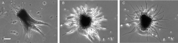
Immediately after dissociation, RPE cells display the typical apical–basal polarity of an epithelial cell (Fig. 1A). When plated on poly-l-lysine coated coverslips, after 1–2 h, cells attach and spread their (formerly) apical projections around the central cell body. Cylindrical pigment granules ranging from 0.5 to 9 μm in length are visible in attached apical projections, and in time-lapse videos, when dispersed into the projections, can be observed to “shuttle” back and forth, over a distance of ca. 4–5 μm (King-Smith et al., Reference King-Smith, Paz, Lee, Lam and Burnside1997; Supplemental Movie S1 and Fig. 1B). Application of nonderivatized cAMP triggers massive unidirectional motility toward the cell center (Fig. 1C and Supplemental Movie S1), which has been shown to be actin- and myosin II-dependent (King-Smith et al., Reference King-Smith, Paz, Lee, Lam and Burnside1997; Barsoum & King-Smith, Reference Barsoum and King-Smith2007).
Dissociated RPE cells from bluegill fish (Lepomis macrochirus). (A) RPE cell immediately after dissociation. (B,C) Dissociated RPE cell after settling and attachment. Slender, elongated pigment granules extend throughout the (formerly) apical projections. Cells in dopamine with dispersed pigment granules (phase bright) (B). Treatment with 1 mM cAMP triggers aggregation of pigment granules out of apical projections into the cell body (C). Bar: 10 μm.

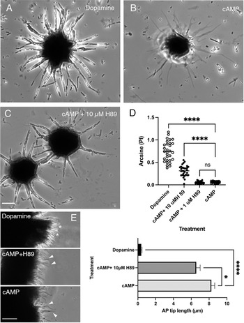
To determine whether cAMP-dependent activation of PKA triggers pigment granule aggregation, cells were treated with the isoquinoline PKA inhibitor H89 or carrier control (Fig. 2). After 30 min pretreatment in 10 μM H-89 while in dopamine to maintain pigment granule dispersion, RPE cells were challenged to aggregate by addition of cAMP, still in the presence of the drug. H89 at 1 μM had no effect on cAMP-stimulated pigment granule aggregation, however 10 uM H89 inhibited full pigment granule aggregation (Fig. 2B–D, P < 0.0001).
Effect of the protein kinase A inhibitor, H89, on pigment granule aggregation. (A) Control cell in 10 μM dopamine with dispersed pigment granules; (B) after cAMP treatment. Both dopamine- and cAMP controls included the carrier, 0.1% DMSO. (C) After cAMP plus 10 μM H-89. (D) Arcsine transformed pigment index data of treated cells. Open circles: dispersed (dopamine) or aggregated (cAMP) plus 0.1% DMSO. Closed circles: cAMP plus 1 or 10 μM H89. Results of a typical experiment; horizontal bar: mean, N = 30 cells per treatment. ****P < 0.0001; n.s., no significant difference (Welch’s ANOVA and Dunnett’s T3 test for multiple comparisons). The experiment was carried out three times. (E, left) Fragments of isolated retinal pigment epithelia sheets treated with dopamine or cAMP in 0.1% DMSO, or cAMP plus 10 μM H89. AP tip length (length of the distalmost part of apical projections that was devoid of pigment granules) was measured for each treatment. Asterisks: pigment granules dispersed at or near the tip of apical projections after dopamine treatment (phase bright regions, AP tip length ≈ 0). Arrowheads: distal portion of apical projections devoid of pigment granules (phase gray). (E, right) AP tip length for RPE sheets treated as in (D), results of a typical experiment. Mean ± S.E.M., N = 100–110 apical projections from 8 to 10 RPE sheet fragments for each treatment. *P < 0.05. The experiment was carried out three times. Bars: 10 μm (A–C), 20 μm (E).

To determine whether H89 similarly inhibited pigment granule migration in RPE that retained apical–basal polarity and cell–cell junctions, experiments were conducted using retinal pigment epithelial sheets. Sheets from 3 to 5 fish were isolated and pooled, then transferred to dopamine- or cAMP-containing solutions with 10 μM H89 or carrier control (0.1% DMSO). Pigment granule position was determined by measuring the length of the distal tip of apical projections devoid of pigment granules (AP tip length, see section “Materials and methods”). In RPE sheets, as in isolated cells, H89 partially blocked pigment granule aggregation (Fig. 2E, right).
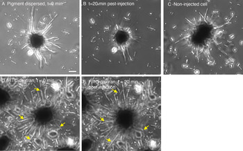
Since H89 has been reported to block other kinases in addition to PKA, including Rho kinases (ROCKs, Leemhuis et al., Reference Leemhuis, Boutillier, Schmidt and Meyer2002; Murray, Reference Murray2008) we confirmed the role of PKA in stimulating pigment granule aggregation by microinjection of PKA catalytic subunit. Injection of the catalytic subunit, rather than the holoenzyme, bypasses the need for cAMP-dependent dissociation of the regulatory and catalytic subunits. Within 20 min of injection, pigment granules in previously dispersed cells aggregated their pigment granules (N = 6 injected cells) while adjacent, noninjected cells remained dispersed (Fig. 3A–C). Similarly, pigment granules in cells injected with FITC dextran alone remained dispersed (Fig. 3D,E).
Effect of PKA catalytic subunit microinjection on isolated RPE cells. (A) RPE cells with dispersed pigment granules, before microinjection. (B) Twenty minutes after injection; pigment granules are mostly aggregated. (C) Neighboring, noninjected cells; pigment granules remain dispersed. (D) Control cell injected with FITC dextran only immediately after injection. (E) Twenty minutes after injection. Arrows indicate pigment granules that remained dispersed within apical projections. Bar: 10 μm.

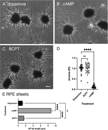
To test a potential role for Epac in pigment granule motility, RPE sheets and isolated cells were treated with dopamine to completely disperse pigment granules (Fig. 4A), followed by either 1 mM cAMP or 5 μm 8CPT-2Me-cAMP (8CPT), an activator of Epac. While pigment granules aggregated normally in cAMP (Fig. 4B, 8CPT failed to induce pigment granule aggregation (Fig. 4C). Similarly, in experiments using RPE sheets treated as above, pigment granules failed to aggregate when treated with 8CPT (Fig. 4E).
Dissociated RPE cells incubated in (A) 10 uM dopamine; (B) dopamine then 1 mM cAMP, (C) dopamine then 8CPT. (D) Arcsine transformed pigment index of treated cells, N = 30 cells per treatment. Horizontal line: mean. ****P < 0.0001; n.s., not significant. Results of a typical experiment. (E) AP tip length for RPE sheet fragments subjected to the same treatments. Mean ± S.E.M., N = 100–110 apical projection measurements for each treatment from 9 to 10 RPE sheet fragments. Results of a typical experiment. Each protocol (on isolated cells or RPE sheets) was carried out twice. Bar: 10 μm.

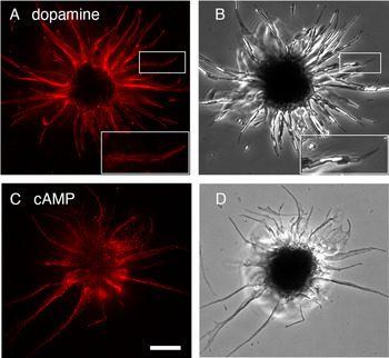
To determine the localization of both PKA and PKA-phosphorylated proteins in isolated RPE, immunofluorescence was carried out using RPE cells with either fully aggregated or dispersed pigment granules. Antibodies against the RIIα regulatory subunit of PKA bound to a band in RPE lysates near the expected molecular weight of 50 kD (data not shown). Cells with either dispersed (Fig. 5A) or aggregated (Fig. 5C) pigment granules that were treated with this antibody demonstrated widespread labeling throughout apical projections and the RPE cell body. RII generally associates with membranes, often through an anchoring protein (A-kinase anchoring protein, AKAP; Skroblin et al., Reference Skroblin, Grossmann, Schäfer, Rosenthal and Klussmann2010, review). Furthermore, PKA has been shown to associate with pigment granule membranes in dermal melanophores transfected with recombinant GFP-PKA (Kashina et al., Reference Kashina, Semenova, Ivanov, Potekhina, Zaliapin and Rodionov2004). However, because the fluorescence in cells with dispersed granules was sometimes quenched by the melanin in the granules (insets in Fig. 5A,B) it was not clear whether RIIα labeling was enhanced on pigment granule membranes.
Immunofluorescence of anti-RIIα labeling in isolated RPE cells having dispersed (A,B) or aggregated (C,D) pigment granules. Insets: rectangles in (A,B). Bar: 10 μm.

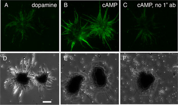
Cells labeled with the anti-PKA phosphorylated substrate antibody similarly displayed diffuse, punctate labeling in the cell body and projections of cAMP-treated RPE cells with aggregated pigment granules (Fig. 6B). In contrast, cells with dispersed pigment granules after incubation in dopamine, in the absence of cAMP (and therefore presumably with lower PKA activity) demonstrated much lower levels of labeling of PKA-phosphorylated substrates.
Fluorescent antibody labeling of PKA-phosphorylated substrates in isolated RPE cells and corresponding phase contrast images. Cells were treated with dopamine to disperse pigment granules (A,D) or cAMP, to aggregate granules (B,C,E,F). Panels C,F no primary antibody. Bar: 10 μm.

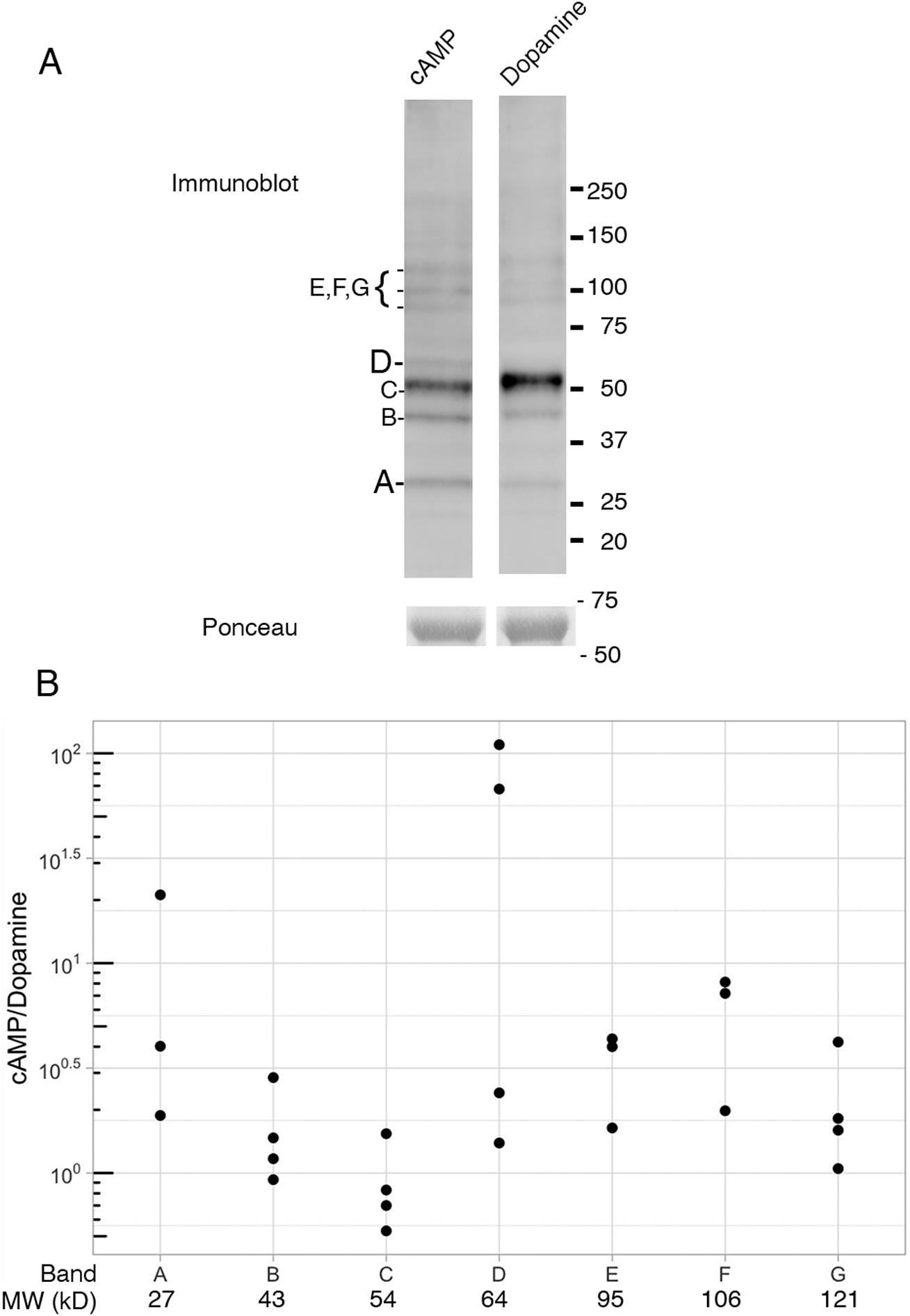
In immunoblots using RPE tissue treated with dopamine or cAMP and probed with the anti-phosphorylated PKA substrate antibody, several bands representing PKA-phosphorylated proteins were apparent in both cAMP-treated and dopamine-treated RPE samples (Fig. 7). Seven bands (A through G, Fig. 7) in each treatment were normalized to the total protein in each lane using a prominent band at ~69 kD in the Ponceau-stained membrane (Fig. 7A, bottom bands). The normalized ratio for cAMP-treated samples relative to dopamine-treated samples was then calculated for bands A through G (Fig. 7B). The intensities of most of these bands was greater overall in cAMP-treated compared to dopamine-treated samples as expected, however the differences in phosphorylation in cAMP-treated cells compared to dopamine-treated tissue varied greatly. Some prominent bands were of near equal intensity for the two treatments (Fig. 7B cAMP/dopamine ≅ 100). In contrast, for two protein bands (A, 27 kD, and D, 64 kD) the intensity of labeling was 20 to over 100 times as intense in cAMP samples compared to dopamine.
Identification of PKA-phosphorylated proteins in RPE. (A, top) Immunoblot of RPE lysates pretreated in dopamine or cAMP and probed with anti-phosphorylated PKA substrate antibodies; (bottom) same lanes as above, Ponceau-stained band showing total protein. Results from one experiment; note the dopamine lane had slightly more total protein than the cAMP lane (compare Ponceau bands for cAMP and dopamine). (B) Ratio of cAMP and dopamine band intensities for bands A–G in panel A normalized to the total protein in the lane using the 69 kD Ponceau-stained band (see section “Materials and methods”). Panel B includes data from four separate experiments; each experiment included both dopamine and cAMP-treated RPE pooled from 2 to 4 fish.

Discussion
Elevated intracellular levels of cAMP, whether produced by forskolin, an activator of adenylyl cyclase, cell-permeant cAMP analogs, or nonderivatized cAMP, induce a dark-adaptive response in fish RPE in vivo and in vitro, triggering pigment granules to aggregate from apical projections into the cell body (Bruenner & Burnside, Reference Bruenner and Burnside1986; García & Burnside, Reference García and Burnside1994; Burnside & King-Smith, Reference Burnside and King-Smith2017, review). Entry of nonderivatized cAMP into the cell likely occurs through organic anion transporters, since pharmacological inhibition of these transporters stops pigment granule aggregation induced by nonderivatized cAMP (García & Burnside, Reference García and Burnside1994). Since cAMP in the retina is known to increase in fish retina in the dark (Burnside et al., Reference Burnside, Evans, Fletcher and Chader1982), cAMP may be the extracellular “dark signal” released by the retina that triggers retinomotor movements, optimizing conditions for maximal light capture in dim light.
Cyclic AMP-dependent phosphorylation by protein kinase A is associated with other aspects of retinomotor motility in the fish eye, specifically the light-induced shortening of rod photoreceptors that accompanies pigment granule aggregation (Pagh-Roehl et al., Reference Pagh-Roehl, Han and Burnside1993). While it had been presumed that RPE pigment granule aggregation is similarly effected through the cAMP-PKA pathway, this had never been demonstrated directly. In addition, the identification of other, noncanonical cAMP signal transduction pathways, such as cAMP-dependent activation of a guanine nucleotide exchange factor for the small GTPase, RAP (Exchange protein activated by cAMP, Epac) had not been ruled out. The current study demonstrates that inhibition of PKA by H89 partially blocks aggregation in both isolated RPE cells and sheets of retinal pigment epithelia. The reason for partial, rather than complete inhibition is not clear. Previous work has shown that inhibition of phosphatases, without the addition of cAMP, is sufficient to stimulate pigment granule aggregation (King-Smith et al., Reference King-Smith, Paz, Lee, Lam and Burnside1997; Barsoum & King-Smith, Reference Barsoum and King-Smith2007). Thus, it could be that even minimal phosphorylation that occurs even in the presence of a PKA inhibitor will result in pigment granule aggregation.
Overall, findings of the current study, including the partial inhibition of pigment granule aggregation by the PKA inhibitor, H89, stimulation of pigment granule aggregation by microinjection of PKA catalytic subunit, and the lack of pigment granule aggregation in response to the Epac agonist, 8CPT-2Me-cAMP, in both isolated RPE cells and sheets of retinal pigment epithelia, together support the conclusion that pigment aggregation in RPE is effected through the canonical cAMP-PKA pathway.
Cyclic AMP triggers pigment granule movement in other pigmented cells, such as the dermal melanophores of fish and amphibians, but in these cells it stimulates pigment granule dispersion along microtubules, while low cAMP concentrations trigger pigment granule aggregation (Nascimento et al., Reference Nascimento, Roland and Gelfand2003, review). Protein kinase A has been shown to associate with microtubule motor complexes on melanophore granule membranes through an AKAP. The AKAP forms complexes with cytoplasmic dynein or kinesin to enable transport along microtubules (Kashina et al., Reference Kashina, Semenova, Ivanov, Potekhina, Zaliapin and Rodionov2004; Semenova et al., Reference Semenova, Ikeda, Ivanov and Rodionov2009). While microtubules act as tracks for transport through the cell body area in fish RPE (Burnside et al., Reference Burnside, Adler and O’Connor1983), in the long apical projections of isolated fish RPE cells, microtubules appear to serve a strictly structural role, while agents that disrupt actin or block nonmuscle myosin II prevent pigment granule aggregation (King-Smith et al., Reference King-Smith, Paz, Lee, Lam and Burnside1997; Barsoum & King-Smith, Reference Barsoum and King-Smith2007). Immunofluorescent labeling of isolated RPE cells using both anti-PKA and anti-phosphorylated PKA substrate antibodies demonstrated widespread and diffuse labeling in apical projections. Yet, since melanin within pigment granules may quench fluorescence, it is not clear if either the RIIα of PKA, or PKA-phosphorylated substrates, localize to granule membranes.
In many systems involving cAMP signaling, there exists cross-talk between pathways stimulated by cAMP and pathways involving another second messenger, calcium (Rasmussen, Reference Rasmussen1981, review). For example, in melanophores of angelfish, cAMP-induced pigment granule motility is associated with an increase in intracellular calcium, even though cAMP is both required and sufficient to trigger motility independent of calcium concentrations (Sammak et al., Reference Sammak, Adams, Harootunian, Schliwa and Tsien1992). In cone photoreceptors of fish, elongation of cone myoids that occurs at dark onset is activated by cAMP, and inhibited by 0.01–10 μM calcium (Gilson et al., Reference Gilson, Ackland and Burnside1986). In contrast, in fish RPE, cAMP-triggered pigment granule aggregation is dependent only on cAMP. Increases in intracellular calcium in cells having dispersed pigment granules (through ionomycin treatment or puncture with a microneedle) neither stimulate pigment granule aggregation, nor inhibit cAMP-triggered pigment granule aggregation (King-Smith et al., Reference King-Smith, Chen, Garcia, Rey and Burnside1996). Thus, although cone elongation appears to be sensitive to calcium, cAMP-dependent pigment granule aggregation is not. Interestingly, isolated fish RPE having aggregated pigment granules will disperse granules partway when stimulated by the muscarinic receptor agonist, carbachol, and partial dispersion is blocked by chelation of intracellular calcium (González et al., Reference González, Crittenden and García2004; Johnson & García, Reference Johnson and García2007) suggesting a cAMP/PKA-independent route contributing to dispersion, possibly through activation of calcium-dependent protein phosphatases such as calcineurin.
As has been observed in fish eyes, cAMP levels in eyes of mammals, including mouse increase in the dark (DeVries et al., Reference DeVries, Cohen, Hall and Ferrendelli1978) raising the question of the role of PKA not only in the pigment granule redistribution in mouse RPE, but also in other metabolic processes in mammalian RPE. RPE contains an isomerase, RPE65, which catalyzes the regeneration of the photoreceptor prosthetic group, trans-retinal, to cis-retinal, part of the classic vertebrate eye visual cycle (Tang et al., Reference Tang, Kono, Koutalos, Ablonczy and Crouch2013). Defects in RPE65 are responsible for a number of retinal degenerative diseases, including Leber Congenital Amaurosis. In mouse RPE, RPE65 localizes to the central portion of the cell in light-adapted eyes, but translocates toward the apical side of the cell in darkness. This dark-adaptive redistribution of RPE65 is abolished in myosin VIIa shaker1 mutant animals (Lopes et al., Reference Lopes, Gibbs, Libby, Aleman, Welch, Lillo and Williams2011). The light-dependent translocation raises the possibility that phosphorylation by PKA could regulate this myosin VIIa-dependent motility. Such a role could be tested by interocular injections of PKA-inhibiting agents, such as H89.
The presence of PKA-phosphorylated substrates in immunoblots in the absence of cAMP suggests a basal level of kinase activity exists even in cells that have not been treated with cAMP. Partial dispersion of pigment granules triggered by microinjection of the PKA inhibitor, PKI5–24 (Garcia, Reference Garcia1993) also supports the idea that PKA phosphorylation promotes aggregation, and that kinases and phosphatases in RPE are in competition. In vivo, light or dark conditions in the retina tip levels of phosphorylation or dephosphorylation in one direction or the other, via cAMP and PKA (in the dark) or via type 2 dopaminergic receptors and associated pathways in the light (Dearry & Burnside, Reference Dearry and Burnside1988).
The identities of the seven (or more) prominent PKA-phosphorylated proteins in cAMP-stimulated RPE are not known. Despite numerous attempts, we were not successful in immunoprecipitating PKA-phosphorylated proteins from fish RPE using the anti-phospho-PKA substrate antibody, which has been used in that capacity previously to isolate and identify phosphoproteins using mass spectrometry (Grønborg et al., Reference Grønborg, Kristiansen, Stensballe, Andersen, Ohara, Mann, Jensen and Pandey2002). In rod photoreceptors, a decrease in light increases cAMP levels and PKA-dependent phosphorylation of a 35 kD protein, the fish homolog of phosducin, and a 33 kD phosphoprotein (Pagh-Roehl et al., Reference Pagh-Roehl, Han and Burnside1993, Reference Pagh-Roehl, Lin, Su and Burnside1995). It is possible that the phosphorylated ~28 kD protein in RPE is actually the 33 or 35 kD phosphoproteins from contaminating rod inner–outer segments in RPE lysates treated with cAMP. However, observations of fixed samples of RPE sheets from the cAMP-treated pool used to prepare lysates for SDS-PAGE revealed very low numbers of rod inner–outer segments compared to RPE cells, reducing the likelihood that the prominent 28 kD protein in RPE lysates was a contaminant from photoreceptors.
Previous research has shown a role for nonmuscle myosin II in RPE pigment granule aggregation (Barsoum & King-Smith, Reference Barsoum and King-Smith2007); treatment with the myosin II disruptor blebbistatin blocks pigment granule aggregation, but not dispersion. These findings, together with results of other studies examining RPE pigment granule aggregation, led to the hypothesis that cAMP-stimulated aggregation of RPE pigment granules is linked in some way to actin retrograde flow. Myosin II is thought to effect retrograde actin flow through formation of a cross-linked network of actin that contracts upon myosin activation, pulling the network centripetally (Verkhovsky et al., Reference Verkhovsky, Svitkina and Borisy1999; Ponti et al., Reference Ponti, Machacek, Gupton, Waterman-Storer and Danuser2004). Based on the known pathways for activation of nonmuscle myosin II by phosphorylation, it is unlikely that cAMP/PKA is directly involved in myosin II activation. Rather, it is more plausible that myosin II-dependent retrograde actin flow is a constitutive process, unrelated to PKA-dependent phosphorylation. The role of PKA may be to phosphorylate proteins that allow RPE pigment granules to associate with F-actin cables moving in the retrograde direction (toward the cell body). This model is similar to what has been proposed in budding yeast, where a “mitochore” of proteins allows retention of mitochondria in the mother cell, by the reversible attachment of mitochondria to actin cables undergoing retrograde movement from daughter back to mother cell (Fehrenbacher et al., Reference Fehrenbacher, Yang, Gay, Huckaba and Pon2004; Peraza-Reyes et al., Reference Peraza-Reyes, Crider and Pon2010, review). Actin in RPE apical projections also forms bundles (see Fig. 2 in King-Smith et al., Reference King-Smith, Vagnozzi, Fischer, Gannon and Gunnam2014). Thus it is possible that PKA-dependent phosphorylation of as yet unidentified target proteins associated with pigment granule membranes allows them to latch onto actin cables, enabling them to be drawn toward the cell body, and disengage upon dephosphorylation, to be carried in the opposite direction via direct interaction with a myosin motor protein. Further investigation of PKA targets in RPE would not only help to better understand several aspects of RPE biology, but would further elucidate the regulation of intracellular organelle motility, a process that occurs in virtually all cells.
Conclusions
We have demonstrated that pigment granule aggregation in fish RPE cells is triggered by cAMP activation of protein kinase A. Immunolocalization of RIIα regulatory subunit of PKA showed diffuse labeling throughout apical projections. Similarly, PKA-phosphorylated substrates were diffusely localized throughout projections, and whether phosphorylated targets exist on pigment granule membranes or other internal membranes remains to be determined. Immunoblots revealed numerous PKA-phosphorylated proteins in RPE treated with cAMP, however some protein bands displayed a much higher degree of phosphorylation than others. While clearly PKA phosphorylation is the switch that ceases the back-and-forth “shuttling” behavior of pigment granules within projections and triggers massive retrograde movement, the specific targets of protein phosphorylation remain to be identified.
Acknowledgments
We thank the many undergraduate students who contributed to this work: Cam Collins, Olivia Connolly, Everett Davis, Elizabeth Del Rio, Audrey Malhotra, Melissa Messalti, Mazvita Nyamucapa, Megan O’Donnell, Joseph Quinlan, and Meghan Quinlan. We thank Dana M. Garcia for reviewing the manuscript, Charlotte Z. Smith for assistance with the figures, Nathan R. King for help catching fish, and Dinesh Rathore for fish care. Preliminary reports were presented at the Annual meeting of the American Society for Cell Biology in 2017 and 2018. This work was supported by the Saint Joseph’s University (SJU) Summer Scholars Program and the Saint Joseph’s University chapter of Sigma Xi. The current address for Nicole E. Leitner is Department of Biology, Washington University in Saint Louis, Saint Louis, Missouri, USA.
Funding statement
This research received no specific grant from any funding agency, commercial, or not-for-profit sectors.
Conflict of Interest
The authors have no competing interest to declare.
Supplementary Materials
To view supplementary material for this article, please visit http://dx.doi.org/10.1017/S0952523821000122.