Introduction
Cisplatin (cis-diamminedichloroplatinum(II)) is a highly effective anticancer drug that is widely used to treat various cancer types including sarcomas, carcinomas, lymphomas and germ cell tumours. However, this drug has a number of side effects that can limit its use, such as nephrotoxicity, neurotoxicity, ototoxicity and myelotoxicity; it also causes electrolyte disturbance, nausea and vomiting. Ototoxicity may be severe and no effective preventive treatment is currently available for this side effect. If this problem cannot be overcome, then cisplatin therapy may need to be proscribed, thus adversely affecting antineoplastic treatment. Cisplatin-induced ototoxicity is characterised by irreversible, progressive, bilateral, high-frequency and sensorineural hearing loss.Reference Rybak, Whitworth, Mukherjea and Ramkumar1–Reference Hellberg, Wallin, Ehrsson and Laurell3 Cisplatin primarily damages outer hair cells in the cochlear base. Thus, distortion product otoacoustic emissions (DPOAE) testing is an appropriate way of detecting cisplatin-induced ototoxicity.Reference Calli, Pinar, Oncel, Alper Bagriyanik and Umut Sakarya4, Reference Daldal, Odabasi and Serbetcioglu5
Many studies have previously demonstrated that intratympanic glucocorticoid administration can improve cisplatin-induced ototoxicity in young people. However, most diseases that require cisplatin treatment are seen in adults. Therefore, young animals may not be a good model for studying cisplatin ototoxicity and developing strategies for its prevention in adult humans. Indeed, the ototoxicity incidence after cisplatin treatment is greater in older cancer patients.Reference Laurell and Borg6 The main goals of this study were to assess and compare the efficacies of intratympanic dexamethasone and intratympanic methylprednisolone in protecting against cisplatin-induced ototoxicity in middle-aged rats and to correlate drug effects with changes in cochlear histology.
Materials and methods
This study was approved by the Animal Experiments Local Ethics Committee (protocol number 2/4-2014), Kocaeli University, Turkey. Adult female Wistar Hannover rats aged 15–17 months and weighing between 420 and 530 g (mean 480 g) were maintained in the central animal laboratory for 10 days under standard laboratory conditions with a 12-hour light–dark cycle at 22 ± 2 °C. These conditions were then maintained throughout the experimental period. Animals had free access to standard laboratory chow and water. Animals with ear disease or the absence of distortion product otoacoustic emission (DPOAE) at any of the ranges test frequencies were not used. Animals were monitored daily for signs of distress, pain and weight loss.
This study was performed in two phases. In phase 1, the optimal cisplatin treatment dose was established. For this, three groups of four rats were treated with a single intraperitoneal injection containing 8, 10 or 12 mg/kg cisplatin (1 mg/ml; Cisplatin Ebewe, Liba, Istanbul, Turkey) followed by the intraperitoneal injection (2 ml) of 0.9 per cent saline using an insulin syringe. All animals treated with 12 mg/kg cisplatin and two animals treated with 10 mg/kg cisplatin died on day 10. All animals treated with 8 mg/kg were alive on day 10 and one died on day 13. Based on these results, a single intraperitoneal injection of 8 mg/kg cisplatin was selected as the optimal dose to achieve a low mortality rate in phase 2 of the study.
In phase 2, the effects of intratympanic dexamethasone and intratympanic methylprednisolone on cisplatin-induced ototoxicity were determined. The mortality rate in this phase was 20 per cent. Data for all animals that died during phase 1 were removed from the analysis and new animals replaced them in phase 2. Eight animals were assigned randomly into four groups and treated with: intratympanic isotonic saline (saline control group); intraperitoneal cisplatin plus intratympanic isotonic saline (cisplatin group); intraperitoneal cisplatin plus intratympanic dexamethasone (dexamethasone group); and intraperitoneal cisplatin plus intratympanic methylprednisolone (methylprednisolone group).
Anaesthesia
Animals were sedated by intraperitoneal injection with a ketamine (40 mg/kg) and xylazine (5 mg/kg) cocktail for DPOAE testing and before intratympanic injection. The depth of anaesthesia was determined using the pedal reflex.
Drug treatment
On days 0, 3, 6 and 9, animals in the saline control and cisplatin groups were intratympanically injected under light microscopy with sufficient isotonic saline to fill the middle-ear cavity using a 28-gauge dental needle. On day 0, 8 mg/kg cisplatin (1 mg/ml) was administered intraperitoneally as a slow infusion into all animals except those in the saline control group, followed by the intraperitoneal administration of a bolus (2 ml) of 0.9 per cent saline via an insulin syringe. On days 0, 3, 6 and 9, the left tympanic membrane was visualised under light microscopy using an aural speculum and an intratympanic injection of dexamethasone (4 mg/ml; Dekort, Deva Holding, Istanbul, Turkey) or methylprednisolone (40 mg/ml; Prednol, Mustafa Nevzat İlaç Sanayi, Istanbul, Turkey) was administered slowly via myringotomy into the anteroinferior quadrant of the middle-ear cavity (approximately 0.2 ml) using a 28-gauge dental needle to the relevant glucocorticoid treatment group. Animals were kept in the same position for 20 minutes after every intratympanic injection.
Distortion product otoacoustic emission testing
Distortion product otoacoustic emission measurements were made under anaesthesia on day 0 (before medication) and day 10 in all animals: testing was performed in a quiet environment using a Madsen Capella DP+TE analyser and software (Otometrics, Miami, Florida, USA). Primary tones were introduced into the outer-ear canal via an insert earphone with a plastic adapter to seal the probe into the outer-ear canal; DPOAE findings are shown as distortion product grams (DP grams). The stimulus consisted of two pure tones (F1 and F2; F2:F1 ratio = 1.22) and DP grams used equilevel primary stimulus levels of 65 dB (L1 = L2); the resulting otoacoustic emissions ranged from approximately 1 to 8 kHz (0.996, 1.416, 2.001, 2.832, 4.003, 4755, 5.654, 6.728 and 7.998 kHz). Distortion product input and output amplitudes were measured by decreasing the primary tones from 75 to 40 dB SPL in 5-dB steps at 2.998, 5.996 and 7.998 kHz.
After the final DPOAE test, rats were anaesthetised with ketamine and ether and were then decapitated; the heads were stored for future histological evaluation. Animal care adhered to the criteria of the Ethics Review Committee for Animal Experimentation and the National Institutes of Health Guidelines for the Care and Use of Laboratory Animals.7, Reference Gross and Tolba8
Histopathological evaluation
Rat heads were fixed in neutral formalin for 24 hours and then placed in ethylene diamine triacetic acid solution for decalcification. After overnight washing under running water, tissues were dehydrated with a graded ethanol series and cleared with xylene. Serial sections (5-μm thick) from the basal turn of cochlea were mounted onto polylysine-coated slides.
Terminal deoxynucleotidyl transferase dUTP nick end labelling
A terminal deoxynucleotidyl transferase dUTP nick end labelling (‘TUNEL’) assay kit (Merck Millipore, Darmstadt, Germany) was used to detect DNA fragmentation and apoptotic cell death in cochlear sections. Apoptotic scoring was independently performed by two researchers blinded to the experimental groups. The average percentage of TUNEL-positive apoptotic cells in 100 cells in random microscopic fields was determined for each sample. Cells in necrotic areas, in areas with poor morphology or at poorly defined borders between sections were not counted.
Immunohistochemical analysis
Antibody staining was performed on the remaining serial sections using standard methodology. Two researchers blinded to the experimental groups independently scored the immunolabeled sections. The staining intensity was scored semi-quantitatively and the H score was calculated using the following equation: H score = Σpi (i + 1), where i is the intensity of staining (1 = weak, 2 = moderate, and 3 = strong), and Pi is the percentage of cells showing staining of each intensity (0–100 per cent).
Statistical analysis
Data were analysed using PASW Statistics version 18 software (SPSS Hong Kong, Quarry Bank, Hong Kong) using paired t-tests to determine differences in DPOAE amplitudes and apoptosis. A p value of less than 0.05 was considered statistically significant.
Results
Distortion product otoacoustic emission and distortion product input and output amplitudes
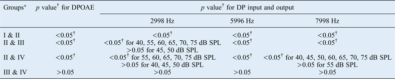
Prior to drug administration, baseline DPOAE and distortion product input and output amplitudes for all frequencies had comparable values in all experimental groups (p > 0.05). The DP gram and distortion product input and output amplitudes for the saline control group had not significantly changed at day 10 (p > 0.05). In contrast, all DP gram and distortion product input and output threshold measurements for the cisplatin-treated group were significantly elevated at day 10 compared with the saline group (p < 0.05). The cisplatin group had significantly higher DP grams for all measurements (p < 0.05) and the p values for distortion product input and output measurements were similar in the dexamethasone and methylprednisolone treatment groups at day 10. All DP gram and distortion product input and output amplitude measurements were comparable between dexamethasone and methylprednisolone treatment groups (p > 0.05; Table I). The DP grams showing results at day 10 for all four experimental groups are shown in Figure 1. Analysis of these results suggests that dexamethasone and methylprednisolone provide a similar level of protection against cisplatin-induced ototoxicity.
Graph showing DP grams for all treatment groups on day 10.

Between-group comparisons of DPOAE thresholds and DP input and output amplitudes on day 10
*I = intratympanic isotonic saline; II = intraperitoneal cisplatin and intratympanic isotonic saline; III = intraperitoneal cisplatin and intratympanic dexamethasone; IV = intraperitoneal cisplatin and intratympanic methylprednisolone. †Statistically significant. DPOAE = distortion product otoacoustic emission; DP = distortion product
Histopathology results
Cochlear sections from rats treated with dexamethasone or saline contained significantly fewer TUNEL-positive cells in the organ of Corti and spiral ganglion neurons at day 10. However, in the cisplatin group, very strongly TUNEL-positive cells were present in both the organ of Corti and spiral ganglion neurons. In the methylprednisolone group, there were few TUNEL-positive cells in the organ of Corti but an increased number of strongly TUNEL-positive spiral ganglion neurons. Figure 2 shows TUNEL-positive cells in rat cochlear sections in all experimental groups.
Photomicrographs showing TUNEL staining of cochlear tissue from rats treated with (a) saline, (b) cisplatin, (c) dexamethasone and (d) methylprednisolone: (1) organ of Corti (magnification ×40; bar = 10 µm); (2) organ of Corti (magnification ×100; bar = 5 µm); and (3) spiral ganglion neuron (magnification ×100; bar = 5 µm). All figure parts show methyl green background staining.  = inner hair cells;
 = outer hair cells;
 = TUNEL-positive ganglion neurons; BM = basilar membrane; SG = spiral ganglion; Slg = spiral ligament; Slm = spiral limbus
In the saline control group, weak caspase-3 immunoreactivity was observed in both the organ of Corti and spiral ganglion neurons. However, in the cisplatin treatment group, very strong staining was observed in both the organ of Corti and spiral ganglion neurons. In the dexamethasone group, weak immunoreactivity was observed in both the inner and outer hair cells of the organ of Corti and some immunopositive spiral ganglion neurons were present. In the methylprednisolone group, varying weak to moderate immunoreactivity was observed in both the inner and outer hair cells of the organ of Corti and some immunopositive spiral ganglion neurons were present. Figure 3 shows caspase-3 immunostaining in cochlear sections from all rat experimental groups.
Photomicrographs showing caspase-3 immunostaining of rat cochlear sections from rats treated with (a) saline, (b) cisplatin, (c) dexamethasone and (d) methylprednisolone: (1) organ of Corti (magnification ×40; bar = 10 µm); (2) organ of Corti (magnification ×100; bar = 5 µm); and (3) spiral ganglion neuron (magnification ×100; bar = 5 µm). All figure parts show Mayer's haematoxylin staining. = inner hair cells; = immunopositive ganglion neurons; BM = basilar membrane; SG = spiral ganglion; Slg = spiral ligament; Slm = spiral limbus
Weak caspase-8 immunoreactivity was observed in the organ of Corti in all experimental groups. Immunopositive spiral ganglion neurons were more common in the cisplatin and methylprednisolone treatment groups than in the saline and dexamethasone groups. Figure 4 shows caspase-8 immunostaining of cochlear sections from all rat experimental groups.
Photomicrographs showing caspase-8 immunostaining of rat cochlear sections from rats treated with (a) saline, (b) cisplatin, (c) dexamethasone and (d) methylprednisolone: (1) organ of Corti (magnification ×40; bar = 10 µm), (2) organ of Corti (magnification ×100; bar = 5 µm); and (3) spiral ganglion neuron (magnification ×100; bar = 5 µm). All figure parts show Mayer's haematoxylin staining. = inner hair cells; = immunopositive ganglion neurons; BM = basilar membrane; SG = spiral ganglion; Slg = spiral ligament; Slm = spiral limbus
In the saline group, weak caspase-9 immunoreactivity was observed in the organ of Corti and spiral ganglion neurons. In the cisplatin treatment group, strong caspase-9 expression was observed in both the inner and outer hair cells of the organ of Corti and some strongly immunopositive spiral ganglion neurons were present. Weak caspase-9 immunoreactivity was observed in the organ of Corti in both the dexamethasone and methylprednisolone treatment groups but moderate immunopositivity was observed in some spiral ganglion neurons of the methylprednisolone group. Figure 5 shows caspase-9 immunostaining of cochlear sections from all rat experimental groups.
Photomicrographs showing caspase-9 immunostaining of rat cochlear sections from rats treated with (a) saline, (b) cisplatin, (c) dexamethasone and (d) methylprednisolone: (1) organ of Corti (magnification ×40; bar = 10 µm); (2) organ of Corti (magnification ×100; bar = 5 µm); and (3) spiral ganglion neuron (magnification ×100; bar = 5 µm). All figure parts show Mayer's haematoxylin staining. = inner hair cells; = immunopositive ganglion neurons; BM = basilar membrane; SG = spiral ganglion; Slg = spiral ligament; Slm = spiral limbus
Figure 6 shows the quantification of apoptotic cells and of caspase-3, caspase-8 and caspase-9 staining in the organ of Corti and spiral ganglion neurons. Statistical analysis of TUNEL-positive cell numbers and the level of caspase-3, caspase-8 and caspase-9 immunostaining in the organ of Corti and spiral ganglion neurons in all experimental groups is shown in Table II.
Bar chart showing the number of apoptotic cells in the organ of Corti (OOC) and spiral ganglion neurons (SGN) as indicated by TUNEL staining and caspase-3, caspase-8 and caspase-9 immunostaining in rats treated with saline, cisplatin, dexamethasone and methylprednisolone on day 10.
Between-group comparisons of TUNEL-positivity and caspase staining in OOC and SGN

*I = intratympanic isotonic saline; II = intraperitoneal cisplatin and intratympanic isotonic saline; III = intraperitoneal cisplatin and intratympanic dexamethasone; IV = intraperitoneal cisplatin and intratympanic methylprednisolone. †Statistically significant. TUNEL = terminal deoxynucleotidyl transferase dUTP nick end labelling; OOC = organ of Corti; SGN = spiral ganglion neuron
Discussion
Cisplatin is a chemotherapeutic drug that is widely used to treat various cancer types; however, it causes at least some ototoxicity in a large percentage of patients. The use of intratympanic erdosteine, vitamin C, Ringer's lactate and N-acetylcysteine to prevent cisplatin-induced ototoxicity has been investigated in animal models, but most studies have focused on intratympanic glucocorticoids.Reference Saliba, El Fata, Ouelette and Robitaille9–Reference Riga, Chelis, Kakolyris, Papadopoulos, Stathakidou and Chamalidou13 Glucocorticoids (intratympanic dexamethasone, in particular) represent a drug class with significant otoprotective potential. Attenuation of cisplatin-induced ototoxicity by intratympanic dexamethasone has been reported in animal models.Reference Calli, Pinar, Oncel, Alper Bagriyanik and Umut Sakarya4, Reference Daldal, Odabasi and Serbetcioglu5, Reference Topdag, Iseri, Gelenli, Yardimoglu, Yazir and Ulubil14–Reference Murphy and Daniel17 However, a few contradictory findings have also been reported.Reference Hughes, Hussain, Pafford and Parham18 In the few studies into the possible protective effect of intratympanic methylprednisolone against cisplatin-induced ototoxicity, contradictory results have been reported. Saliba et al. reported that intratympanic methylprednisolone failed to demonstrate protective efficacy against cisplatin-induced ototoxicity.Reference Saliba, El Fata, Ouelette and Robitaille9 In the present study, poor TUNEL reactivity was observed in the organ of Corti and spiral ganglion neurons in intratympanic dexamethasone treated rats. In intratympanic methylprednisolone treated rats, poor TUNEL reactivity was observed in the organ of Corti but an increased number of strongly positive spiral ganglion neurons was observed. Weak caspase-3, caspase-8 and caspase-9 immunostaining was observed in both the inner and outer hair cells in the organ of Corti and spiral ganglion neurons in the intratympanic dexamethasone treatment group, and weak to moderate staining of the same cell types was observed in the intratympanic methylprednisolone treatment group. Distortion product otoacoustic emission (DPOAE) and distortion product input and output amplitudes were comparable between intratympanic dexamethasone and intratympanic methylprednisolone treatment groups, but a slight increase in distortion product input and output thresholds was noted in the intratympanic methylprednisolone treatment group. Therefore, intratympanic dexamethasone seems to have a greater protective effect than intratympanic methylprednisolone against cisplatin-induced ototoxicity in this study.
Many mechanisms have been suggested to explain cisplatin-induced ototoxicity. Cisplatin generates reactive oxygen species that seem to be involved in the ototoxic process.Reference Kim, Lee, Kim, Oh, Moon and Kwon19 Corticosteroids have been shown to limit the formation of reactive oxygen species in the inner ear.Reference Kolls, Xie, LeBlanc, Malinski, Nelson and Summer20, Reference Nagura, Iwasaki, Wu, Mizuta, Umemura and Hoshino21 Unfortunately, corticosteroids also down-regulate apoptotic gene expression in tumour cells.Reference Herr, Ucur, Herzer, Okouoyo, Ridder and Krammer22 Therefore, the use of systemic corticosteroids to protect against cisplatin-induced ototoxicity may decrease the tumoricidal activity of cisplatin. Thus, intratympanic delivery of steroids may be a better treatment option. Intratympanic dexamethasone and intratympanic methylprednisolone are widely used to treat hearing loss, especially sudden sensorineural hearing loss. Although most studies have reported favourable results in animal models, the use of these drugs to treat cisplatin-induced ototoxicity in humans is not generally accepted. Only a limited number of studies into treatments for cisplatin-induced ototoxicity have been performed in humans. For example, prior to each session of cancer treatment (which included cisplatin), Marshak et al. injected intratympanic dexamethasone into 26 patients suffering from a neoplastic disease; they concluded that intratympanic dexamethasone may reduce cisplatin-induced ototoxicity.Reference Marshak, Steiner, Kaminer, Levy and Shupak23
• Intratympanic glucocorticoid may protect against cisplatin-induced ototoxicity
• Intratympanic dexamethasone and methylprednisolone effects on DPOAE thresholds and cochlear histology were evaluated
• Dexamethasone and, to a lesser extent, methylprednisolone protected against cisplatin-induced ototoxicity
• Dexamethasone use might provide the best clinical treatment against cisplatin ototoxicity
A study into the cochlear pharmacokinetics of cisplatin in an animal model reported that the perilymph-to-blood ratio of cisplatin had increased 60 minutes after administration and that delayed cisplatin elimination from the scala tympani perilymph compared with blood might correlate with the cisplatin-induced loss of outer hair cells in the cochlear base.Reference Hellberg, Wallin, Ehrsson and Laurell3 Indeed, in the present study, cisplatin also had toxic effects on outer hair cells, mainly in the cochlear base, but the protective effect of intratympanic dexamethasone and intratympanic methylprednisolone was independent of the applied frequency.
It is theoretically possible that because of age-related depletion, older cochlea may not have sufficient antioxidant enzyme reserves that can be activated by intratympanic rescue drugs such as dexamethasone to preserve hearing after cisplatin challenge. Indeed, older cancer patients treated with cisplatin show a greater incidence of ototoxicity.Reference Laurell and Borg6 Although most studies have been performed in young animals (generally 3 months of age), cisplatin is commonly used in middle-aged or older patients. Parham demonstrated that intratympanic dexamethasone has a protective effect against age-related hearing loss on the high-frequency region of the cochlea in 24-month-old cisplatin-treated mice.Reference Parham24 In the present study, 16-month-old rats with normal hearing were used to model middle-aged adult patients. The results revealed that intratympanic dexamethasone and intratympanic methylprednisolone had protective effects on both the low- and high-frequency regions of the cochlea.
Conclusion
Intratympanic dexamethasone seems to have a greater protective effect than intratympanic methylprednisolone against cisplatin-induced ototoxicity in a middle-aged animal model. The safety and effectivity of intratympanic steroid injections for preventing cisplatin-induced ototoxicity in humans should be investigated in the future. It might be preferable to use dexamethasone for this purpose in clinical studies in adults.
Acknowledgements
We thank the Si-Ser Hearing Center for their support for distortion product-evoked otoacoustic emission testing. Financial support for this work was provided by Kocaeli Derince Research and Training Hospital, Turkey (grant number 30278912).