Psoriasis is a chronic skin disease affecting about 2% of the Western population. It is characterised by raised red scaly plaques that preferentially occur on the scalp, buttocks and extensor sites of the knees and elbows. In severe cases, the whole body may be involved: this serious condition is called erythroderma. First symptoms usually occur between 20 and 30 years of age. Around 20–30% of patients with psoriasis suffer from psoriatic arthritis. Psoriasis is a multifactorial disease resulting from polygenic predisposition and environmental triggering factors, such as medication, infection or trauma (Ref. Reference Schon and Boehncke1). Several local and systemic therapies are available for psoriasis. However, there is a need for better treatment because the long-term use of currently available drugs is limited by their side effects, and 40% of patients with psoriasis are not satisfied with the available care (Ref. Reference Nijsten2). Over the past few decades there has been huge growth in the understanding of the cellular and molecular processes driving the inflammation observed in psoriasis. This knowledge has revealed similarities with other (organ-specific) autoimmune diseases, such as Crohn disease, rheumatoid arthritis and multiple sclerosis.
Biologics are a new treatment entity for autoimmune diseases that have been developed based on novel insights into the cellular interactions that occur during pathogenesis of these diseases. Successful clinical introduction of biologics has boosted research towards more specific targeted therapies. However, unexpected side effects, such as the activation of latent viral infections or a worsening of disease activity during treatment, indicate the need for a better understanding of the mode of action of these therapies and the pathogenesis of the disease.
The epidermal compartment in psoriasis: the regenerative or wound-healing phenotype
The epidermis is a stratified squamous epithelium consisting of keratinocytes, which undergo a tightly regulated differentiation programme to form a mechanical, permeable and antimicrobial barrier to the external environment (Ref. Reference Fuchs and Horsley3). The skin barrier function is enhanced by incorporation of proteins with bactericidal or bacteriostatic properties, which are constitutively or inducibly expressed (Ref. Reference Gallo and Huttner4). The importance of an intact skin barrier is supported by accumulating data suggesting that genetic deficiencies in skin barrier function are associated with an increased risk of inflammatory skin disease (Ref. Reference Palmer5).
The epidermal balance between proliferation and differentiation is disturbed in psoriasis (Refs Reference Bernerd, Magnaldo and Darmon6, Reference Bernard7). The large, silvery scales observed in the lesions are a consequence of altered differentiation (hyper- and parakeratosis), whereas thickening of the epidermis is due to a strongly increased pool of proliferating keratinocytes. Altered differentiation of psoriatic keratinocytes is characterised by focal absence of the granular layer and the consequent downregulation of the late keratinocyte differentiation markers filaggrin (FLG), loricrin (LOR) and caspase-14 (CASP14), and upregulation of the early differentiation markers involucrin (IVL) and small proline-rich proteins (SPRRs). These alterations in keratinocyte differentiation lead to impairment of the skin barrier function, which is shown to correlate with the severity of disease (Ref. Reference Ghadially, Reed and Elias8).
Differentiation of epidermal cells is controlled by Notch and Wnt signalling pathways, and by the transcription factors peroxisome proliferator-activated receptor-α (PPARA), AP2 and CCAAT-enhancer-binding protein-α and -β (CEBPA and CEBPB). Proliferation of epidermal stem cells is maintained by p63, Myc, β1 integrin (ITGB1) and transforming growth factor-α (TGF-α/TGFA) signalling pathways, and negatively regulated by TGF-β signalling (Ref. Reference Fuchs and Horsley3). Signalling through insulin-like growth factor receptor (IGFR) and epidermal growth factor receptor (EGFR) also regulate proliferative behaviour in the epidermis (Ref. Reference Fuchs and Horsley3). Notably, all of these pathways are differentially expressed in lesional psoriatic skin (Refs Reference Thelu, Rossio and Favier9, Reference Mossner10, Reference Komine11, Reference King12, Reference Shen13, Reference Miura14, Reference Elder15), indicating that there is a complex network of regulatory pathways in which the primary pathogenic switch is difficult to pinpoint.
Psoriasis is often considered to be a result of genetically determined dysregulation of innate and adaptive immunity (Refs Reference Gaspari16, Reference Bos17, Reference Buchau and Gallo18). Dysregulation of the innate detection and response system can result in the epidermal phenotype and abnormal T cell function that characterise the disease (Ref. Reference Buchau and Gallo18). Examples of abnormalities in the innate response system in psoriasis are the increased expression of antimicrobial peptides (Ref. Reference Hollox19), activation of type I interferon (IFN) system (Refs Reference van der Fits20, Reference Nestle21), and dysregulation of the expression of the interleukin-1 (IL1) family of cytokines (Ref. Reference Cooper22). In addition, psoriasis is often compared with the normal skin-wounding response, or the regenerative epidermal phenotype (Refs Reference Hertle23, Reference Nickoloff24). As part of this regenerative or ‘stress’ response, some antimicrobial peptides such as elafin (PI3), psoriasin (S100A7), LL-37 (CAMP) and β-defensin 2 (DEFB4), are highly induced. In addition, neutrophils, macrophages, dendritic cells (DCs) and natural killer cells initiate an innate immune response to defend the organism from invading pathogens.
Several molecular processes drive this epidermal regenerative or stress response, both during wound healing and in psoriasis: signal transducer and activator of transcription 3 (STAT3) and the mitogen-activated protein kinase (MAPK) cascade being the most important. STAT3 is activated in healing wounds, whereas epidermis-specific disruption of STAT3 in mice leads to delayed wound healing (Refs Reference Sano25, Reference Dauer26). In psoriatic patients, STAT3 is activated in lesional keratinocytes (Ref. Reference Sano27), and transgenic mice with constitutively active STAT3 in keratinocytes spontaneously develop skin lesions resembling human psoriasis (Ref. Reference Sano27). These data indicate that activation of STAT3 has a role in the development of the regenerative epidermal phenotype observed in psoriasis. MAPK-activated protein kinase 2 (MAPKAPK2) and mitogen- and stress-activated protein kinase 1 (MSK1) regulate the expression of TNF-α and other proinflammatory mediators in psoriatic lesions (Refs Reference Johansen28, Reference Funding29). Integrin-induced activation of MAPK is thought to be responsible for epidermal hyperproliferation in psoriasis and wound healing, either directly or through increased IL-1α production (Ref. Reference Haase30). More recently, expression of IL-20, a cytokine with high expression in psoriasis, was shown to be induced by p38 MAPK in keratinocytes (Refs Reference Otkjaer31, Reference Otkjaer32). IL-20 induces the regenerative epidermal phenotype that is characteristic of psoriasis (Ref. Reference Wolk33).
It has been widely debated whether epidermal alterations in psoriasis occur in response to skin inflammation or whether keratinocytes themselves have a role in the initiation and propagation of psoriasis. Data arguing in favour of intrinsic disturbances in keratinocytes in psoriasis include findings of genetic association studies. Genetic variations within loci containing epidermal defence genes DEFB4 (encoding human β-defensin 2) and LCE3B/LCE3C (which encode late cornified envelope proteins 3B and 3C, repectively) have been reported to be associated with psoriasis, but need broader confirmation (Refs Reference Hollox19, Reference de Cid34). Genetic alterations in the epidermal differentiation complex on chromosome 1q21 are associated with psoriasis (Refs Reference de Cid34, Reference Zhang35).
Two experimental models in mice add further evidence for the central role of the epidermis in the initiation of psorasis. First, psoriasis-like skin inflammation may be induced by changing the expression of just one gene in keratinocytes, as is observed in several keratinocye-specific transgenic mice (Refs Reference Sano27, Reference Stratis36, Reference Pasparakis37, Reference Xia38). Second, in all known murine xenograft psoriasis models, whereby the induction of the psoriasis phenotype is dependent on the interaction between autologous leukocytes and transplanted skin, the full psoriasis phenotype will only develop in skin from patients with psoriasis, and not in skin samples from healthy controls (Refs Reference Boyman39, Reference Wrone-Smith and Nickoloff40). The latter indicates a specific response of skin genetically predestined for psoriasis. There is thus strong evidence that intrinsic alterations in the epidermis contribute to the pathogenesis of psoriasis.
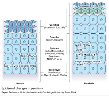
In summary, psoriasis is characterised by hyperproliferation and disturbed differentiation of epidermal keratinocytes. Keratinocytes have an activated phenotype in psoriasis, which is similar to their condition during wound healing (Fig. 1). Keratinocytes produce mediators that activate cells of the immune system and endothelial cells in the dermis. However, leukocyte-derived cytokines might activate keratinocytes, as discussed below.
Epidermal changes in psoriasis. In psoriasis (right panel) signalling pathways that are associated with keratinocyte differentiation, such as Notch and PPAR-α signalling, are suppressed, whereas proliferation-associated pathways, such as Myc, β-integrin and Wnt5a, are induced. STAT3, MEK1 and IL-1 signalling pathways are responsible for the regenerative epidermal phenotype, involving an increased expression of antimicrobial peptides, such as β-defensin 2 and LL-37.

The contribution of leukocytes to the pathogenesis of psoriasis
The immune system is strongly implicated in the pathogenesis of psoriasis, including both acquired immunity via T cells, and innate immunity, involving antigen-presenting cells (APCs), neutrophils, mast cells and keratinocytes. In psoriasis lesions, the number of T cells and CD11c+ DCs is markedly increased (Ref. Reference Bos41). T cells, especially CD8+ T cells, migrate into all layers of the epidermis. CD11c+ DCs also infiltrate the epidermis, but remain confined to the lower cell layers (Ref. Reference Zaba42).
The number of immature Langerhans cells are only slightly reduced; however, mobilisation of these cells in response to stimuli that normally induce migration (chemical allergen, TNF-α and IL-1β) is largely absent (Ref. Reference Cumberbatch43). Neutrophils accumulate in small aggregates called Munro abscesses beneath the corneal layers (Refs Reference Beurskens44, Reference van de Kerkhof and Lammers45). As a consequence of the activation of leukocytes, levels of the proinflammatory cytokines IFN-α, IFN-γ, TNF-α, IL-1, IL-6, IL-23, IL-22 and IL-17 are high in psoriasis lesions (Refs Reference Nestle21, Reference Wolk46, Reference Uyemura47, Reference Lee48, Reference Johansen49). These cytokines drive the activation of leukocytes, as well as contribute to the epidermal alterations described above. Chemokines, which are responsible for the chemoattraction of leukocytes from the circulation into the skin, form a bridge between the epidermal compartment and the immunological components of the psoriatic pathology.
In noninflamed human skin, there are three main DC populations: epidermal Langerhans cells, (mature) dermal myeloid (CD11c+) DCs, and sporadically, dermal plasmacytoid DCs (pDCs) (Ref. Reference Zaba, Krueger and Lowes50). Although pDCs have the capacity to present antigen, they are characterised by their ability to produce large amounts of type I IFN during viral infection, and in autoimmune diseases. One of the earliest events driving the inflammatory eruption in psoriasis is the secretion of IFN-α, a type I IFN, from pDCs (Ref. Reference Nestle21). pDCs are only sporadically seen in normal skin, but are clearly increased in psoriasis lesions (Ref. Reference Wollenberg51). We have previously shown that the type I IFN pathway is activated in psoriatic lesions (Ref. Reference van der Fits20). Treatment of non- or prepsoriatic patients with IFN-α can induce or exacerbate psoriasis (Refs Reference Downs and Dunnill52, Reference Funk53, Reference Ketikoglou54, Reference Pauluzzi55). A functional role of IFN-α or IFN-β in the initiation of psoriasis has been demonstrated using a xenograft murine model of psoriasis. Blockade of IFN-α signalling prevented the T-cell-dependent development of psoriatic lesions in nonlesional skin transplanted onto immunocompromised mice (Ref. Reference Nestle21). Recently, LL-37 complexed to self-DNA, was shown to trigger pDCs to produce large amounts of IFN-α (Ref. Reference Lande56), providing the first illustration of the autoimmune nature of psoriasis. Further studies are required to unravel the exact contribution of pDCs to the different stages of psoriasis.
CD11c+ myeloid DCs are dramatically increased in the psoriatic dermis (with numbers around the same as T cell numbers) (Ref. Reference Zaba57). This change is not due to an increase in DCs that are also present in a steady state, but to the occurrence of a novel, immature population of DCs (Ref. Reference Zaba42). Thus, psoriatic lesional skin has two populations of dermal DCs: (1) CD11c+ BDCA-1+ cells, which are phenotypically similar to those contained in normal skin and (2) CD11c+ BDCA-1– cells, which are phenotypically immature and produce inflammatory cytokines. The second group might be derived from circulating DC precursors (e.g. monocytes or pre-DCs) migrating into the skin as a result of inflammatory and chemotactic signals (Ref. Reference Zaba, Krueger and Lowes50). Importantly, an increase in the number of activated DCs was observed in perilesional, normal-appearing skin of psoriatic patients, suggesting a role of these cells in the provocation or initiation of epidermal inflammation (Ref. Reference Komine11).
CD11c+ BDCA1– myeloid dermal DCs in psoriasis are called ‘inflammatory’ DCs, or TNF- and iNOS-producing DCs (TIP-DCs) (Ref. Reference Lowes58). In addition, these inflammatory DCs produce IL-20, a cytokine that induces the regenerative epidermal phenotype observed in psoriasis (Refs Reference Zaba57, Reference Wang59), and IL-23, which is important for induction of IL-17 and IL-22 in T cells. Myeloid dermal DC populations induce T cell proliferation, and polarise T cells to become T helper 1 (Th1) and T helper 17 (Th17) cells (Ref. Reference Zaba42).
In normal skin, a considerable number of T cells are found in the dermis (~2 × 10⁶ cells/cm2, which means that there are about twice as many T cells in the skin than in the circulation (Ref. Reference Clark60)), but there are almost no T cells located in the epidermis (Ref. Reference Clark60). In psoriatic skin lesions, both CD4+ and CD8+ T cell subsets are present, with CD8+ cells predominant in the epidermis (Ref. Reference Albanesi, De Pita and Girolomoni61). T cells are thought to be critical pathogenic triggers of psoriasis, and therapies that inhibit T cell activation and trafficking are effective in treating psoriasis. Immunosuppressive drugs, such as cyclosporine, with preferential activity towards T cells have been shown to be particularly effective, and are widely used for systemic therapy of psoriasis. In addition, psoriasis can be induced in xenograft murine models by the injection of T cells from human skin lesions. The genetic association of psoriasis with certain major histocompatibility complex (MHC) alleles including HLA-Cw6 also suggests a pathogenic role of T cells (Ref. Reference Nair, Stuart and Nistor62). Furthermore, psoriasis might develop for the first time after bone marrow transplantation from a donor with psoriasis, or may not recur in patients with psoriasis after bone marrow transplantation from healthy donors (Refs Reference Eedy63, Reference Gardembas-Pain64).
Activated T cells secrete proinflammatory cytokines, such as IFN-γ, IL-17, IL-21, IL-22, TNF-α, IL-4 or IL-13, depending on their phenotype. Cytotoxic CD8+ T cells in psoriasis produce cytotoxic granule components Tia-1 (TIA1), granzyme B (GZMB) and perforin (PRF1), although their pathogenic action is mostly related to cytokine secretion (Ref. Reference Gaspari16). HLA-Cw6 is the product of the psoriasis susceptibility gene HLA-C and it might be involved in the activation of CD8+ T cells by DCs. Activated skin-homing CD8+ T cells might recognise keratinocyte antigens presented in the context of HLA-Cw6 (Ref. Reference Johnston65). Thus, the preferential usage of certain T-cell-receptor β-chain families by T cells in psoriatic lesions implicates a role for (microbial) antigens or superantigens in triggering the disease (Refs Reference Skov and Baadsgaard66, Reference Travers67, Reference Lin68, Reference Prinz69). However, the exact antigenic specificity of these cytotoxic T lymphocytes in psoriasis has not been identified (Ref. Reference Gaspari16). CD4+ Th cells with a Th1 or Th17 phenotype accumulate in psoriasis lesions (Refs Reference Lowes70, Reference Pene71). When activated, Th1 cells produce IFN-γ and TNF-α, whereas Th17 cells secrete IL-17 or IL-22 (and also some IFN-γ). Th1 and Th17 cells exist side by side in psoriatic lesions, and even synergise in inducing human β-defensin 2 (Ref. Reference Kryczek72); however, regulatory T cells are deficient in psoriasis, resulting in a defective brake on the inflammatory process (Ref. Reference Sugiyama73).
Infiltrating macrophages in psoriatic plaques accumulate along the basal membrane of the epidermis (‘lining cells’) (Ref. Reference Boehncke74). The importance of these cells in the maintenance of psoriatic inflammation has been demonstrated in different psoriasis-like dermatitis mouse models, where depletion of macrophages led to significant attenuation of psoriasis-like skin changes (Refs Reference Stratis36, Reference Wang75).
Leukocytes are recruited to the skin by transmigration through activated endothelial cells. In addition, resident skin leukocytes might also expand to create the dense infiltrates seen in psoriatic lesions (Ref. Reference Lowes, Bowcock and Krueger76). Under the influence of T-cell-derived proinflammatory cytokines, keratinocytes express a broad array of mediators, thereby amplifying the inflammatory response. Such keratinocyte-derived chemokines have a key role in the recruitment of the inflammatory infiltrate. Several chemokines and their corresponding receptors are associated with the psoriatic phenotype and might support leukocyte recruitment (Ref. Reference Homey and Meller77). CCL27 is exclusively produced by epidermal keratinocytes, and binds CCR10. More than 90% of skin-infiltrating lymphocytes in psoriasis and in atopic and contact dermatitis express CCR10, suggesting a pivotal role of the interaction of CCR10 with CCL27 during the pathogenesis of T-cell-mediated skin inflammation (Ref. Reference Homey78). Chemokines that differentiate psoriasis from atopic dermatitis are CCL4, CCL20, CXCL2 and CXCL8/IL-8 (Ref. Reference Nomura79). Neutrophils are a defining feature of psoriasis and are an important source of IL-8, CXCL10 and the S100 proteins S100A8 and S100A9.
Leukocyte-derived cytokines that can induce the epidermal changes seen in psoriatic plaques are IL-21, IL-22 and IFN-γ (Refs Reference Wolk46, Reference Caruso80, Reference Wolk81, Reference Sa82, Reference Nograles83, Reference Wei84). The prototypic Th17 cytokine IL-22 stimulates keratinocyte proliferation and the production of antimicrobial peptides; therefore, it is a key effector cytokine in the pathogenesis of psoriasis. Elevated levels of IL-22 are found in the blood of patients with psoriasis (Ref. Reference Wolk81). In addition, IL-22 inhibits terminal differentiation of keratinocyte, induces STAT3 upregulation, and causes psoriasis-like morphological changes in human skin models (Ref. Reference Wolk33). IL-22 secretion is induced by IL-23, which has emerged recently as a key cytokine in the pathogenesis of psoriasis (Ref. Reference Blauvelt85). IL-23 is produced in great quantities in psoriasis lesions by DCs and keratinocytes, and it stimulates Th17 cells to produce Th17 cytokines (Refs Reference Lee48, Reference Kryczek72, Reference Piskin86). The importance of this immunological pathway is underlined by the fact that polymorphisms in genes encoding the IL23 receptor and IL-12/IL-23p40 are associated with a susceptibility to psoriasis (Refs Reference Cargill87, Reference Nair88, Reference Garcia89). In addition, it is noteworthy that monoclonal antibodies targeting the p40 subunit of IL-12 and IL-23 are highly effective in treating psoriasis (Ref. Reference Krueger90). A psoriasis-like dermatitis in mice induced by the TLR7 and TLR8 agonist imiquimod has been shown to be critically dependent on IL-23 and IL-17 (Ref. Reference van der Fits91). Cyclosporine A, which is a standard treatment for psoriasis, appears to modulate the Th17 pathway in psoriatic skin within 2 weeks of the start of treatment (Ref. Reference Haider92). Downmodulation of this pathway correlated well with the clinical improvement of the disease (Ref. Reference Haider92). In addition, etanercept (a TNF-receptor–immunoglobulin fusion protein), which is another effective treatment for psoriasis, was shown to inhibit the Th17 pathway (Ref. Reference Zaba57).
IFN-γ can induce the regenerative psoriatic phenotype in nonlesional psoriatic skin and normal healthy skin (Ref. Reference Wei84). Interestingly, the TNF-α inhibitor infliximab inhibits the synthesis of IFN-γ by Th1 cells (Ref. Reference Haider93). Thus, secretion of IFN-γ might be downstream of TNF-α. TNF-α has been called a sentinel cytokine, or the body's ‘fire alarm’, because it initiates the defence response to local injury (Ref. Reference Tracey94). Many different immune and nonimmune cell types can produce TNF, including macrophages, T cells, mast cells, granulocytes, natural killer (NK) cells, fibroblasts, neurons, keratinocytes and smooth muscle cells. At low concentrations, TNF is thought to have beneficial effects, such as the augmentation of host defence mechanisms against infection. In disease states, TNF mediates a variety of direct pathogenic effects and induces the production of other mediators of inflammation and tissue destruction, placing it in a key position in the inflammatory cascade within an inflammatory network (Ref. Reference Tracey94).
Recently, in addition to the known psoriasis susceptibility gene HLA-C (Ref. Reference Nair, Stuart and Nistor62), several novel genetic regions have been identified that confirm the involvement of the immune system in the pathogenesis of psoriasis: IL23A, which encodes the p19 subunit of IL-23, TNFAIP3 and TNIP1, which are regulators of TNF-α signalling, and IL4 and IL13 – genes whose products support development of Th2 cells (Refs Reference Nair95, Reference Chang96).
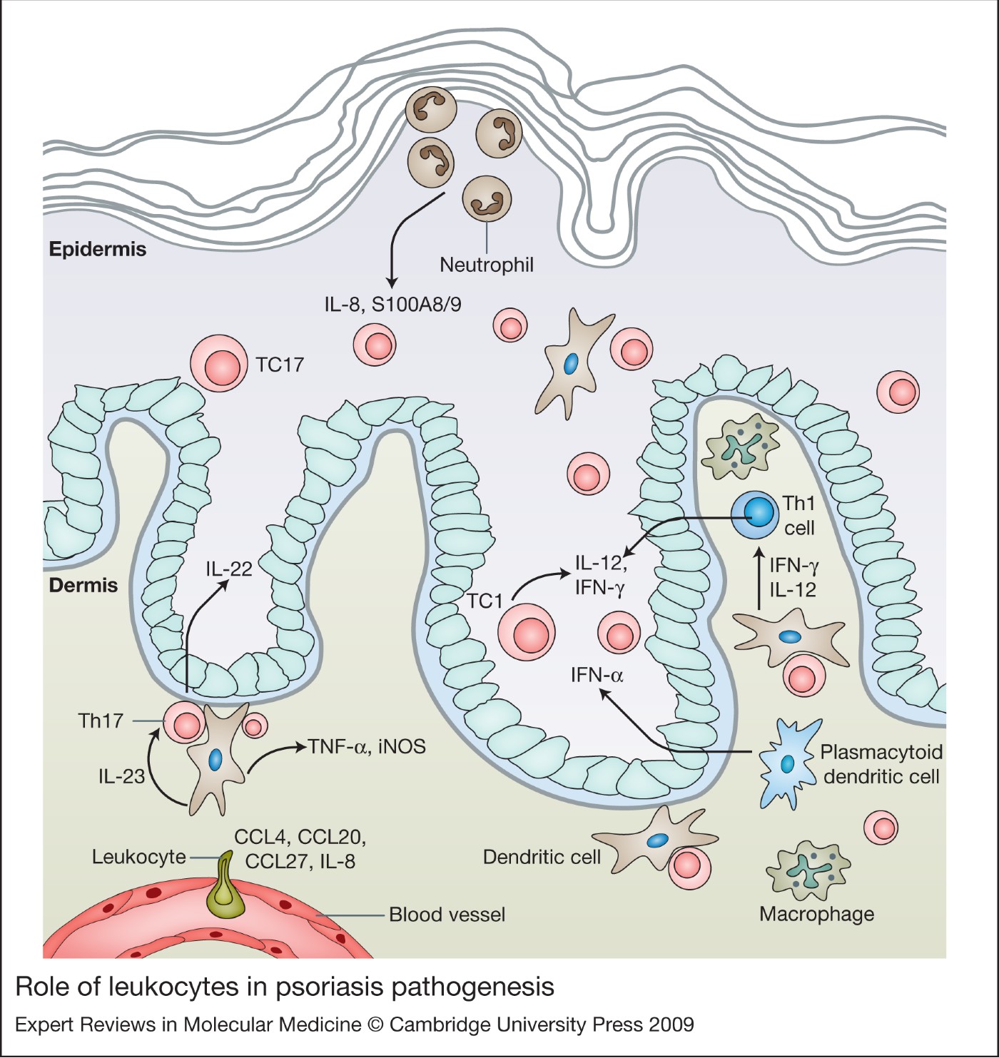
In conclusion, components of innate and adaptive immunity are activated in psoriasis (Fig. 2). An early event is the production of IFN-α by pDCs, which activates keratinocytes and cells of the immune system. Dermal DCs acquire an inflammatory phenotype and produce IL-23, TNF-α and iNOS, resulting in the activation of Th1 and Th17 cells. Active Th1 and Th17 cells synthesise IFN-γ and IL-22, respectively, and these cytokines stimulate the activation and proliferation of epidermal keratinocytes, thus contributing to the vicious circle of psoriatic skin inflammation.
Role of leukocytes in psoriasis pathogenesis. A chemotactic gradient formed by different chemokines, such as IL-8, CCL20 and CCL27, aids the extravasation of leukocytes into the skin. Myeloid dendritic cells (DCs) produce IL-23, TNF-α and iNOS, thereby activating T helper (Th) cells to produce IL-22, IL-12 and IFN-γ. Plasmacytoid DCs (pDCs) produce large amounts of IFN-α, which has a role in the initiation of psoriatic plaque formation. Neutrophils in the stratum corneum are a source of chemokines. Cytotoxic T cells (TC1, TC17) infiltrate the epidermis, where they release cytotoxic molecules, as well as proinflammatory cytokines.

Vascular and endothelial cell activation
A hallmark of psoriatic skin is the transformation of the local microvascular system, characterised by dilation and tortuosity of capillaries, increased permeability, and high endothelial venule formation, which is usually seen in lymph nodes. Extravasation of leukocytes into the dermis is aided by this transformation of the capillaries, as well as by the increased expression of adhesion molecules, such as intracellular adhesion molecule-1 (ICAM1), vascular cell adhesion molecule-1 (VCAM1) and E-selectin (SELE), by endothelial cells (ECs).
Accumulating evidence indicates that many of the cells involved in inflammatory processes release factors that act directly or indirectly on vascular endothelial cells. The most important of these factors might be nitric oxide (NO), a well-established inflammatory agent produced by the activation of iNOS in inflammatory DCs. NO stimulates vessel dilation and permeability, a feature required for leukocyte extravasation (Ref. Reference Costa, Incio and Soares97). In addition, cyclo-oxygenase-2 (COX2), an inducible enzyme responsible for the increase in prostaglandin biosynthesis during inflammation, also contributes to increased angiogenesis by upregulating the production of vascular endothelial growth factor (VEGF) (Ref. Reference Costa, Incio and Soares97). VEGF has a crucial role in the neoangiogenesis that occurs in psoriasis (Ref. Reference Costa, Incio and Soares97), where it induces migration, survival and proliferation of ECs. VEGF acts by engaging with its tyrosine kinase receptors VEGFR1 and VEGFR2 in endothelial cells. Although VEGF binds to both receptors, it appears that most of its biological functions are mediated via VEGFR2.
VEGF is secreted by keratinocytes, and VEGF serum levels are increased in patients with psoriasis (Ref. Reference Costa, Incio and Soares97). Distinct single-nucleotide polymorphisms of VEGF have been described in patients with psoriasis, although the association has not been confirmed in larger patient populations (Ref. Reference Detmar98). Furthermore, overexpression of VEGF in mouse skin results in a psoriasis skin phenotype, which implies a complex interplay between blood vessels, keratinocytes and leukocytes (Ref. Reference Xia38).
Although vascular remodelling is a hallmark of many chronic inflammatory disorders, antivascular strategies for treating these conditions have received little attention. In psoriasis, selective photothermolysis of the dermal microvasculature using a 585 nm pulsed-dye laser, resulted in partial or complete disappearance of psoriasis plaques (Refs Reference Katugampola, Rees and Lanigan99, Reference Ros100, Reference Hern101, Reference Zelickson102). The antiangiogenic agents paclitaxel and AE-941 (Neovastat) demonstrated therapeutic activity in patients with severe psoriasis (Refs Reference Ehrlich103, Reference Sauder104). Recently, a VEGFR antagonist that is already in clinical use for the treatment of malignancies and inhibition of retinal neovascularisation was shown to inhibit psoriasis-like skin inflammation in mice (Ref. Reference Halin105).
Genome-wide expression studies in psoriasis
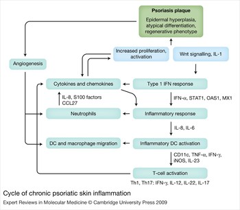
Several studies have been published on the gene expression profile of lesional psoriatic skin. Some of these compared lesional skin with nonlesional skin (Refs Reference Bowcock106, Reference Oestreicher107, Reference Zhou108, Reference Kulski109, Reference Reischl110, Reference Yao111, Reference Quekenborn-Trinquet112, Reference Gudjonsson113), others compared psoriatic lesional skin with other inflammatory skin disorders (Refs Reference Nomura79, Reference Guttman-Yassky114, Reference Ito115, Reference de Jongh116, Reference Wenzel117), and yet another group of studies followed patients receiving different treatments for psoriasis and compared gene expression profiles at different time points during treatment (Refs Reference Haider92, Reference Haider118, Reference Hochberg119, Reference Koczan120, Reference Jung121). These studies describe the genomic signature of psoriasis lesions, thereby helping to better understand molecular pathophysiology of the disease (Fig. 3).
Cycle of chronic psoriatic skin inflammation. The activated epidermis produces chemokines, cytokines and angiogenic factors. In the dermis, plasmacytoid dendritic cells (pDCs) produce IFN-α, which triggers an adaptive immune response. Inflammatory myeloid DCs activate T cells to produce proinflammatory cytokines, such as IL-22 and IFN-γ, which in turn stimulate the regenerative phenotype of epidermal keratinocytes. Neutrophils are attracted to the epidermis by keratinocyte-derived chemokines, and are themselves a source of chemoattractant molecules. Modified from Ref. Reference Kulski109.

Studies comparing lesional psoriatic skin with nonlesional skin and normal skin confirmed increased proliferation, altered epidermal differentiation and barrier function, and an activated (innate) immune response in psoriatic skin (Refs Reference Bowcock106, Reference Oestreicher107, Reference Zhou108, Reference Kulski109, Reference Reischl110, Reference Yao111, Reference Gudjonsson113). This work has provided new insights into the pathogenesis of psoriasis, such as the large contribution of CD11c+ myeloid DCs, as well as the activation of interferon- and Wnt-signalling pathways. The psoriasis transcriptome does not depend on body site location, and it correlates well with disease severity (Ref. Reference Quekenborn-Trinquet112).
The psoriatic transcriptome was compared with cell-specific gene expression profiles to identify the contribution of different cellular subsets to the pathogenesis. With this approach, a surprisingly large contribution of myeloid DCs to the psoriatic transcriptome was found (Refs Reference Guttman-Yassky114, Reference Haider122). Gene expression profiling of IL-1α-stimulated keratinocytes identified a set of psoriasis genes, among them mainly antimicrobial peptides and other components of the epidermal barrier, emphasising the anomalies of innate immunity in psoriasis (Refs Reference Mee123, Reference Mee124).
Gene expression profiles in psoriasis and atopic dermatitis have been compared by several research groups (Refs Reference Nomura79, Reference Guttman-Yassky114, Reference Ito115, Reference de Jongh116, Reference Wenzel117). An advantage of this approach is that only genes that are disease specific will be identified, and molecules that are secondary to the chronic inflammatory and hyperproliferative state will not be found (Ref. Reference de Jongh116). One major difference between the two conditions is the elevated expression of antimicrobial peptides seen in psoriasis (Ref. Reference de Jongh116). The spectrum of inflammatory mediators produced by dermal myeloid DCs in psoriasis clearly differs from that produced in atopic dermatitis: TNF-α and iNOS are the dominant inflammatory mediators in psoriasis (Ref. Reference Guttman-Yassky114). Chemokine expression also differentiates psoriasis from atopic dermatitis. Chemokines with high expression in psoriasis are CCL4, CCL20, CXCL2, CXCL8 and the chemokine receptor CXCR2 (Ref. Reference Nomura79). This comparative approach applied to other dermatological conditions such as lichen planus, atopic dermatitis and psoriasis also identified the type I IFN signature as being specific for psoriasis (Ref. Reference Wenzel117).
Psoriasis has also been compared with squamous cell carcinoma using microarray technology (Ref. Reference Haider125). Here, a set of common upregulated genes was identified, including, surprisingly, molecules with immunoregulatory roles (Ref. Reference Haider125).
Genome-wide expression analysis of lesional skin in psoriasis patients receiving established effective therapies, such as cyclosporine A (Ref. Reference Haider92), UVB phototherapy (Ref. Reference Hochberg119) and dithranol (Ref. Reference Koczan120) has generated valuable new information on the molecular pathways involved. Such knowledge of the genomic signature of successful treatments of psoriasis might aid in the design and development of new therapies.
Molecular targets of current treatments of psoriasis
Most established treatments for psoriasis have been developed empirically. Biologics, however, specifically target key mechanisms of psoriasis pathogenesis. Most of these compounds are monoclonal antibodies, anti-cytokines or fusion proteins that block T cell migration and adhesion, or antagonise or neutralise the effects of cytokines.
Established empirical treatments for psoriasis include topical and systemic therapies. In the majority of patients with psoriasis (approximately 60%), the disease is managed by topical therapy alone, although most patients are dissatisfied with this type of treatment. Corticosteroids and Vitamin D3 are the most frequently used topical treatments and can be very effective in controlling mild to moderate psoriasis lesions. A recent study on the effects of corticosteroids on keratinocytes showed that in addition to regulation of proinflammatory genes, corticosteroids regulate keratinocyte motility and metabolism, suppress the expression of essentially all IFN-γ-regulated genes, including IFNGR1 (IFN-γ receptor) and STAT1, promote terminal epidermal differentiation while simultaneously inhibiting early stage differentiation (Ref. Reference Stojadinovic126). Because of their side effects upon prolonged use, corticosteroids are combined with other topical treatments in psoriasis. Vitamin D3 analogues (calcipotriol and tacalcitol) and the topical retinoid tazarotene, all of which affect keratinocyte functions and the immune response, are now more widely used than either anthralin or coal tar (Ref. Reference Schon and Boehncke1). Moderate-to-severe psoriasis can be treated with UV phototherapy or systemic drugs. The mode of action of traditional systemic therapies is incompletely understood. Some of these, such as cyclosporine A and fumarates, inhibit immune functions, whereas others, such as retinoids and methotrexate, also target keratinocyte functions.
Novel therapeutic agents are characterised by more specific targeting of defined molecules in the pathological pathways. The T-cell-targeted drug alefacept, a lymphocyte function-associated antigen (LFA3)–immunoglobulin fusion protein interferes with LFA3–CD2 interactions on T cells and antigen-presenting cells. Alefacept is an effective therapy in selected patients and might induce relatively long remissions. In responding patients, alefacept induces a reduction in CD11c+ DCs as well as the number of T cells in the skin (Ref. Reference Chamian127). T cells are the primary target for therapy, but DCs and a spectrum of Th1 inflammatory genes are coordinately suppressed (Ref. Reference Haider118).
Infliximab, adalimumab and etanercept target TNF-α; all are indicated for several immune-mediated inflammatory diseases. All three TNF antagonists are parenterally administered protein therapeutics (biologics); infliximab and adalimumab are monoclonal antibodies (mAbs) that specifically bind TNF; and etanercept is a TNF-receptor fusion protein that binds TNF and lymphotoxin (LT) family members. Infliximab not only blocks TNF-α, but also (indirectly) inhibits IFN-γ synthesis by T cells, thereby blocking the Th1 pathway (Ref. Reference Haider128); in this aspect it differs from etanercept (Ref. Reference Haider129). Adalimumab is a fully human recombinant anti-TNF antibody, and, theoretically, has similar actions and effects to infliximab, which still contains a considerable murine component. Etanercept downregulates Th17 responses and induces apoptosis of dermal DCs in patients with psoriasis (Refs Reference Zaba57, Reference Malaviya130). The response of psoriasis to three distinct TNF inhibitors certainly suggests that this cytokine has a key role in disease pathogenesis. New TNF-α blockers include golimumab, a fully human anti-TNF-α antibody, and certolizumab pegol. As certolizumab pegol does not have an Fc region, unlike infliximab and adalimumab, it should not fix complement or cause antibody-dependent cell-mediated cytotoxicity in vitro (Ref. Reference Rozenblit and Lebwohl131).
Ustekinumab is a fully human monoclonal antibody against the p40 subunit of IL-12 and IL-23 (Refs Reference Papp132, Reference Leonardi133). It binds with high affinity and specificity to the p40 subunit, preventing the cytokines from binding to IL-12 and IL-23 receptor 1 (IL12R1) on the surface of leukocytes. Ustekinumab appears to be as effective as the anti-TNF antibodies during induction therapy, and the available long-term data indicate a stable clinical response over time, with no rebound after withdrawal of the drug (Ref. Reference Reich, Yasothan and Kirkpatrick134). Another monoclonal antibody against the p40 subunit is ABT-874, which has also been tested in Phase II clinical trials, where it was found to be effective and comparable to ustekinumab (Ref. Reference Kimball135). The clinical efficacy of anti-p40 antibodies is much longer than its serum half-life, which suggests that this intervention collapses a key population of cells. This long-lasting effect makes these drugs highly practical, because they require only four to five injections per year. At present, information on the safety of ustekinumab is based on smaller patient numbers and shorter duration of exposure compared with the TNF antagonists.
Meta-analysis of trials with new and established therapies showed that these novel, specific treatments are more effective than traditional systemic treatments (Ref. Reference Schmitt136). Infliximab appears to be the most effective treatment for moderate-to-severe psoriasis, followed by adalimumab. Etanercept induces stable long-term response rates (Ref. Reference Schmitt136).
Several other approaches for the treatment of psoriasis are currently under clinical investigation. The PPAR-γ (PPARG) antagonist pioglitazon enhanced the efficacy of acitretin in clearing psoriasis (Ref. Reference Mittal137). Janus kinase (JAK) inhibitors lestaurtinib (CEP-701) and INVB18424 aim to block proinflammatory JAK–STAT signalling pathways and the pan-selectin antagonist Bimosiamose prevents leukocytes from entering tissues from the circulation. Use of recombinant α-fetoprotein (MM-093) is based on the observation that psoriasis, in common with most autoimmune diseases, tends to improve during pregnancy (Ref. Reference Murase138).
Clinical implications and outstanding research questions
Improved understanding of psoriasis pathogenesis has led to the development of targeted therapies that have proved very effective. Studies in patients treated with biologics provide further novel and often surprising results on the mode of action, and contribute to better understanding of molecular interactions in vivo. Unexpected in vivo results, such as the inhibition of IFN-γ synthesis by the TNF-α inhibitor infliximab, underline the importance of further research on molecular pathways and interactions in the pathogenesis and treatment of psoriasis. Adverse effects of biologics encountered in the past, such as the activation of the John Cunningham (JC) virus in four patients by the T-cell-targeted antibody efalizumab (now withdrawn from the European market) (Ref. Reference Sterry139), mean that administration of these novel immunosuppressive drugs to patients warrants extreme care and long-term safety follow-up.
Despite intense research, identification of the initiating event in the pathogenesis of psoriasis remains elusive. Although psoriasis is often referred to as an autoimmune disorder, the autoantigen that stimulates T cell activation still has to be identified. In addition, the functional consequences of genomic alterations that have been described in the epidermal genes DEFB4 and LCE3 need to be defined, as does the role of these genes in pathogenesis. In the field of therapeutics, improved forms of established treatments are being developed, such as novel calcineurin and TNF-α inhibitors, as well as novel targeted therapies, such as anti-IL-12 and anti-IL-23 antibodies. Extended long-term studies on the efficacy and safety of these biologics are imperative. Basic research has identified possible novel therapeutic targets, such as IL-22, MAPK and JAK–STAT signalling. In the coming years, evaluation of these molecules as valid therapeutic targets is expected.
Acknowledgements
We would like to thank the reviewers for their constructive and useful suggestions, and thank Tar van Os for the preparation of the figures.