Significant outcomes
-
High plasma sTNF-R1 level was associated with low volumes of brain subregions in BD-I.
-
Plasma YKL-40, not MCP-1and FKN, was the chemokine associated with the reduction of BD-related brain subregion volumes.
Limitations
-
Inflammatory markers in the CSF were not assessed.
-
Medications with potential differential effects on brain structure and inflammatory response were not controlled in the analysis.
Introduction
Bipolar disorder (BD) is associated with aberrant structural and functional abnormalities in the brain (Hallahan et al., Reference Hallahan, Newell, Soares, Brambilla, Strakowski, Fleck, Kieseppä, Altshuler, Fornito, Malhi, McIntosh, Yurgelun-Todd, Labar, Sharma, MacQueen, Murray and McDonald2011; Selvaraj et al., Reference Selvaraj, Arnone, Job, Stanfield, Farrow, Nugent, Scherk, Gruber, Chen, Sachdev, Dickstein, Malhi, Ha, Ha, Phillips and McIntosh2012; Hanford et al., Reference Hanford, Nazarov, Hall and Sassi2016). The predominant pattern of reduced grey matter volumes in adults with BD may result from neurotoxicity caused by elevated inflammatory activity in these structures (Phillips & Swartz, Reference Phillips and Swartz2014). For example, increased excitotoxicity and glial markers have been observed in the frontal cortex of patients with BD (Rao et al., Reference Rao, Harry, Rapoport and Kim2010). Neuroinflammation may contribute to the etiopathogenesis of BD and play a role in its pathophysiology (Goldsmith et al., Reference Goldsmith, Rapaport and Miller2016; Isgren et al., Reference Isgren, Sellgren, Ekman, Holmén-Larsson, Blennow, Zetterberg, Jakobsson and Landén2017). The search for immune biomarkers in psychiatric disorders has primarily focused on proinflammatory cytokines. However, brain-specific immune mechanisms, beyond systemic inflammatory processes, may also be involved in the pathophysiology of BD (Zhang et al., Reference Zhang, Song, Isgren, Jakobsson, Blennow, Sellgren, Zetterberg, Bergen and Landén2020). Chemokines are a special type of cytokine that are crucial for direct chemotaxis induction, leukocyte and macrophage migration, and inflammatory response propagation (Milenkovic et al., Reference Milenkovic, Stanton, Nothdurfter, Rupprecht and Wetzel2019). Chemokines may be involved in the development of psychiatric disorders through numerous neurobiological processes, beyond their classical chemotactic functions, including neuromodulator effects, neurotransmitter-like effects, and regulation of neurogenesis (Stuart & Baune, Reference Stuart and Baune2014). However, few studies on BD have investigated chemokines.
Macrophages are responsible for innate immune response activation in the periphery, whereas microglia are tissue-resident macrophages of the central nervous system (CNS), responsible for homeostasis and synaptic modulation (Ascoli et al., Reference Ascoli, Géa, Colombo, Barbé-Tuana, Kapczinski and Rosa2016). Microglia and astrocytes, the resident innate immune cells of the CNS, are essential cellular mediators of neuroinflammation. Microglia are activated in response to peripheral inflammation and are involved in most inflammatory processes of the CNS (Szepesi et al., Reference Szepesi, Manouchehrian, Bachiller and Deierborg2018). It has been suggested that defects in microglia–neuron activities contribute to neurodevelopmental alterations resulting in BD and depression (Réus et al., Reference Réus, Fries, Stertz, Badawy, Passos, Barichello, Kapczinski and Quevedo2015; Ascoli et al., Reference Ascoli, Géa, Colombo, Barbé-Tuana, Kapczinski and Rosa2016; Pinto et al., Reference Pinto, Passos, Librenza-Garcia, Marcon, Schneider, Conte, da Silva, Lima, Quincozes-Santos, Kauer-Sant and Kapczinski2018). Mounting evidence suggests that BD may involve microglial activation and alterations in peripheral cytokines. Therefore, it has been suggested that the neuron–glia interaction could be a pathophysiological mechanism underlying BD and a link between neuroinflammation and peripheral toxicity (Pinto et al., Reference Pinto, Passos, Librenza-Garcia, Marcon, Schneider, Conte, da Silva, Lima, Quincozes-Santos, Kauer-Sant and Kapczinski2018).
Elevated levels of chemokines, namely monocyte chemoattractant protein 1 (MCP-1, also known as C-C motif chemokine ligand 2 (CCL2) and chitinase-3-like protein 1 (CHI3L1, also known as YKL-40), have been detected in the serum and cerebrospinal fluid (CSF) of patients with BD (Jakobsson et al., Reference Jakobsson, Bjerke, Sahebi, Isgren, Ekman, Sellgren, Olsson, Zetterberg, Blennow, Palsson and Landén2015; Isgren et al., Reference Isgren, Sellgren, Ekman, Holmén-Larsson, Blennow, Zetterberg, Jakobsson and Landén2017; Sahin et al., Reference Sahin, Inanli, Calıskan and Uysal2019). MCP-1 is produced by endothelial and macrophage-like cells, including astrocytes, monocytes, and microglial cells. MCP-1 regulates the migration and infiltration of monocytes and microglia to sites of inflammation (Szepesi et al., Reference Szepesi, Manouchehrian, Bachiller and Deierborg2018). YKL-40 is mainly secreted by activated macrophages and expressed by astrocytes and, to a lesser degree, microglia. YKL-40 transcription in astrocytes is associated with cell migration and morphological changes that are characteristic of reactive gliosis (Bonneh-Barkay et al., Reference Bonneh-Barkay, Bissel, Kofler, Starkey, Wang and Wiley2012). Neurons release a variety of inhibitory factors to limit the activated role of microglia under resting and basal conditions in the brain, such as fractalkine (FKN, also known as C-X3-C motif chemokine ligand 1 [CX3CL1]), which is a membrane-bound chemokine present in the central and peripheral circulation. Soluble FKN is a potential serologic marker of disease activity and organ damage in patients with neuropsychiatric manifestations (Yajima et al., Reference Yajima, Kasama, Isozaki, Odai, Matsunawa, Negishi, Ide, Kameoka, Hirohata and Adachi2005). Transforming growth factor-β1 (TGF-β1) is a suppressor of inflammatory cytokines and a key regulator of astrocyte differentiation and function. Patients with BD have lower CSF levels of TGF-β1, which may indicate an underlying proinflammatory imbalance and astrocyte dysfunction (Kim et al., Reference Kim, Myint, Lee, Han, Lee, Leonard and Steinbusch2004; Wang et al., Reference Wang, Lee, Chen, Chang, Wang, Chen, Chen, Chu, Huang, Tzeng, Li, Chung, Hsieh, Lee, Chen, Yang, Hong and Lu2016). These immunological studies demonstrate the potentially critical role of chemokines and TGF-β1 in the pathophysiology of psychiatric disorders (Milenkovic et al., Reference Milenkovic, Stanton, Nothdurfter, Rupprecht and Wetzel2019).
Immune system involvement in BD has mainly been evidenced by blood sample analyses of circulating inflammatory markers, including cytokines and cytokine receptors. However, because of the relative impermeability of the blood-brain barrier, elevated inflammatory markers in the blood cannot be presumed to reflect inflammatory processes in the CNS (Rolstad et al., Reference Rolstad, Jakobsson, Sellgren, Isgren, Ekman, Bjerke, Blennow, Zetterberg, Pålsson and Landén2015). Blood is a valuable source for biomarker targets because of the availability of sample collection and the relatively low burden to patients. CSF levels of MCP-1 and YKL-40 are correlated with serum levels in patients with BD (Jakobsson et al., Reference Jakobsson, Bjerke, Sahebi, Isgren, Ekman, Sellgren, Olsson, Zetterberg, Blennow, Palsson and Landén2015). The chemokines that are widely expressed in the CNS and peripheral circulation have the potential to become practical markers of brain ischaemia (Kjaergaard et al., Reference Kjaergaard, Bojesen, Johansen and Nordestgaard2010) and mood disorders (Milenkovic et al., Reference Milenkovic, Stanton, Nothdurfter, Rupprecht and Wetzel2019). In patients with BD, peripheral cytokines and chemokines secreted in chronic inflammation may cross the blood–brain barrier and reach the CSF, spreading an inflammatory signal to different regions of the CNS (Patel & Frey, Reference Patel and Frey2015). The potential contribution of peripheral cytokines and chemokines to brain changes in BD deserves further exploration. Neuroimaging studies investigating cerebral morphology in patients with BD may have underestimated the effect of inflammatory markers on brain volume, including the marker of microglial inflammatory reaction MCP-1, the astroglial activation marker YKL-40, and the neuron-microglia communication marker FKN.
Markers of activity in tumor necrosis factor (TNF) and interleukin-1 (IL-1) system have been correlated with severity of affective symptoms in patients with BD but not in schizophrenia (Hope et al., Reference Hope, Dieset, Agartz, Steen, Ueland, Melle, Aukrust and Andreassen2011). Given the evidences and aforementioned knowledge gaps for peripheral macrophage and macrophage-like cells in brain of BD, we investigated the plasma levels of sTNF-R1, IL-1β, TGF-β1, MCP-1, YKL-40, and FKN. Most studies have reported consistent cortical thinning in the frontal and temporal regions among individuals with BD, suggesting a common neuropathology (Hanford et al., Reference Hanford, Nazarov, Hall and Sassi2016). We hypothesised that peripheral inflammatory markers and illness severity may be associated with volume abnormalities in subregions of limbic, frontal, and temporal lobes known to be highly relevant in BD. Thus, as the state-dependent effect of symptomatic severity on inflammatory marker measurement may exist, the present study attempted to investigate the association between peripheral inflammatory markers related to macrophage or microglia activation and brain regions in each hemisphere separately in euthymic BD patients.
Materials and methods
Participants
Patients who met the following inclusion criteria were recruited: (1) a diagnosis of bipolar I disorder (BD-I) according to the Diagnostic and Statistical Manual of Mental Disorders (DSM-5), (2) age 20–45 years, (3) symptomatic remission, (4) clinically stable outpatient without change in medication within the past 3 months, and (5) ability and willingness to provide informed consent. Exclusion criteria were (1) psychiatric disorder, mental disorder associated with general medical conditions, or substance or alcohol use disorder except for tobacco use; (2) trauma or acute medical disease requiring treatment; (3) a history of autoimmune disease, current infection or allergy, or the use of medication that may affect immunity or endocrine levels; and (4) inability to undergo brain imaging. The project has been approved by the Joint Institutional Review Board of Taipei Medical University, Taiwan. Written informed consent for study participation was obtained from all patients. The authors assert that all procedures contributing to this work comply with the ethical standards of the relevant national and institutional committees on human experimentation and with the Helsinki Declaration of 1975, as revised in 2008.
Data collection
Diagnoses were based on patient interviews, performed by two experienced psychiatrists using the Structured Clinical Interview of the DSM-5, patient edition. Demographic data, clinical course, concurrent medical morbidity, and duration of psychopharmacological treatment were extracted from the medical record and by interviews of patients and their reliable informants. Mood symptom severity was assessed with the Young Mania Rating Scale (YMRS) (Young et al., Reference Young, Biggs, Ziegler and Meyer1978) and the 21-item Hamilton Depression Rating Scale (HAMD-21) (Hamilton, Reference Hamilton1967). Blood samples of participants in full remission, that is, those with YMRS scores <5 and HAMD-21 scores <7 continuously for >8 weeks (Strakowski et al., Reference Strakowski, DelBello, Fleck, Adler, Anthenelli, Keck, Arnold and Amicone2005), were collected on the day of the brain scan.
Brain images were obtained for each participant using a 1.5-T MR scanner (Signa contour, GE-Yokogawa Medical Systems, Tokyo, Japan) with three different pulse sequences: (1) 124 contiguous, 1.2-mm-thick axial plane three-dimensional T1-weighted images (spoiled gradient recalled acquisition in steady state: repetition time [TR], 40 ms; echo time [TE], 7 ms; flip angle [FA], 90°; voxel size, 0.86 mm × 0.86 mm × 1.2 mm); (2) 58 contiguous, 3-mm-thick axial plane images of proton density (spin echo [SE]: TR, 2860 ms; TE, 15 ms; voxel size, 0.86 mm × 0.86 mm × 3 mm); (3) 58 contiguous, 3-mm-thick axial plane T2-weighted images (SE: TR, 2860 ms; TE, 120 ms; voxel size, 0.86 mm × 0.86 mm × 3 mm). The MR images were converted into the ANALYZE format using MRIcro software (www.mccauslandcenter.sc.edu/CRNL/) before performing the computational procedures.
Image analysis was performed using the FMRIB software library (FSL 3.3). Image segmentation was performed based on a hidden Markov random field model and an associated expectation-maximisation algorithm. Brain tissue was then classified into different tissue types (e.g. grey matter and white matter) while corrected for spatial intensity variations (i.e. bias field). The individually segmented grey matter mask was automated and categorised into 116 different cortical regions. The volume of each region was calculated using the Individual Brain Atlases using Statistic Parametric Mapping Software toolbox. For inter-individual comparisons of different brain regions, each brain region was divided by the individual’s total intracranial volume to obtain a volume percentage. This protocol for image collection and analysis has been successfully employed in previous studies conducted by this team (Tsai et al., Reference Tsai, Gildengers, Hsu, Chung, Chen and Huang2019, Reference Tsai, Sajatovic, Hsu, Chung, Chen and Huang2020).
The anatomical region of the prefrontal lobe was defined as the dorsolateral superior frontal gyrus and middle frontal gyrus, the opercular part of the inferior frontal gyrus, and the triangular part of inferior frontal gyrus. The anatomical region of the orbitofrontal lobe was defined as the gyrus rectus, olfactory lobe, and orbital part of the superior frontal gyrus, middle frontal gyrus, and inferior frontal gyrus. The anatomical region of the medial frontal lobe was defined as the medial part of the superior frontal gyrus, supplementary motor area, and paracentral lobule (Tzourio-Mazoyer et al., Reference Tzourio-Mazoyer, Landeau, Papathanassiou, Crivello, Etard, Delcroix, Mazoyer and Joliot2002).
Inflammatory parameter measurements
Patients provided fasting blood samples between 08 : 30 and 09 : 30 a.m. on the day of neuroimaging to control for circadian effects. Freshly drawn venous blood was collected at baseline in tubes containing ethylenediaminetetraacetic acid, which were then centrifuged. For long-term storage, a carrier protein (0.1% human serum albumin) was added to the plasma. The plasma samples were stored in polypropylene tubes at −80 °C until use. The frozen plasma samples were thawed <3 times.
The plasma levels of IL-1β, TGF-β1, sTNF-R1, MCP-1, YKL-40, and FKN were automatically measured in duplicate using commercially available kits (Research and Diagnostic Systems, Minneapolis, MN, USA) for enzyme-linked immunosorbent assays, following the manufacturer’s instructions. The minimum detectable doses in plasma samples were 0.033 pg/mL for IL-1β, 4.61 pg/mL for TGF-β1, 0.77 pg/mL for sTNF-R1, 1.7 pg/mL for MCP-1, 3.55 pg/mL for YKL-40, and 0.018 ng/mL for FKN. The intra-assay and interassay coefficients for all assays were <8% and <10.7%, respectively.
Statistical analyses
Data were visually inspected for outliers before processing and analysis. The normality of data distribution was verified using the Shapiro–Wilk test because of the small size, and transformations were performed if necessary. T-tests were used to compare the levels of inflammatory markers and the volumes of specific brain regions with a dichotomous independent variable, using Levene’s test for equality of variances. Pearson correlations were used to examine the relationships between the demographic and clinical variables and the volumes of the regions of interest. The correlations between predictor variables were examined to identify multicollinearity problems before running the regressions. Regression models were repeated using the backward stepwise method to examine the stability of the models. Both methods provided identical results, but only the forward method is reported. Given the exploratory nature of this study, univariate analyses are presented without Bonferroni corrections. A statistical significance threshold of p < 0.05 using a family-wise error rate threshold was employed to correct for multiple testing of the volumes of the regions of interest.
Results
We recruited 15 women and 16 men with a mean age of 29.5 years (SD = 6.6) and a mean onset age of 21.3 years (SD = 5.5). Demographic, inflammatory, and clinical data are presented in Table 1. Five patients (16.1%) were active smokers. Based on international criteria, two patients were obese (6.5%; body mass index [BMI] ≥ 30 kg/m2) and 13 were overweight (41.9%; BMI = 25–29.9 kg/m2). Based on the Taiwanese definition (Pan, Reference Pan2019), seven patients were obese (22.6%; BMI ≥ 27) and ten were overweight (32.3%; 24–27 kg/m2). BMI was significantly positively correlated with plasma YKL-40 level (γ = 0.40, p = 0.026) and sTNF-R1 level (γ = 0.55, p = 0.001). At study entry, 14 patients were treated with lithium, 10 with valproate, and 7 with the combination of lithium and valproate. Four patients were treated with the combination of lithium and risperidone, and three with valproate and quetiapine. No significant difference was observed in the mean volumes of the regions of interest between men and women (data not shown). The mean plasma levels of sTNF-R1 (1094.0 ± 350.1 pg/mL) and TGF-β1 (20907.4 ± 13133.8 pg/mL) in men were significantly higher than those in women (sTNF-R1 : 877.2 ± 142.0 pg/mL, t = 2.29, p < 0.05; TGF-β1 : 12652.3 ± 7018.9 pg/mL, t = 2.16, p < 0.05). There was no significant difference in mean plasma level of any inflammatory marker between non-smoker and active smokers (data not shown).
Demographic and clinical data of 31 young bipolar patients

MCP-1: monocyte chemoattractant protein 1.
YKL-40: chitinase-3-like protein 1.
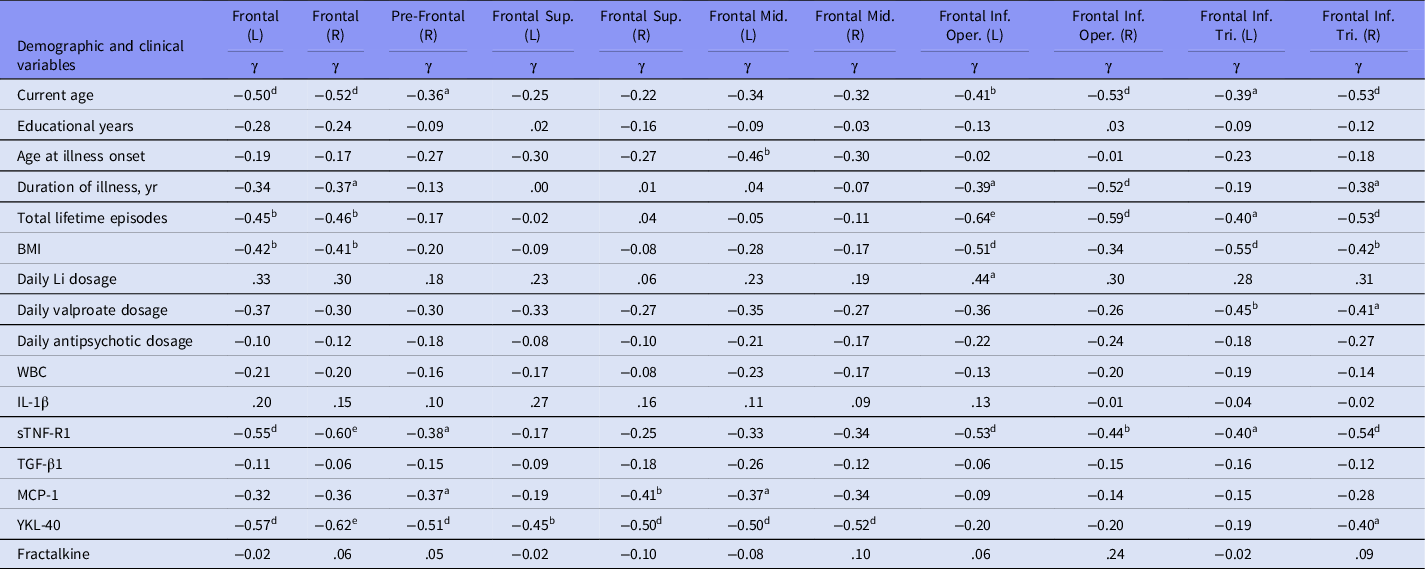
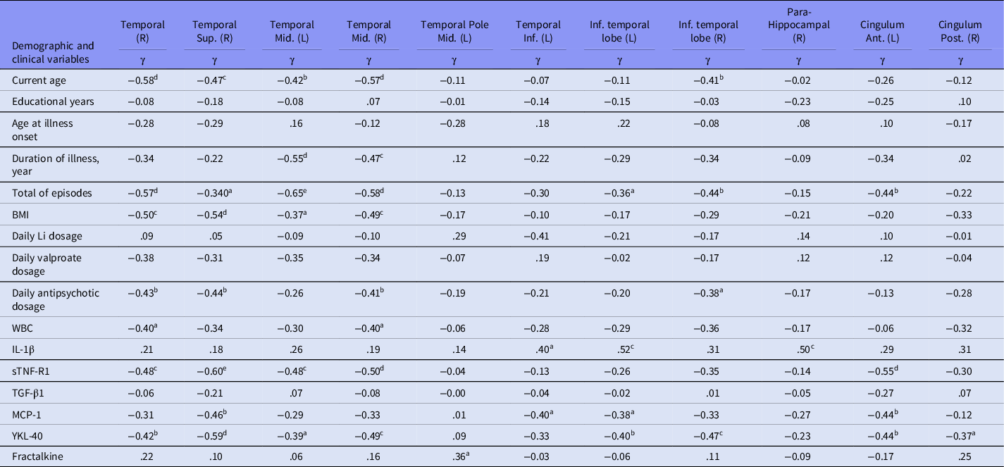
Tables 2 and 3 present significant clusters of correlations between volumes of various brain regions of interest, inflammatory biomarkers, and clinical variables. Among the inflammatory markers, only plasma IL-1β levels always exhibited a positive relationship with brain subarea volumes. However, the volumes of the bilateral amygdala and hippocampus were not significantly correlated with any inflammatory parameters (data not shown). With respect to medication status, current daily lithium dosage was positively correlated with volume of the left pars opercularis of the inferior frontal gyrus (γ = 0.44, p = 0.043) (Table 2) and of the right middle temporal pole (γ = 0.43, p = 0.045). Current daily valproate dosage was negatively correlated with volume of the bilateral pars triangularis of the inferior frontal gyrus (left: γ = −0.45, p = 0.023; right: γ = −0.41, p = 0.039) and of the right superior temporal pole (γ = −0.42, p = 0.035). Antipsychotic dosage (chlorpromazine equivalents) was negatively correlated with the volume of the right middle cingulum (γ = −0.39, p = 0.029), of the right inferior temporal lobe (γ = −0.38, p = 0.037), of the right superior (γ = −0.44, p = 0.014), and of the middle temporal gyrus (γ = −0.41, p = 0.023).


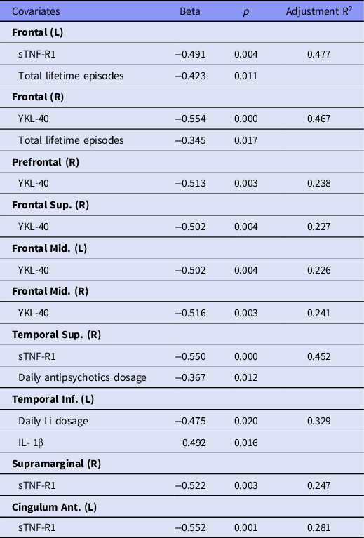
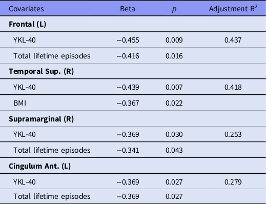
Stepwise multiple regression analyses were performed to assess the association of inflammatory markers, clinical features, BMI, age, and medication status with brain region measures. As displayed in Table 4, elevated plasma YKL-40 level was the most frequent inflammatory marker and the only chemokine among the inflammatory parameters that remained significantly associated with lower volumes of various brain subareas. Higher plasma YKL-40 levels were associated with lower volumes of the following brain subregions: the prefrontal cortex, superior frontal gyrus, and superior temporal gyrus of the right hemisphere; the anterior cingulum, inferior temporal gyrus, and inferior temporal lobe of the left hemisphere; and the bilateral middle frontal gyrus and frontal lobes. Higher plasma levels of IL-1β were associated with higher volumes of the left inferior temporal gyrus and lobe. Higher sTNF-R1 levels were also significantly associated with lower volumes of the left anterior cingulum, left frontal lobe, right superior temporal gyrus, and right supramarginal gyrus. Higher YKL-40 levels replaced sTNF-R1 and remained significantly associated with lower volumes of all four sTNF-R1-related areas when only chemokines were entered into the model (Table 5). With respect to BD-I illness course, greater number of total lifetime mood episodes and higher plasma either YKL-40 or sTNF-R1 level were collectively associated with lower volumes of supramarginal gyrus of the right hemisphere and bilateral frontal lobes (Tables 4 and 5).
Regression for subregions of brain volumes in 31 young bipolar patients (including cytokine molecules)

Regression after removing sTNF-R1 for subregions of brain volumes in 31 young bipolar patients

Discussion
This cross-sectional analysis of brain MRI, inflammatory markers, and clinical variables in euthymic BD-I adults found that higher plasma YKL-40 and sTNF-R1 levels were associated with lower volumes in many brain regions of interest. This finding suggests that increased peripheral YKL-40 and sTNF-R1 levels may be involved in a neuroprogression pathway. Further, plasma YKL-40 was the most common inflammatory marker and the only chemokine among the immune parameters in this study associated with reduced volumes in numerous brain subregions, particularly those regions that have been implicated in BD-I. The volume changes in brain regions involved in severe mental illness, mainly atrophy, are thought to be the results of a reduction in the neuronal and glial density and changes in cell body size and shape (Uranova et al., Reference Uranova, Vostrikov, Orlovskaya and Rachmanova2004). Peripheral YKL-40 is mainly secreted by macrophages, which are involved in the inflammatory response to tissue injury. The bidirectional interaction between the immune system of the brain and the immune system of peripheral organs could help explain the neuroprogressive changes related to chronic peripheral inflammation, particularly the monocyte/marcophage activation in BD-I.
It has been reported that YKL-40 levels in CSF are astrocyte activation markers involved in longitudinal brain macro- and microstructural changes in cognitively unimpaired adults (Falcon et al., Reference Falcon, Monté-Rubio, Grau-Rivera, Suárez-Calvet, Sánchez-Valle, Rami, Bosch, Haass, Gispert and Molinuevo2019). YKL-40 is crucial in the astrocyte response to disease-related environmental conditions (Bonneh-Barkay et al., Reference Bonneh-Barkay, Wang, Starkey, Hamilton and Wiley2010). Although astrocytes are mainly neuroprotective, they also may help perpetuate a self-destructive environment by secreting brain-derived YKL-40 and TNF-α into the blood (Bélanger and Magistretti Reference Bélanger and Magistretti2009; Fakhoury, Reference Fakhoury2018). Plasma YKL-40 can be elevated years before a patient presents clinical signs of an ischaemic stroke (Kjaergaard et al., Reference Kjaergaard, Bojesen, Johansen and Nordestgaard2010). Plasma levels of YKL-40 in euthymic BD patients are both positively correlated with CSF levels and are known to be higher than those of normal controls (Jakobsson et al., Reference Jakobsson, Bjerke, Sahebi, Isgren, Ekman, Sellgren, Olsson, Zetterberg, Blennow, Palsson and Landén2015). Overall, our findings suggest that elevated YKL-40 in the plasma of patients with BD-I may be an indicator of the degree of inflammation and subsequent brain tissue loss. The release of FKN from apoptotic cells appears to guide the immune response, thereby minimising further tissue damage and preserving tissue function. However, that neither YKL-40 nor any brain subarea volume was associated with FKN may reflect a sign of the neuroinflammatory imbalance. This study recruited euthymic patients and thus we could not exclude the possibility of an association between brain volume and plasma MCP-1 and FKN levels during acute affective episodes. MCP-1 has neuroprotective and neurotoxic functions, and its effects may thus be more complex than those of YKL-40. The peripheral YKL-40 levels of healthy subjects do not exhibit a significant diurnal variation and exhibit relatively low variation over up to three years; thus, peripheral YKL-40 can serve as a relatively stable and reliable marker of inflammation (Johansen et al., Reference Johansen, Lottenburger, Nielsen, Jensen, Svendsen, Kollerup and Christensen2008). Serum YKL-40 levels have been considered a useful biomarker of disease severity, prognosis, and survival rate in patients with diseases characterised by chronic inflammation, such as cardiovascular disease and diabetes (Rathcke & Vestergaard, Reference Rathcke and Vestergaard2009; Kastrup, Reference Kastrup2012). Plasma levels of YKL-40 are also elevated in first-episode psychotic patients compared with healthy controls (Orhan et al., Reference Orhan, Schwieler, Fatouros-Bergman, Malmqvist, Cervenka, Collste, Flyckt, Farde, Sellgren and Piehl2018). YKL-40 is a marker of core immune-mediated pathophysiology of severe mental disorders, and elevated YKL-40 level is associated with subsequent type 2 diabetes development in young psychotic disorder victims (da Fonseca AC, Matias D, Garcia C, Amaral R, Geraldo LH, Freitas C, Lima FR. 2014 The impact of microglial activation on blood-brain barrier in brain diseases. Frontiers in Cellular Neuroscience 8: 362.Dieset et al., Reference Dieset, Mørch, Hope, Hoseth, Reponen, Gran, Aas, Michelsen, Reichborn-Kjennerud, Nesvåg, Agartz, Melle, Aukrust, Djurovic, Ueland and Andreassen2019). Further investigations are necessary to clarify the origin of increased YKL-40 production in the peripheral circulation and the effects of YKL-40 on the cardiovascular and metabolic systems of patients with BD-I.
Both TNF-α and IL-1β appear to stimulate YKL-40 synthesis in macrophages, following peripheral inflammatory activation (Bonneh-Barkay et al., Reference Bonneh-Barkay, Bissel, Kofler, Starkey, Wang and Wiley2012). Similarly, a positive correlation was observed between sTNF-R1 and YKL-40 levels in our analyses. The volumes of all sTNF-R1-associated brain subregions were also associated with the YKL-40 level in the multivariate models. TNF-α is a proinflammatory cytokine associated with the inhibition of adult neurogenesis (Keohane et al., Reference Keohane, Ryan, Maloney, Sullivan and Nolan2010). Our findings support an inverse association between TNF family molecule levels in the plasma and brain structure volumes in patients with BD-I, and results are consistent with other investigators (Hoseth et al., Reference Hoseth, Ueland, Dieset, Birnbaum, Shin, Kleinman, Hyde, Mørch, Hope, Lekva, Abraityte, Michelsen, Melle, Westlye, Ueland, Djurovic, Aukrust, Weinberger and Andreassen2017). Although TNF-α levels may decrease after an acute episode is treated, sTNF-R1 level may be persistently elevated during the euthymic period (Hope et al., Reference Hope, Dieset, Agartz, Steen, Ueland, Melle, Aukrust and Andreassen2011; Doganavsargil-Baysal et al., Reference Doganavsargil-Baysal, Cinemre, Aksoy, Akbas, Metin, Fettahoglu, Gokmen and Davran2013). Moreover, sTNF-R1 does not exhibit a significant circadian rhythm and can thus serve as a relatively stable and reliable marker of inflammatory activity in TNF systems of bipolar patients (Hope et al., Reference Hope, Dieset, Agartz, Steen, Ueland, Melle, Aukrust and Andreassen2011).
In the CNS, TNF-α and IL-1β are produced by activated microglia when the homeostasis of the microenvironment is disturbed, resulting in neuroinflammation. TNF-α and IL-1β may impair the blood–brain barrier by altering the expression of molecules related to endothelial cell junctions (da Fonseca et al., Reference da Fonseca, Matias, Garcia, Amaral, Geraldo, Freitas and and Lima2014). With a model of blood–brain barrier dysfunction in BD, a persistent or transient loss of blood–brain barrier integrity is associated with reduced CNS protection and increased permeability of proinflammatory mediators from peripheral blood into the brain, triggering microglial activation and promoting damage (Patel & Frey, Reference Patel and Frey2015). Our findings raise questions regarding whether TNF signalling and increased YKL-40 levels in peripheral blood is a primary pathophysiological process or whether it is secondary to microglia activation mechanisms to neuronal loss. However, causal relationships between structural changes and plasma levels of YKL-40 and TNF family molecules cannot be determined based on a cross-sectional study such as ours. The main outcome of the study is the identification of longitudinal volumetric reduction in BD-related regions associated with the plasma levels of YKL-40 in euthymic BD-I patients.
Obesity-induced chronic inflammation is supported by our findings of a significant positive relationship between BMI and plasma levels of YKL-40 and sTNF-R1. BMI was negatively correlated with volumes of frontal and temporal lobe but did not remain significant in the regression model. Our findings also highlight the importance of inflammatory pathophysiology of BD-I and suggest that, after controlling for medication status, BMI, and other inflammatory markers, increased plasma YKL-40 and sTNF-R1 levels are associated with brain parenchyma loss in the following regions: the prefrontal cortex, superior frontal gyrus, and superior temporal gyrus of the right hemisphere; the anterior cingulum, inferior temporal gyrus, and inferior temporal lobe of the left hemisphere; and the bilateral middle frontal gyrus and frontal lobes.
The heterogeneous symptoms common in BD and use of multiple pharmacological therapies may at least partially explain why region-based and voxel-based neuroanatomical studies on BD have yielded inconsistent results (Altamura et al., Reference Altamura, Maggioni, Dhanoa, Ciappolino, Paoli, Cremaschi, Prunas, Orsenigo, Caletti, Cinnante, Triulzi, Dell’Osso, Yatham and Brambilla2018). However, our findings on YKL-40 related brain subregions align with a large-scale coordinated analysis in a heterogeneous worldwide population of patients with BD-I that reported consistently widespread cortical thinning across the frontal, temporal, and parietal regions of both brain hemispheres (Hibar et al., Reference Hibar, Westlye and Doan2018). Reduced volume of the left anterior cingulate was associated with either higher plasma sTNF-R1 level alone or higher plasma YKL-40 level along with a higher number of lifetime affective episodes. Reduced left anterior cingulate thickness is one of the most consistent differences between patients with BD and healthy controls (Hanford et al., Reference Hanford, Nazarov, Hall and Sassi2016).
Highly elevated numbers of microglial cells have also been observed in the anterior cingulate of patients with schizophrenia (Steiner et al., Reference Steiner, Bielau, Brisch, Danos, Ullrich, Mawrin, Bernstein and Bogerts2008), and volumetric anomalies appear to be particularly extensive in patients with chronic schizophrenia and those with poor outcomes (Dietsche et al., Reference Dietsche, Kircher and Falkenberg2017). Our earlier work also suggested that inflammatory activity in IL-1 and IL-2 systems is involved in grey matter loss in the anterior cingulate cortex of patients with schizophrenia (Tsai et al., Reference Tsai, Sajatovic, Hsu, Chung, Chen and Huang2020). As most inflammation-related brain regions are not BD specific, the present study provides additional evidence that the inflammatory processes of various major psychiatric disorders may involve the same structural abnormalities of key emotional processing regions, such as the anterior cingulate cortex. In contrast to sTNF-R1, higher plasma IL-1β levels were associated with a higher volume of the left inferior temporal gyrus and lobe. This finding might be partially explained by the pleiotropic effects of IL-1β. IL-1β is involved in neuroprotection, tissue remodelling, and tissue repair, despite having potent proinflammatory functions (Hewett et al., Reference Hewett, Jackman and Claycomb2012). However, the CSF IL-1β level fluctuates depending on the BD state (Söderlund et al., Reference Söderlund, Olsson, Samuelsson, Walther-Jallow, Johansson, Erhardt, Landén and Engberg2011). The role of IL-1β in monocyte activation and its interaction with TNF-α and YKL-40 in BD-I warrant further investigation.
A higher total number of lifetime mood episodes appear to negatively affect volume of the bilateral frontal lobes. This finding aligns with the notion that neuronal loss developing over the course of the illness is influenced by prior mood episodes (Bonneh-Barkay et al., Reference Bonneh-Barkay, Bissel, Kofler, Starkey, Wang and Wiley2012), which may reflect a stepwise neuroprogressive effect in the brain. The biological mechanisms driving this declining course suggest that monocyte and macrophage activation play a critical role. In our correlational analyses of medication and brain volumes, only current daily lithium dosage was positively related to volumes of several brain subregions. In patients with BD, increased cortical thickness associated with lithium treatment has been consistently noted and is hypothesised to be driven by its neurotrophic effect on grey matter, while anticonvulsants and antipsychotics are negatively associated with cortical thickness and brain surface area (Hibar et al., Reference Hibar, Westlye and Doan2018). However, it must be noted that our statistical model showed that current daily dosages of lithium, valproate, and antipsychotics were all negatively associated with various brain subregion volumes. Different drug combinations across individuals’ lifetime and the small number of patients in our sample being treated with monotherapy make it difficult to examine medication-related effects on brain volume and inflammation. Studies with larger sample size and using longitudinal design in patient with BD-I are needed to specifically examine how treatment and illness course over time affects the brain.
Limitations
Several limitations should be considered. First, the study was a cross-sectional design and had small sample size. Therefore, as the BMI was positively correlated to the level of YKL-40, the effect of BMI on the level of YKL-40 cannot be entirely excluded. Second, the cytokine and chemokine profiles of the brain may differ from those of the periphery. The pathophysiological changes detected in the periphery may not reliably indicate changes in the CNS. Without measuring the present inflammatory markers in the CSF of the same patients, we cannot determine the effect of glial activation on brain volume. Third, we did not control for medications that may have differential effects on brain structure and inflammatory response. No associations between the use of psychotropic medication and blood levels of YKL-40 (Sahin et al. Reference Sahin, Inanli, Calıskan and Uysal2019) and other inflammatory markers (Mørch et al., Reference Mørch, Dieset, Færden, Reponen, Hope, Hoseth, Gardsjord, Aas, Iversen, Joa, Morken, Agartz, Melle, Aukrust, Djurovic, Ueland and Andreassen2019) in BD were reported. However, the effects of medications may be difficult to determine without a long-term and accurate record of life-time exposure to antipsychotics and mood stabilisers. Finally, though the mean levels of all inflammatory marker are comparable between non-smokers and smokers in our sample, cigarette smoke-induced inflammation and the effect of smoking on increasing YKL-40 level (Majewski et al., Reference Majewski, Tworek, Szewczyk, Kiszałkiewicz, Kurmanowska, Brzeziańska-Lasota, Jerczyńska, Antczak, Piotrowski and Górski2019) should be considered.
Conclusions
These findings provide additional evidence supporting the relationship between neuroinflammatory and systemic inflammatory processes in the pathophysiology of BD-I. The negative correlations between BD-specific brain regions and the YKL-40 and sTNF-R1 levels reflect that macrophage and macrophage-like cells may be at least partially responsible for brain volume changes. Further investigation is required to clarify the factors that induce YKL-40 expression in the presence of BD pathophysiology and how increases in YKL-40 expression influence the disease process and neuroprogression. Identifying the chemokines associated with BD in the CSF and peripheral circulation may enable further understanding of microglia–neuron communication in BD. Furthermore, peripheral YKL-40 may be an additional marker of brain volume reduction in patients with BD-I.
Acknowledgements
The authors also thank Ruei-Siang Shen for assistance in data management and statistical analysis.
Author contributions
SYT designed the study, oversaw data analyses, drafted the manuscript, and edited the manuscript. MS oversaw data analyses, drafted the manuscript, and edited the manuscript. JLH undertook image analyses and carried out the statistical analysis. KHC, PHC, and YJH analysed and provided assistance with data collection. All authors participated in interpreting data and approved the final manuscript.
Financial support
This work was supported by the Ministry of Science and Technology, Taiwan (MOST 106-2314-B-038−050-MY3), Taipei Medical University, Taiwan, and Case Western Reserve University, USA (2019/20 TMU-CWRU Pilot Program 107-3805-005-400).
Conflict of interest
MS has received research grants from Nuromate, Otsuka, Alkermes, Janssen, International Society for Bipolar Disorders, Reuter Foundation, Woodruff Foundation, Reinberger Foundation, National Institutes of Health (NIH), Centers for Disease Control and Prevention (CDC); has been a consultant to Alkermes, Bracket, Otsuka, Janssen, Neurocrine, Health Analytics, Frontline; and has received royalties from Springer Press, Johns Hopkins University Press, Oxford Press, and UpToDate.
Other authors declare no conflict of interest.
Ethical standards
The authors assert that all procedures contributing to this work comply with the ethical standards of the relevant national and institutional committees on human experimentation and with the Helsinki Declaration of 1975, as revised in 2008.