Primary cardiac tumours are rare in children.Reference Beghetti, Gow and Haney1 When found, up to one-sixth are myxomas and, depending on their location, size, and mobility,Reference Heath2 patients may suffer one or more signs from the triad of embolism, intracardiac obstruction, and constitutional symptoms. Occasionally, nonetheless, the myxomas may not produce symptoms. They are found most frequently in the left atrium. In this report, we illustrate the value of recently developed non-invasive techniques for imaging to enhance the diagnosis of such tumours when found in atypical forms or locations.
Case report
An asymptomatic girl, aged 12 years, was referred for assessment of a recently discovered systolic cardiac murmur, grade at 2 from 6, and best heard along the left parasternal border. There was no direct or familial history of note, especially regarding the heart. Transthoracic echocardiographic interrogation, using a General Electrics Vingmed Vivid 5 machine, demonstrated a mobile hyperechogenic mass within the right ventricular outflow tract. The mass 23 by 25 millimetres, and was causing mild obstruction, albeit that neither pulmonary nor tricuspid regurgitation was seen. When compared to the first echocardiogram performed by the referring cardiologist, we noted that the mass had increased in size over a period of 3 months from 15 to 23 millimetres.
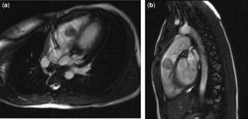
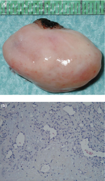
Investigation using dynamic magnetic resonance imaging, in a Sonata Siemens machine is 1.5 Tesla, confirmed the presence of a round mass, measuring 21 by 24 by 22 millimetres, which was attached to the ventricular septum by a short pedicle (Fig. 1). Concomitant with cardiac contraction, the mass moved within the right ventricular outflow tract to occupy a position beneath the leaflets of the pulmonary valve, resulting in moderate obstruction. The lesion was excised surgically through a right ventriculotomy, and the patient had an uneventful postoperative course. The specimen, a yellowish, oval, polypoid lesion with a smooth surface, measured 27 by 20 by 9 millimetres (Fig. 2a). Histologic examination revealed polygonal, non-mitotic cells, scattered in a myxoid matrix, as expected for a cardiac myxoma (Fig. 2b). The state of the margins of the specimen showed that it had been completely resected. After a period of 6 months following the surgical procedure, the patient was well, and echocardiography showed no residual or recurrent tumour. We are currently screening all members of her family to exclude any familial incidence.
Cross sectional (a) and sagittal (b) magnetic resonance cuts revealed a round mass in the right ventricular outflow tract, attached to the ventricular septum by a short pedicle, and located just beneath the leaflets of the pulmonary valve.

Gross examination of the tumour (a) showed a yellowish, oval, polypoid lesion. Histology (b) showed polygonal, non-mitotic cells scattered in a myxoid matrix, consistent with a typical, benign myxoma (X 200, blue alcian).

Discussion
Our case is unusual in both the type and the location of the lesion, with primary cardiac tumours themselves being rare during childhood. When seen, the majority of tumours found in children are rhabdomyomas,Reference Beghetti, Gow and Haney1 in contrast with the adult population, in which about half of primary cardiac tumours are myxomas.Reference Larrieu, Jamieson and Tyers3 The histological findings, therefore, were completely unexpected. While patients may suffer from various symptoms, depending on the location, size and structural dysfunction caused by the tumour, these are not absolutely diagnostic for the type of lesion. The indications for surgical resection are significant symptoms, continuing growth, haemodynamic compromise, or, in the case of rhabdomyomas, tumours which have not resolved by 6 years of age.Reference George, Przygodzki, Jonas and Di Russo4 When resection is complete, the risk of recurrence is very low, at no more than 3% for sporadic tumours, but this should be contrasted with familial cases, which show a higher risk of recurrence.Reference Waller, Ettles, Saunders and Williams5
The second unusual feature in our patient was the location of the tumour in the right ventricular outflow tract. When found, three-quarters of myxomas are in the left atrium, with up to one-fifth found in the right atrium, and less than one-tenth in the ventricles.Reference Reynen6 When found in the right ventricular outflow tract, complications can include pulmonary embolism and infundibular obstruction, as well as systemic symptoms.Reference Hirota, Akiyama and Taniyasu7 In our patient, magnetic resonance imaging provided useful information concerning the composition of the tumour, as well as its origin, extent, mobility, and relation to the leaflets of the pulmonary valve.Reference Rahmanian, Castillo and Sanz8 All this information helped to plan the surgical resection, and may be useful for future follow-up. Given its atypical features, and notwithstanding the histological findings, we considered it advisable to screen the other family members so as to exclude a familial form, or Carney’s complex.Reference Mahilmaran, Seshadri and Nayar9