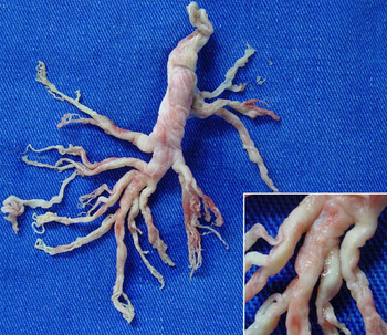
A 4-year-and-9-month-old girl underwent Rashkind septostomy, pulmonary artery banding and ligation of a persistently patent arterial duct for double inlet left ventricle with ventriculo arterial concordance at 1 month of age, followed by total cavopulmonary anastomosis using an intracardiac lateral tunnel at 3 years. Three months after definitive palliation, she developed anasarca with a serum albumin of 1.7 milligrams per decilitre. Faecal alpha-1 antitrypsin, 52 milligrams per gram, confirmed a diagnosis of protein-losing enteropathy. She initially improved with dietary measures, but relapsed 11 months later and began to expectorate bronchial moulds of 10–20 centimetres in length (Fig 1), which had been interpreted as “bronchiolitis” on high-resolution thoracic computed tomography. Histology revealed that these moulds were not gelatinous plugs of mucus ordinarily observed in airways, but rather compact tissue, composed mainly of fibrin and lymphocytes, and consistent with plastic bronchitis. The patient improved and has remained stable for 1 year with airway humidification, furosemide, captopril, spironolactone, and sildenafil.
Figure 1

Both protein-losing enteropathy and plastic bronchitis are rare, but ominous complications of the Fontan circulation. They may occur with increased or, as in this patient, normal central venous pressure, and both pulmonary and systemic lymphatic disorders are implicated in their pathogenesis. Once a haemodynamic compromise of the Fontan circulation is excluded, treatment with diuretics, pulmonary and systemic vasodilators, high molecular weight heparinReference Stiller, Riedel, Paul and van Landeghem1, and careful airway management, in the first instance, often relievesymptoms. Thoracic duct ligation, fenestration of the Fontan circuit, conversion to extracardiac cavopulmonary connections, and cardiac transplantation are advocated when conservative therapy fails.Reference Stiller, Riedel, Paul and van Landeghem1