Introduction
The Miocene/Pliocene transition is known as a key event in the renewal of terrestrial vertebrate faunas in Eurasia (van de Weerd and Daams, Reference van de Weerd and Daams1978; van der Meulen and van Kolfschoten, Reference van der Meulen and van Kolfschoten1986; Aguilar et al., Reference Aguilar, Clauzon and Michaux1989; Cerling et al., Reference Cerling, Harris, MacFadden, Leakey, Quade, Eisenmann and Ehleringer1997; Geraads, Reference Geraads1998; Rouchy et al., Reference Rouchy, Orszag-Sperber, Blanc-Valleron, Pierre, Rivière, Combourieu-Nebout and Panayides2001; Vislobokova et al., Reference Vislobokova, Sotnikova and Dodonov2003; Eronen and Rook, Reference Eronen and Rook2004; Fortelius et al., Reference Fortelius, Eronen, Liu, Pushkina, Tesakov, Vislobokova and Zhang2006; Montuire et al., Reference Montuire, Maridet and Legendre2006; Pierre et al., Reference Pierre, Caruso, Blanc-Valleron, Rouchy and Orzsag-Sperber2006; de Bruijn et al., Reference de Bruijn, Ünay and Hordijk2013; Koufos, Reference Koufos2013, Reference Koufos2016; Koufos and Vasileiadou, Reference Koufos and Vasileiadou2015). Concerning mammals, which are to some extent more sensitive than many other terrestrial vertebrates to environmental changes, this boundary is known by the progressive impoverishment of late Miocene mammals, which displayed a great diversity in large territories in Eurasia, and finally the extinction of many groups that flourished during the middle−late Miocene. Most late Miocene elements of the so-called ‘Pikermian biome’ became extinct toward the end of Miocene (Kostopoulos, Reference Kostopoulos2009; de Bruijn et al., Reference de Bruijn, Ünay and Hordijk2013), but at the same time, new mammalian taxa arose to encompass the Miocene/Pliocene transition, and became diversified during the Pliocene. These new appearing elements play a part in the origins of the extant mammalian faunas of Eurasia.
Late Miocene mammals are well documented in eastern Mediterranean regions, in particular in Greece, Bulgaria, and Turkey, whereas those dated to the Miocene/Pliocene transition and to early Pliocene are much less known. For small mammals, the discoveries of the last two decades in several localities in Greece (e.g., Maramena, Ano Metochi, Monasteri, Silata, Kessani, Tomea Eksi, Ptolemais, Maritsa) allowed a better picture of small mammalian communities in this interval (Koufos, Reference Koufos2016), whereas our knowledge in Turkey is still fragmentary (de Bruijn et al., Reference de Bruijn, Ünay and Hordijk2013). The large mammal assemblages of latest Miocene and early Pliocene ages in this region are characterized by a low diversity and occurrence of genera that have survived to the present. Contrary to this, small mammalian associations are more diverse and their composition displays local differences probably under the influence of regional physiographic and climatic conditions. For the Turkish side of this region, small mammalian faunas of the latest Miocene (mammalian zone MN 13) are only known from preliminary lists (Kangal 1 and Süleymanlı 2; Wessels, Reference Wessels2009; de Bruijn et al., Reference de Bruijn, Ünay and Hordijk2013). For the following zone MN 14 (earliest Pliocene), we can enumerate Igdeli, Babadat and Develi localities, which are documented by rather limited material and species, and/or insufficiently studied fossil assemblages. In this context, rodents and lagomorphs from three localities in the Çankırı Basin, recorded in a controlled stratigraphy that encompasses the Miocene/Pliocene boundary, provide new insight on the time range of several taxa.
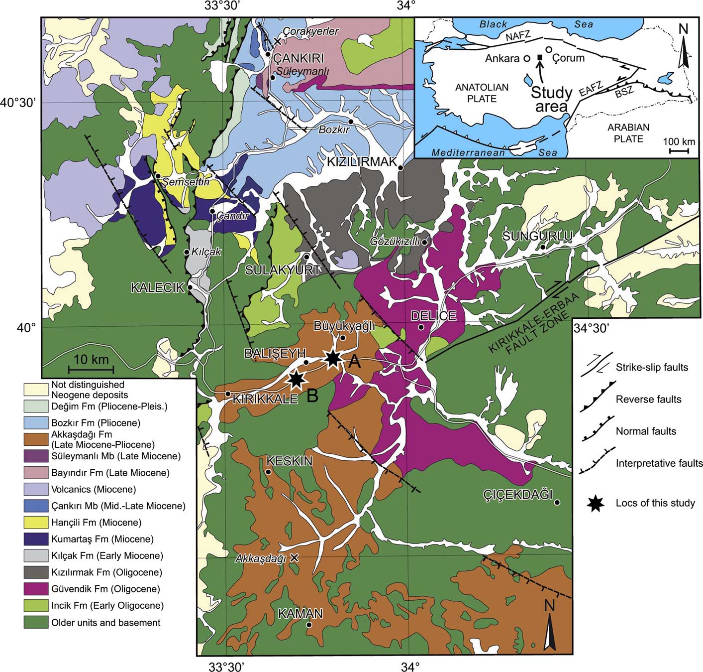
The Çankırı Basin is situated in Central Anatolia, 100− 200 km east of the capital Ankara, and between the main cities of Çankırı and Çorum to the north and Kırıkkale and Kirşehir to the south (Fig. 1). It is one of the largest Anatolian Tertiary basins, covering an area>25,000 km2. It is surrounded by the ophiolitic belt of the Izmir-Ankara-Erzincan suture zone to the west, north, and east, and by the metamorphic rocks of the Kırşehir Massif to the south. Its sedimentary infill is estimated as >3,500 m thick (Karadenizli, Reference Karadenizli1999, Reference Karadenizli2011; Kaymakcı, Reference Kaymakçı2000; Kaymakcı et al., Reference Kaymakçı, White and Van Dijk2003a, Reference Kaymakçı, Duermeijer, Langereis, White and Van Dijkb, Reference Kaymakçı, Özçelik, White and Van Dijk2009; Kazancı et al., Reference Kazancı, Karadenizli, Seyitoğlu, Sen, Alçiçek, Varol, Saraç and Hakyemez2005). The last marine deposits in this basin are dated to the Bartonian, whereas later deposits are entirely formed in continental depositional environments (Kaymakcı, Reference Kaymakçı2000; Kaymakcı et al., Reference Kaymakçı, Özçelik, White and Van Dijk2009; Karadenizli, Reference Karadenizli2011).
Geological map of the central part of the Çankırı Basin (modified after Karadenizli, Reference Karadenizli2011). Kale Tepe (A) and Mahmutlar (B) mammal localities are indicated by black stars; other mammal localities indicated by 'x'. The italicized place names on the map correspond to the major mammal localities already known in this basin. The inset shows the present Turkey with major continental blocks separated by major fault zones, and also the study area.

Terrestrial deposits in the Çankırı Basin cover the late Eocene−Pleistocene interval. However, their mammalian faunas are still insufficiently documented to correlate all stratigraphic units accurately, and their patchy outcrops are often grouped into lithostratigraphic units based on their lithological characters. Some key mammal localities, such as Gözükızıllı for the late Oligocene (Ünay et al., Reference Ünay, de Bruijn and de Saraç2003; Antoine et al., Reference Antoine, Karadenizli, Saraç and Sen2008), Kılçak for the early Miocene (de Bruijn and Saraç, Reference de Bruijn and Saraç1991, Reference de Bruijn and Saraç1992; de Bruijn et al., Reference de Bruijn, Fahlbusch, Saraç and Ünay1993; Ünay, Reference Ünay1994), Çandır for the middle Miocene (Sickenberg et al., Reference Sickenberg, Becker-Platen, Benda, Berg, Engesser, Gaziry, Heissig, Hünermann, Sondaar, Schmidt-Kittler, Staesche, Staesche, Steffens and Tobien1975; Güleç et al., Reference Güleç, Begun and Geraads2003), Akkaşdağı and Çorakyerler for the late Miocene (Sen et al., Reference Sen, Seyitoglu, Karadenizli, Kazancı, Varol and Araz1998; Kazancı et al., Reference Kazancı, Sen, Seyitoğlu, Bonis, de, Bouvrain, Araz, Varol and Karadenizli1999; Sen, Reference Sen2005; Ünay et al., Reference Ünay, de Bruijn and Suata-Alpaslan2006; Güleç et al., Reference Güleç, Sevim, Pehlevan and Kaya2007), and Büyükyağlı for the latest Pliocene deserve to be mentioned. In this context, the discovery of latest Miocene−early Pliocene small mammalian faunas in three localities in the southern Çankırı Basin is of considerable importance for dating isolated outcrops and for the chronology of lithostratigraphic units. Moreover, these new small mammalian assemblages provide new insight for the evolution of mammalian biodiversity in Central Anatolia around the Miocene/Pliocene transition, and finally they bring to light paleogeographic relationships of this region with the surrounding bioprovinces.
Geologic setting
The Çankırı Basin is one of the basins formed following the collision of the Afro-Arabian landmass with the Eurasian continent. The Izmir-Ankara-Erzincan suture zone is one of the major tectonic structures of Anatolia. It was formed by the closure of the northern branch of Neotethys during the late Cretaceous−Eocene interval (Şengör and Yılmaz, Reference Sengör and Yilmaz1981; Koçyiğit, Reference Koçyiğit1991; Tüysüz and Dellaloğlu, Reference Tüysüz and Dellaloğlu1992; Erdoğan et al., Reference Erdogan, Akay and Ugur1996; Görür et al., Reference Görür, Tüysüz and Şengör1998; Kaymakcı et al., Reference Kaymakçı, Özçelik, White and Van Dijk2009) as a consequence of compressive tectonic regime due to northeastward motion of the Afro-Arabian plate and collision of the Taurus and Kırşehir blocks against the Eurasian continent. The suture zone is marked by an ophiolitic belt that extends all along the southern margin of the Sakarya block and Pontid belt, which are parts of the Eurasian continent. Consecutive to this collision, a large marine foreland depression formed south of this suture zone during the Paleogene, and this depression became fragmented during the late Paleogene and Neogene under the neotectonic regime to form the main Central Anatolian basins—from west to east Beypazarı, Haymana-Polatlı, Tuzgölü, Çankırı, and Sivas basins (Görür et al., Reference Görür, Tüysüz and Şengör1998; Görür and Tüysüz, Reference Görür and Tüysüz2001; Huvaz, Reference Huvaz2009; Karadenizli et al., Reference Karadenizli, Varol, Saraç and Gedik2016; Poisson et al., Reference Poisson, Vrielynck, Wernli, Negri, Bassetti, Büyükmeriç, Özer, Guillou, Kavak, Temiz and Orszag-Sperber2016).
In this frame (Fig. 1), the Çankırı Basin occupies a central position and is characterized by its Ω shape. Its sedimentary infill starts in late Cretaceous times. Kaymakcı et al. (Reference Kaymakçı, Özmutlu, Van Dijk and Özçelik2010) distinguished five different cycles of sedimentation: (1) Upper Cretaceous to Paleocene volcaniclastics and regressive shallow marine deposits, (2) Paleocene to Oligocene regressive flysch to molasse sequence referred to as the ‘Tertiary clastics,’ (3) fluviolacustrine clastics deposited in the early−late Miocene, (4) late Miocene−Pliocene fluviolacustrine deposits that frequently alternate with evaporites, and (5) Pliocene−Quaternary alluvial fan deposits. The small mammalian faunas of the present study are found in the fourth unit.
In the southern part of the Çankırı Basin, late Miocene and Pliocene fluviolacustrine deposits occupy quite large areas, and they unconformably cover Paleogene or earlier sedimentary and metamorphic basement units (Kazancı et al., Reference Kazancı, Sen, Seyitoğlu, Bonis, de, Bouvrain, Araz, Varol and Karadenizli1999, Reference Kazancı, Karadenizli, Seyitoğlu, Sen, Alçiçek, Varol, Saraç and Hakyemez2005; Kaymakcı, Reference Kaymakçı2000; Karadenizli et al., Reference Karadenizli, Seyitoğlu, Sen, Arnaud, Kazancı, Saraç and Alçiçek2005; Seyitoğlu et al., Reference Seyitoğlu, Karadenizli, Kazancı and Sen2005; Karadenizli, Reference Karadenizli2011). These deposits are named the Akkaşdağı Formation (Karadenizli et al., Reference Karadenizli, Seyitoğlu, Sen, Arnaud, Kazancı, Saraç and Alçiçek2005; Kazancı et al., Reference Kazancı, Karadenizli, Seyitoğlu, Sen, Alçiçek, Varol, Saraç and Hakyemez2005; Seyitoğlu et al., Reference Seyitoğlu, Karadenizli, Kazancı and Sen2005), and they crop out around the towns of Kırıkkale, Balışeyh, Keskin, Kaman, and Akpınar. They are dispersed as patches in an area measuring ~100 km north− south and ~60 km east−west (Fig. 1). This formation is composed of massive mudstones, gravelly sandstones, bedded limestones, bedded gypsums, laminated claystones, gypsum roses, and tuffs (Kazancı et al., Reference Kazancı, Sen, Seyitoğlu, Bonis, de, Bouvrain, Araz, Varol and Karadenizli1999, Reference Kazancı, Karadenizli, Seyitoğlu, Sen, Alçiçek, Varol, Saraç and Hakyemez2005; Karadenizli et al., Reference Karadenizli, Seyitoğlu, Sen, Arnaud, Kazancı, Saraç and Alçiçek2005). The tuffs at Akkaşdağı yielded a rich mammalian fauna correlated to middle Turolian (Sen, Reference Sen2005), and the age of the tuffs was determined as 7.1±0.1 Ma by single-cristal Ar/Ar dating (Karadenizli et al., Reference Karadenizli, Seyitoğlu, Sen, Arnaud, Kazancı, Saraç and Alçiçek2005). Although the Akkaşdağı Formation displays several facies potentially favorable for the preservation of vertebrate fossils, it was poorly explored, and our knowledge is limited to the Akkaşdağı fauna, which was the subject of detailed studies (Sen, Reference Sen2005), and some elephantid remains from Kale Tepe 4 (Büyükyağlı in Albayrak, Reference Albayrak2017).
The mammalian localities of Kale Tepe 1, Kale Tepe 2, and Mahmutlar were discovered in late 2000s in the course of the Turkish-French joint project, ‘The Neogene evolution of the Çankırı Basin.’ Researchers from the General Directorate of Mineral Research and Exploration (here after MTA, Ankara), the Natural History Museum of Paris (MNHN), and the Université de Montpellier were involved in this project. The aim was primarily biostratigraphy, and consequently we sampled some key horizons favorable for small mammals to date the lithostratigraphic units of this basin rather than sampling tons of sediment to obtain large fossil samples from a few localities. As noted above, the Akkaşdağı Formation displays many other horizons apparently favorable to the preservation of fossil vertebrates, in particular in the area that yielded the small mammals of the present study. Limited time and finances did not allow further field work.
The fossiliferous horizons of Kale Tepe and Mahmutlar are situated on the highway that connects the cities of Kırıkkale and Sungurlu (Fig. 1). The coordinates and altitudes above sea level of these localities are: KT1: 39°56’13.19”N, 33°55’16.74”E, altitude 847 m; KT2: 39°56’13.97”N, 33°55’06.74”E, altitude 863 m; Mahmutlar: 39°52’39.05”N, 33°39’36.98”E, altitude 860 m.
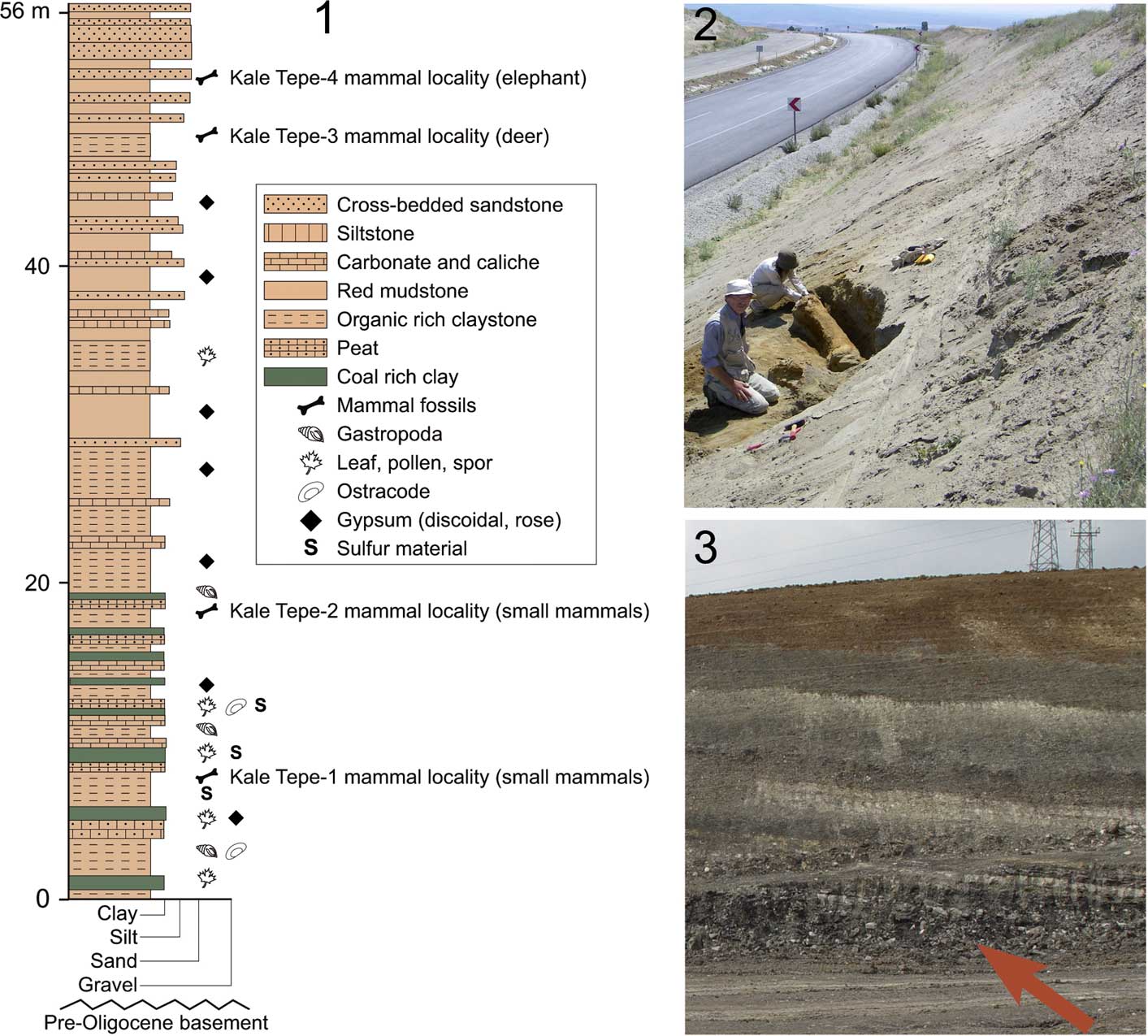
The Kale Tepe section (hereafter KT), which yielded mammalian fossils in four horizons, is situated between the towns of Balışeyh and Delice, and was logged along the new highway. Its lower part is mainly composed of organic-rich clays formed in more-or-less permanent lakes and ponds, interbedded with carbonate-rich horizons; the localities KT1 and KT2 are included in this part (Fig. 2). The lower part of this section was also sampled for pollen and yielded a dry grassland flora with shrubs and riparian forest elements (Kayseri-Özer et al., Reference Kayseri-Özer, Karadenizli, Akgün, Oyal, Saraç, Sen, Tunoglu and Tuncer2016); the same study also showed that ostracods from the same horizons indicate brackish environments. The upper part of the Kale Tepe section is dominated by floodplain marls and sands and cross-bedded sandstones; the locality KT3, which yielded a lower jaw of deer and some bone fragments, is included in this part, but is not yet studied.
(1) Lithologic characteristics of the Kale Tepe section (locality A in Fig. 1). The stratigraphic position of mammal localities is indicated by a bone symbol; (2) locality with elephant bones (KT4) at the upper part of the section; (3) locality KT1 at the lower part of the section.

Upward in the Kale Tepe section, the locality KT4 (39°56’21.5”N, 33°53’41.5”E) yielded remains of a primitive elephantid, consisting of a complete mandible bearing left m1−m3 and right m2−m3, a tusk, a humerus, and several ribs. These remains apparently belong to a single individual. Albayrak (Reference Albayrak2017) identified this elephant as cf. Elephas planifrons Falconer and Cautley, Reference Falconer and Cautley1845 and suggested an MN 16a correlation for this locality. This mammal age is calibrated as 2.6−3.2 Myr in the Geologic Time Scale 2012 of Gradstein et al. (Reference Gradstein, Ogg, Schmitz and Ogg2012). It can be added that in the chronology of Siwaliks of the Indian subcontinent, the Elephas planifrons Interval Zone is dated as 2.9–1.5 Myr (Nanda, Reference Nanda2013).
The locality of Mahmutlar is located 23 km WSW of the Kale Tepe section, in a road cut between the towns of Balışeyh and Kırıkkale, 1.4 km east of Mahmutlar village. This outcrop cannot be directly correlated to the Kale Tepe section. According to local stratigraphy, it is in the middle part of the Akkaşdağı Formation. All three fossiliferous horizons are in organic-rich dark-colored clays. The faunal content of these localities will be summarized in the Conclusions (below).
Materials and methods
McKenna and Bell (Reference McKenna and Bell1997) grouped the family Muridae into several groups of muroid rodents previously having family rank, in particular the Cricetidae, Arvicolidae, and Gerbillidae. These authors considered these groups and many others as subfamilies of the family Muridae. Paleomammalogists are divided on this issue. Some follow closely the classification of McKenna and Bell (Reference McKenna and Bell1997), whereas many others reject this oversimplification of the family Muridae and consider that most of the groups included in this family by McKenna and Bell (Reference McKenna and Bell1997) are sufficiently diversified to deserve family rank. This is also the opinion here.
We follow Schaub (Reference Schaub1938) and de Bruijn et al. (Reference de Bruijn, Saraç, van den Hoek Ostende and Roussiakis1999) in the definition of the genus Apodemus Kaup, Reference Kaup1829: majority of M1 and M2 with a distinct t7, except Micromys Dehne, Reference Dehne1841 (differentiated with a t1bis on M1−M2 and weak labial cingulum on m1) and Rhagapodemus Kretzoi, Reference Kretzoi1959 (robust and subhypsodont molars). We must note that Martín-Suárez and Mein (Reference Martín-Suárez and Mein1998) have another concept of the genus Apodemus, which includes species previously referred to Parapodemus Schaub, Reference Schaub1938. Nevertheless, in our opinion, the acquisition of a t7 is a derived character strong enough, in addition to other characters, to distinguish the genera Apodemus, Rhagapodemus, and Micromys from Parapodemus.
For biometric comparisons of the species studied in this work, the senior author constructed tables of comparative measurements with Excel software. These tables include, for each occurrence of a species, the species name, locality, age, number of specimens, range, and the average values for length and width, and for some groups, the crown height (e.g., Arvicolidae) for premolars and molars. These measurements are either taken from the literature, and in that case the reference is included, or taken by the senior author for some collections of fossil and extant species. These biometric comparison tables are available upon request from the senior author.
We collected 15 sacks (each sack ~25−30 kg) of sediment from KT1, 15 sacks from KT2, and 30 sacks from Mahmutlar. The sediment was wet-screened in a set of sieves (10, 3, and 0.5 mm) and the senior author sorted all of the residue with a front loupe (>3 mm) or under a stereomicroscope (<3 mm).
Dental terminologies follow López Mártínez (Reference López Mártínez1989) for lagomorphs, Martín-Suárez and Freudenthal (Reference Martín-Suárez and Freudenthal1993) for murids, Mein and Freudenthal (Reference Mein and Freudenthal1971) for cricetids and gerbillids, and Daams (Reference Daams1981) for glirids. Photographs were taken with a Surface Electronic Microscope TESCAN Vega2 at MNHN and retouched with Adobe Photoshop Elements 6.0.
Anatomical abbreviations and measurements
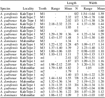
Upper cheek teeth are abbreviated with capital letters (P, M) and lower cheek teeth with small letters (p, m). Measurements were taken using a Mitutoyo caliper, with a precision of 0.01 mm (see Table 1). Length (L) and width (W) correspond to maximum diameter along the longitudinal axis and perpendicular to it, respectively.
Measurements of Apodemus molars from the localities of Kale Tepe 1, Kale Tepe 2, and Mahmutlar in the Çankırı Basin, Turkey. N=number of specimens.

Repositories and institutional abbreviations
The studied specimens are deposited at the Natural History Museum of the General Directorate of Mineral Research and Exploration (well known in Turkey as MTA) in Ankara under the labels of KT1 (Kale Tepe 1), KT2 (Kale Tepe 2), and MAH (Mahmutlar). The studied specimens were compared with the collections housed at the Instituto Nacional de Geología (INC), Madrid, Spain; Institut Català de Paleontologia Miquel Crusafont (ICP), Sabadell, Spain; Muséum National d’Histoire Naturelle (MNHN), Paris, France; Museum of Natural History of Verona (MNHV), Italy; Natural History Museum (NHMW), Vienna, Austria; Hungarian Geological Institute (HGI), Budapest; Pamukkale University (PUD), Denizli, Turkey; and MTA, Ankara, Turkey.
Systematic paleontology
Order Rodentia Bowdich, Reference Bowdich1821
Family Muridae Illiger, Reference Illiger1811
Genus Apodemus Kaup, Reference Kaup1829
Type species
Mus agrarius Pallas, Reference Pallas1771; Recent, Eurasia.
Apodemus gorafensis Ruiz Bustos et al., Reference Ruiz Bustos, Sesé, Dabrio, Peña and Padial1984
Figures 3.8−3.13, 4.7−4.11, 5.7−5.9, 6.11−6.12
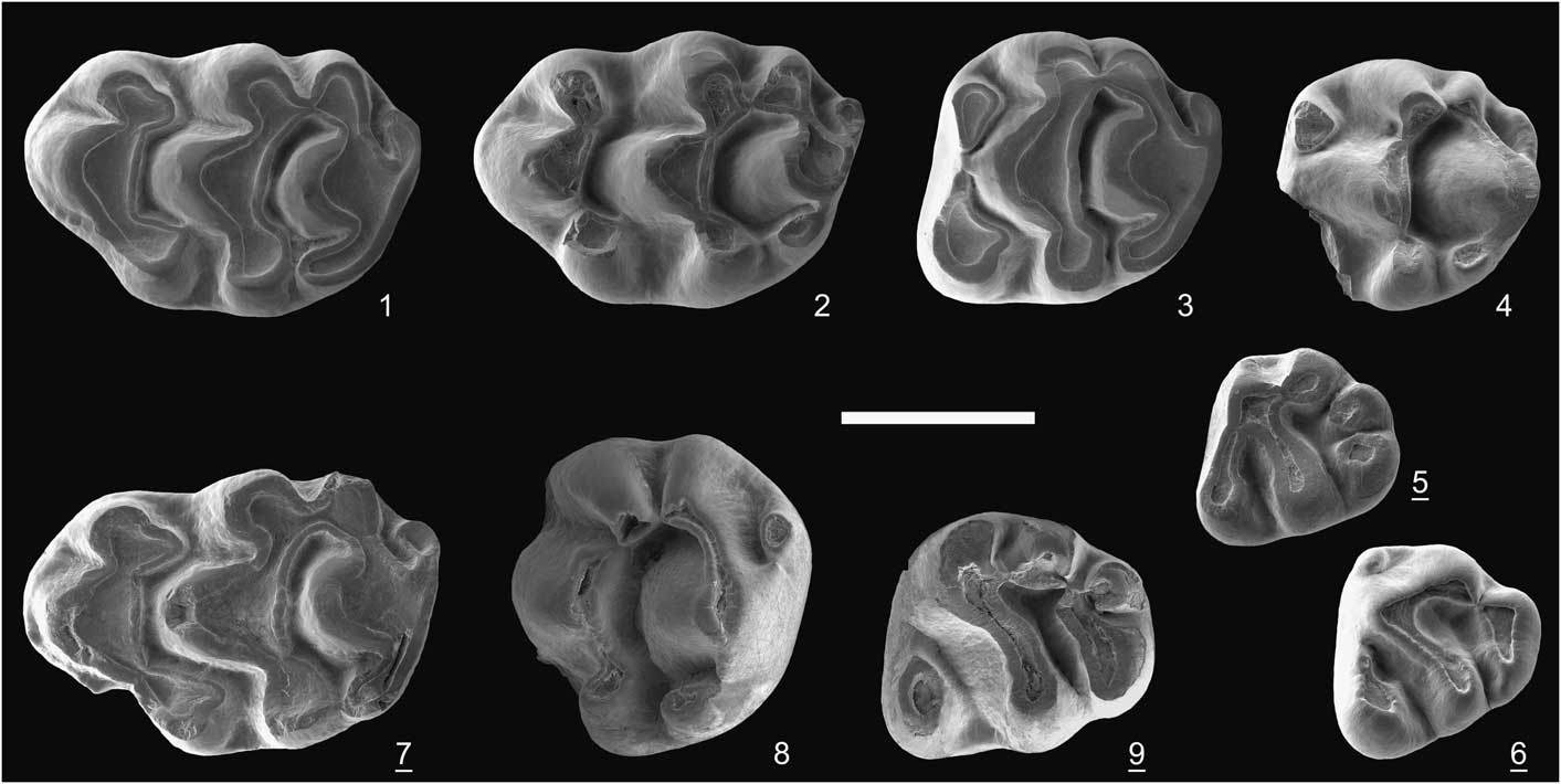
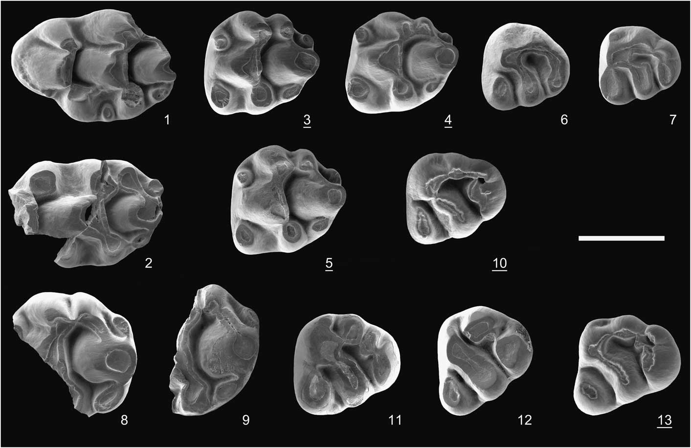
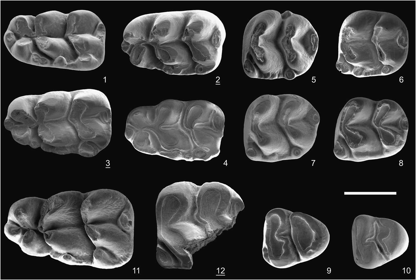
Apodemus upper molars from Kale Tepe 1. Apodemus gudrunae: (1) M1, KT1-17; (2) damaged M1, KT1-18; (3) M2, KT1-21; (4) M2, KT1-22; (5) M2, KT1-23; (6) M3, KT1-27; (7) M3, KT1-28. Apodemus gorafensis: (8) distal part of M1, KT1-1; (9) distal part of M1, KT1-2; (10) M3, KT1-3; (11) M3, KT1-4; (12) M3, KT1-7; (13) M3, KT1-5. All molars are illustrated as left; the underlined numbers indicate reversed pictures. Scale bar=1 mm.
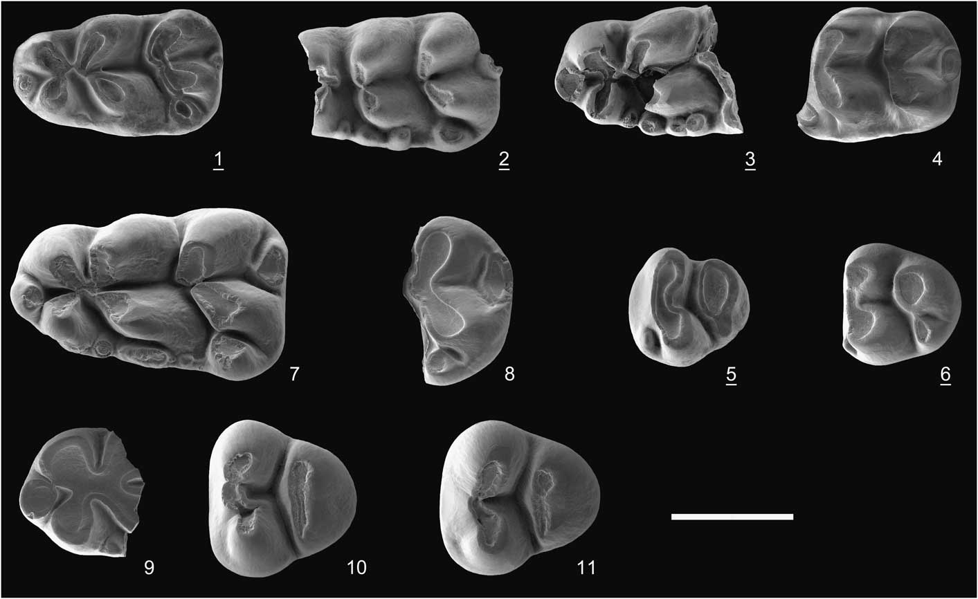
Apodemus lower molars from Kale Tepe 1. Apodemus gudrunae: (1) m1, KT1-30; (2) damaged m1, KT1-31; (3) damaged m1, KT1-32; (4) m2, KT1-33; (5) m3, KT1-36; (6) m3, KT1-37. Apodemus gorafensis: (7) m1, KT1-9; (8) distal part of m1, KT1-56; (9) mesial part of m1, KT1-10; (10) m3, KT1-12; (11) m3, KT1-13. All molars are illustrated as left; the underlined numbers indicate reversed pictures. Scale bar=1 mm.
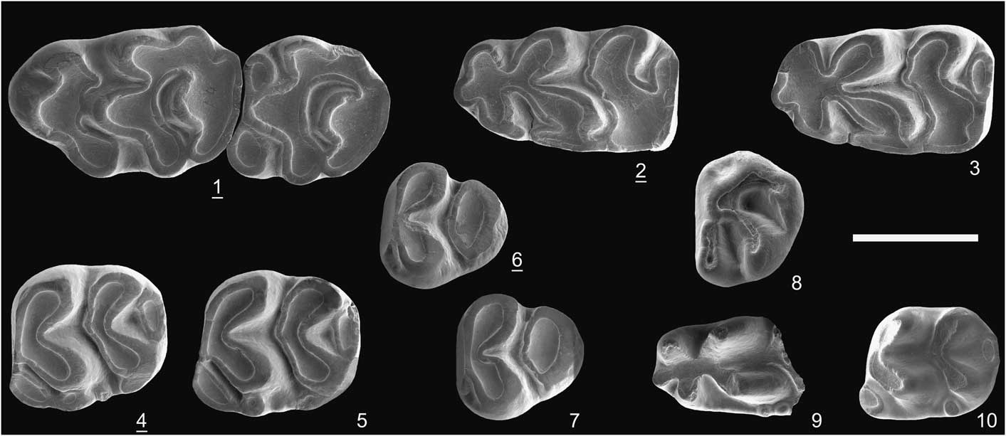
Apodemus upper molars from Kale Tepe 2. Apodemus gudrunae: (1) M1, KT2-18; (2) M1, KT2-19; (3) M2, KT2-24; (4) damaged M2, KT2-25; (5) M3, KT2-30; (6) M3, KT2-29. Apodemus gorafensis: (7) M1, KT2-20; (8) distal part of M1, KT2-1; (9) M3, KT2-3. All molars illustrated as left; the underlined numbers indicate the reversed pictures. Scale bar=1 mm.

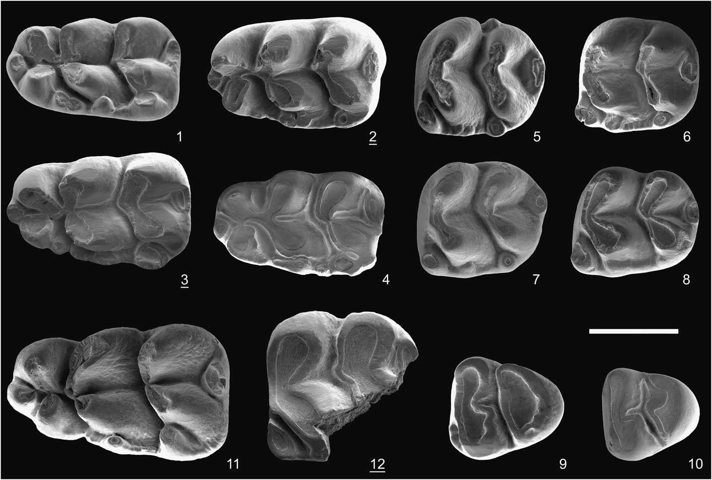
Apodemus lower molars from Kale Tepe 2. Apodemus gudrunae: (1) m1, KT2-32; (2) m1, KT2-33; (3) m1, KT2-34; (4) worn m1, KT2-31; (5) m2, KT2-37; (6) m2, KT2-38; (7) m2, KT2-39; (8) m2, KT2-40; (9) m3, KT2-43; (10) m3, KT2-44. Apodemus gorafensis: (11) m1, KT2-5; (12) damaged m2, KT2-9. All molars are illustrated as left; the underlined numbers indicate reversed pictures. Scale bar=1 mm.
Holotype
Left M1 (INC G-8) from the locality Gorafe-A in the depression of Guadix-Baza (Granada Basin, SE Spain), early Pliocene (Mammalian Neogene zone MN 14).
Occurrence
From the Iberian Peninsula to Greece in southern Europe and Turkey, during the late Miocene and early Pliocene.
Description
M1 with occlusal outline elongated. Cusp t1 far distal to t2 and t3; t3 smaller than t1, with short posterior spur; t4 and t6 almost symmetrical; t6 and t9 closely situated or connected by short ridge; t7 oval, weakly connected to t8; t12 cusp-shaped, connected to base of t8. Three roots; lingual one large, compressed. M2 with t1 large, isolated; t3 small, situated next to base of t2; t4 slightly posterior to t6; four roots. M3 with a large t1 but without t3; t4 and t5 fused to form oblique loph. Connection between t5 and t6 weak or absent; t9 present in all specimens except one (KT1-7; Fig. 3.12); three roots.
The m1 distally wide, much more than usually observed in Apodemus spp.; tma large, isolated, situated on longitudinal axis; lingual anteroconid larger than labial one; central connection between anteroconid and protoconid/metaconid complex; lingual cusps slightly displaced mesially; c1 large, situated next to hypoconid; labial cingulum with several cuspids; posterior heel oval, lingually displaced. The m2 with large, isolated anterolingual cusp; posterior heel oval, lingually situated. The m3 with triangular occlusal outline; anterolabial cusp as tiny cingulum; protoconid and metaconid equal in size, symmetrical; hypoconid and entoconid fused. All lower molars with two roots.
Material from KT1
Two fragmentary M1 (KT1-1, 2; Fig. 3.8, 3.9), five M3 (KT1-3 to 7; Fig. 3.10−3.13), three m1 of which one is complete (KT1-9, 10, 56; Fig. 4.7–4.9), four m3 (KT1-12 to 15; Fig. 4.10, 4.11).
Material from KT2
One complete and one fragmentary M1 (KT2-1, 20; Fig. 5.7, 5.8), one complete and two fragments of M2 (KT2-2, 24, 73), two M3 (KT2-3, 4; Fig. 5.9), one complete and three fragments of m1 (KT2-5 to 8; Fig. 6.11), one m2 (KT2-9; Fig. 6.12), one m3 (KT2-43).
Measurements
See Table 1.
Remarks
The size of these molars from KT1 and KT2 is clearly larger than those of Apodemus atavus Heller, Reference Heller1936, A. dominans Kretzoi, Reference Kretzoi1959, and A. gudrunae van de Weerd, Reference van de Weerd1976, which are the common Apodemus species in latest Miocene and Pliocene localities of Europe and the eastern Mediterranean. The size of these specimens compares better to that of A. jeanteti Michaux, Reference Michaux1967 from Seynes (type locality, southern France), and A. gorafensis from Spain, Greece, and Turkey. Apodemus jeanteti has M1-M2 with lingual cusps straightened and small t12, M1 with the cusp t1 close to t2, and m1 with incipient or absent tma and with narrow angles between the two cusps. These characters were not observed on the specimens from KT1 and KT2.
The molars from KT1 and KT2 are similar in size to those of Apodemus gorafensis from the type locality Gorafe A (Spain; Ruiz Bustos et al., Reference Ruiz Bustos, Sesé, Dabrio, Peña and Padial1984). According to the NOW Database (NOW Community, 2017), this species has been recorded in 33 localities in Spain. Lastly, Mansino et al. (Reference Mansino, Crespo, Montoya and Ruiz-Sánchez2017) and Piñero et al. (Reference Piñero, Agustí, Oms, Blain, Laplana, Ros-Montoya and Martínez-Navarro2017) provided an update of the Spanish record. On morphological grounds, all characters of the molars from KT1 and KT2 are within the range of morphological variation observed in latest Miocene and earliest Pliocene assemblages from Spain. Aside from Spain, this species has been reported from Greece (Maramena, Silata, Tomea Eksi 1 and 2; de Bruijn, Reference de Bruijn1995a; Vasileiadou et al. Reference Vasileiadou, Koufos and Syrides2003; Hordijk and de Bruijn, Reference Hordijk and de Bruijn2009) and from Turkey (Amasya and Süleymanlı 2; Sarica-Filoreau, Reference Sarıca-Filoreau2002; de Bruijn et al., Reference de Bruijn, Ünay and Hordijk2013). All of these localities are correlated to the latest Miocene or earliest Pliocene. The assemblages from Tomea Eksi 1 and 2 that Hordijk and de Bruijn (Reference Hordijk and de Bruijn2009) assigned to A. gorafensis exhibit size overlap with both A. gorafensis and A. gudrunae and some molars display characters that rather fit A. gudrunae, e.g., t1 close to t2 and anterolingual notch not marked on M1. In other words, the material from Tomea Eksi 1 and 2 could include both species. Sarica-Filoreau (Reference Sarıca-Filoreau2002, p. 26) also noted that the material from Amasya (Turkey) could also include “two different Apodemus species in Amasya as evidenced by the morphology of the M1. The limited material from Amasya does not permit the description of two species, based on the small differences in size and morphology.” In summary, the co-occurrence of A. gorafensis and A. gudrunae in the eastern Mediterranean region is obviously a common feature of rodent faunas around the Miocene/Pliocene transition.
Apodemus gudrunae van de Weerd, Reference van de Weerd1976
Holotype
An isolated M1 (ICP VDC3-352) from the locality Valdecebro 3, Teruel Basin, Spain. This locality is correlated to late Turolian, MN 13 (van de Weerd, Reference van de Weerd1976).
Occurrence
From the Iberian Peninsula to Greece in southern Europe and Turkey, during the late Miocene and early Pliocene.
Description
M1 moderately elongated with shallow anterolingual notch between t1 and t2, thus t1 situated close to t2; t1 with short posterior spur (2/4 specimens at KT1; 1/4 specimen at KT2); t3 smaller than other cusps, with posterior spur (2/4 specimens at KT1 and KT2); t3−t5 connected on one specimen (KT1-19); one specimen (KT1-17) with cusp-shaped enterostyle between t1 and t4; t7 always present, drop-shaped, connected to t8, but never to t4; t6 and t9 situated close to each other and weakly connected; distal cingulum cusp-shaped; three or four roots. M2 with t1 twice as large as t3; t3 with posterior spur on one specimen (KT2-24); t7 ridge-shaped, always separated from t4; posterior cingulum always present, either ridge- or cusp-shaped; three or four roots. M3 with large t1 isolated or firmly connected to t5; other cusps organized in continuous garland; three roots.
The m1 elongated, distally narrow, with tma large and isolated; lingual connection between anteroconid and metaconid; labial cingulum thick, with three or more cuspules; posterior heel lingually situated. The m2 with large anterolabial cusp connected by low ridge to anterior base of protoconid; cusp rows with distal angle more open than on m1; labial cingulum thick with two or more cuspules. The m3 with incipient anterolabial cusp, very large entoconid, and reduced hypoconid. Lower molars with two roots.
Material from KT1
Four M1 (KT1-17 to 20; Fig. 3.1, 3.2), six M2 (KT1-21 to 26; Fig. 3.3−3.5), two M3 (KT1-27, 28; Fig. 3.6, 3.7), four m1 (KT1-29 to 32; Fig. 4.1−4.3), three m2 (KT1-33 to 35; Fig. 4.4), four m3 (KT1-36 to 39; Fig. 4.5, 4.6).
Material from KT2
Five M1 (KT2-18 to 22; Fig. 5.1, 5.2), five M2 (KT2-24 to 28; Fig. 5.3, 5.4), two M3 (KT2-29, 30; Fig. 5.5, 5.6), fragment of right mandibular ramus with m1-m2 (KT2-71), six m1 (KT2-31 to 35, KT2-72; Fig. 6.1−6.4), seven m2 (KT2-36 to 42; Fig. 6.5−6.8), three m3 (KT2-43 to 46; Fig. 6.9, 6.10).
Measurements
See Table 1.
Remarks
As observed by Ruiz Bustos et al. (Reference Ruiz Bustos, Sesé, Dabrio, Peña and Padial1984), Sarica-Filoreau (Reference Sarıca-Filoreau2002), and Colombero et al. (Reference Colombero, Bonelli, Kotsakis, Pavia, Pavia and Carnevale2013), Apodemus gudrunae differs from A. gorafensis by its smaller size and some morphological characters. The material from KT1 and KT2 includes two clearly different size groups without overlap. The smaller matches that of A. gudrunae. The larger species A. gorafensis differs in having M1 with a large t1 separated from t2 by a marked notch on the lingual side, whereas on the specimens of KT1 and KT2 referred to A. gudrunae, t1 is smaller than t2, situated rather close to this cusp, the anterolingual notch not marked as in A. gudrunae from its type locality. In addition, the molars from KT1 and KT2 have M1 and M2 with weak, short connections between t6 and t9, and a large, cusp-shaped t12 as in A. gudrunae. Another distinctive feature separating A. gudrunae and A. gorafensis not noted previously concerns the organization of cusps on M3. In A. gorafensis, cusps t4 and t5 are strongly fused to form an oblique loph, separated from or weakly connected to cusp t6. In A. gudrunae, cusps t4, t5, t6, t9, and t8 form a garland. In addition, M3 of A. gorafensis has cusps t6 and t8 often totally isolated, and t9 occasionally present and small. In A. gudrunae, as noted above, these cusps form a garland, and t9 is present in most specimens. In summary, based on these morphological features and size, the smaller molars from KT1 and KT2 are referred to A. gudrunae.
Apodemus gudrunae is a common species of latest Miocene and Pliocene localities in Spain (in 20 localities according to NOW Community, 2017, but certainly many more), but also in southern France (Castelnou 3, Celleneuve, La Tour, Lissieu, Pigeonnier d’Ange 2, Vives 2; see Aguilar et al., Reference Aguilar, Calvet and Michaux1995), Italy (Brisighella, Borro Strolla, Moncucco Torinese, Verduno; see Colombero et al., Reference Colombero, Pavia and Carnevale2014a, Reference Colombero, Angelone, Bonelli, Carnevale, Cavallo, Delfino, Guintelli, Mazza, Pavia, Pavia and Repettob), Greece (Ano Metochi-2 and 3, Monasteri and possibly Tomea Eksi-1 and 2; see Koufos, Reference Koufos2016), and Turkey (Amasya and Süleymanli-2; see de Bruijn et al., Reference de Bruijn, Ünay and Hordijk2013).
Martín-Suárez and Mein (Reference Martín-Suárez and Mein1998) suggested the existence of an anagenetic lineage from Parapodemus barbarae van de Weerd, Reference van de Weerd1976 to Apodemus gudrunae to A. gorafensis, spanning in time from middle Turolian to early Ruscinian. The increase in size and reduction of the longitudinal connections between the cusps of upper molars are given as the main arguments to support their hypothesis. However, because A. gudrunae and A. gorafensis are associated in several late Turolian and early Ruscinian localities from Spain to Turkey, direct ancestry is unlikely.
Apodemus dominans Kretzoi, Reference Kretzoi1959
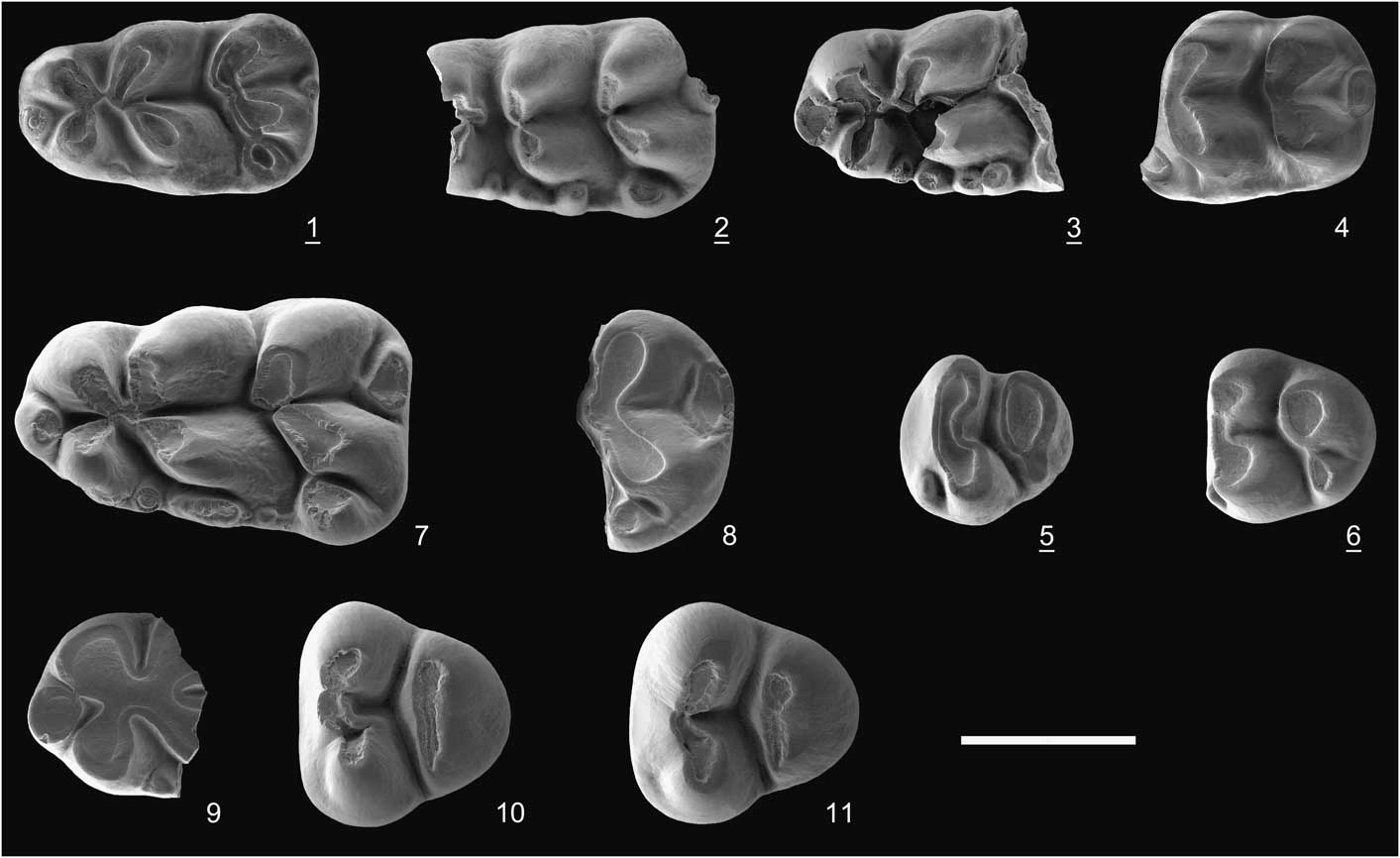
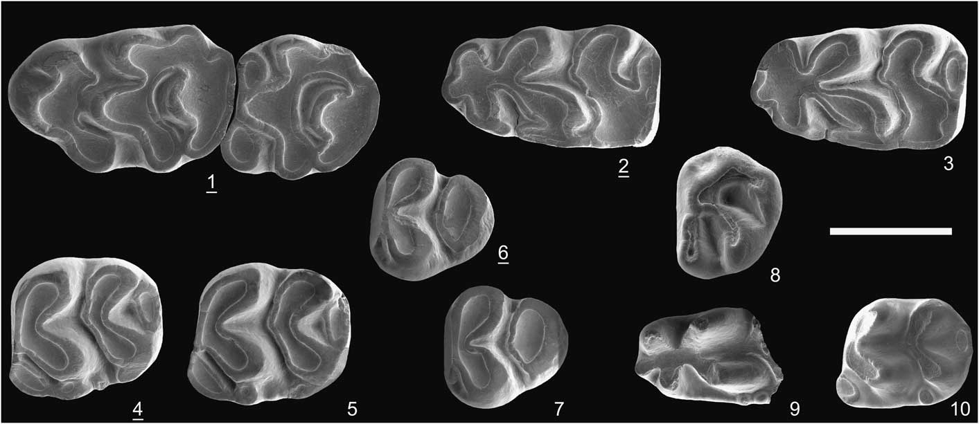
Apodemus dominans molars from Mahmutlar: (1) M1-M2, MAH-1; (2) m1, MAH-4; (3) m1, MAH-5; (4) m2, MAH-6; (5) m2, MAH-7; (6) m3, MAH-8; (7) m3, MAH-9. Micromys sp. indet. molars from Kale Tepe 2: (8) M3, KT2-13; (9) damaged m1, KT2-10; (10) m2, KT2-12. All molars are illustrated as left; the underlined numbers indicate reversed pictures. Scale bar=1 mm.
Lectotype
Left M1 of Kretzoi (Reference Kretzoi1962, pl. 4, fig. 4) from Csarnota-2 in Hungary correlated to Mammalian Neogene zone MN 15. Kretzoi (Reference Kretzoi1959) named this species with a short description and without illustration. In a second paper, Kretzoi (Reference Kretzoi1962) described with more detail small and large mammals from Csarnota-2 and, for A. dominans, he illustrated four molars (left M1, left M2, left M3, and right m1) without catalog number. In accordance with the International Code of Zoological Nomeclature (ICZN, 1999, Art. 74.1), we designate the most characteristic specimen, the left M1 (Kretzoi, Reference Kretzoi1962, pl. 4, fig. 6), as the lectotype, preserved at the HGI (no catalog number).
Occurrence
Apodemus dominans is recorded in Pliocene localities all over Europe, Turkey, Afghanistan, and Moghreb.
Description
Molars sufficiently homogeneous in size and dental pattern to be securely referred to one species. M1 with a large t1 slightly distal to t3; posterior spur on t3 directed toward angle t5-t6; t4 separated from t7; t6 and t9 strongly connected; large t12. M2 with t4 separated from t7. On m1, tma central and bent labially; protoconid-metaconid connected to lingual anteroconid; posterior heel large, situated lingually; c1 strong on labial margin, fused with hypoconid; labial cingulum interrupted between protoconid and labial anteroconid. The m2 with large, ellipsoid anterolabial cusp; posterior heel large, situated lingually; labial cingulum weak but with strong cuspule next to protoconid. The m3 with incipient anterolabial cusp; protoconid and metaconid symmetrical; distal lobe reduced to single large cusp. All lower molars with two roots.
Material from Mahmutlar
An upper jaw fragment with M1-M2 (MAH-1; Fig. 7.1), two fragments of upper molars, two m1 (MAH-4, 5; Fig. 7.2, 7.3), two m2 (MAH-6, 7; Fig. 7.4, 7.5), and two m3 (MAH-8, 9; Fig. 7.6, 7.7).
Measurements
See Table 1.
Remarks
Identification of small to medium-sized Pliocene and early Pleistocene species of Apodemus has been confused during the last three decades. Fejfar and Storch (Reference Fejfar and Storch1990) synonymized A. dominans (type locality Csarnota-2, Hungary) with A. atavus (type locality karstic fissure fillings near Gundersheim, Germany). Several paleontologists, in particular Martín Suárez and Mein (Reference Martín-Suárez and Mein2004), Minwer-Barakat et al. (Reference Minwer-Barakat, García-Alix, Martín-Suárez and Freudenthal2005), García-Alix et al. (Reference García-Alix, Minwer-Barakat, Martín-Suárez and Freudenthal2008), and Colombero et al. (Reference Colombero, Pavia and Carnevale2014a, Reference Colombero, Angelone, Bonelli, Carnevale, Cavallo, Delfino, Guintelli, Mazza, Pavia, Pavia and Repettob), followed this opinion and considered A. dominans a junior synonym of A. atavus. On the contrary, many other paleontologists retained both species as valid, and assigned Apodemus remains of the same age, same regions, and even the same localities to either one or the other species (Storch and Dahlmann,Reference Storch and Dahlmann1995; Ünay and de Bruijn, Reference Ünay and de Bruijn1998; Vasileiadou et al., Reference Vasileiadou, Koufos and Syrides2003, Reference Vasileiadou, Konidaris and Koufos2012; Hordijk and de Bruijn, Reference Hordijk and de Bruijn2009; Suata Alpaslan et al., 2010; Hellmund and Ziegler, Reference Hellmund and Ziegler2012; van den Hoek Ostende et al., Reference van den Hoek Ostende, Diepeveen, Tesakov, Saraç, Mayhew and Alçiçek2015a, Reference van den Hoek Ostende, Gardner, van Bennekom, Alçiçek, Murray, Frank, Wesselingh, Alçiçek and Tesakovb). According to the latter authors, A. atavus has molars smaller than those of A. dominans, t7 more frequently isolated, t3 with a strong posterior spur, a low t1-t2 connection on M1, and a large tma on m1. For Hellmund and Ziegler (Reference Hellmund and Ziegler2012), A. atavus occurred in late Pliocene−early Pleistocene faunas (MN 15 to 17) in Europe, whereas A. dominans existed during the early Pliocene (MN 14, 15) in a much larger area in Europe and probably in Asia (Sen, Reference Sen1983; Zhang and Zheng, Reference Zhang and Zheng2001; Zheng and Zhang, Reference Zheng and Zhang2001). According to Colombero et al. (Reference Colombero, Pavia and Carnevale2014a), who discussed the similarities and differences between the species, A. atavus from Gundersheim has smaller molars compared to A. dominans, but the tooth morphology is variable in the assemblages referred to A. atavus and A. dominans and does not allow clear morphological distinction to retain both species valid. Indeed, the descriptions and illustrations given by Fejfar and Storch (Reference Fejfar and Storch1990) for A. atavus from Gundersheim and by Kretzoi (Reference Kretzoi1962) for A. dominans from Csarnota-2 do not allow a clear morphological distinction between these species. The main difference remains the larger size of the molars from Csarnota-2, based on the measurements of van de Weerd (Reference van de Weerd1976) and from other localities where A. dominans is recognized, ranging from Spain to Ukraine to Afghanistan. This problematic issue cannot be solved without a detailed study of the type materials of A. dominans from Csarnota-2 and A. atavus from Gundersheim, and a detailed comparison of their molar morphologies to document their differences or their synonymy. Pending the revision of the systematic status of these two species and taking into account the similarities in size and morphology of the molars from Mahmutlar with those of Csarnota-2, we conclude that the Mahmutlar material is best referred to to A. dominans.
Genus Micromys Dehne, Reference Dehne1841
Occurrence
The genus Micromys is known all over Eurasia since the late Miocene.
Description
M3 with large, tranversally elongated t1 connected to t5; t3 reduced to tiny anterolabial ridge; t4−t6 and a posterior cusp (? t9) organized in continuous crescent surrounding deep sinus. First lower molar gracile, narrow in occlusal outline; tma small, firmly connected to anteroconid; anterior and medial cusps connected to form X-pattern; posterior heel oval, low, lingually situated; labial cingulum weak, bearing small c1 labial to hypoconid and one or two cuspules next to protoconid. The m2 with large anterolabial cusp connected to base of protoconid, without labial cingulum except small c1 in labial sinusid; posterior heel low, lingually displaced. The m1 and m2 with two roots.
Material from KT2 and measurements
Left M3 (KT2-13: 0.78 mm L x 0.89 mm W; Fig. 7.8), anterior part of a left m1 (KT2-10; Fig. 7.9), posterior part of a left m1 (KT2-11: 0.94 mm W), left m2 (KT2-12: 1.18 mm L x 0.94 mm W; Fig. 7.10).
Remarks
These molars from KT2 are attributed to the genus Micromys because of their small size, the presence of a tma on the m1, weak labial cingulum, low posterior heel displaced lingually on the lower molars, and reduced posterior part on the M3. These molars are larger than those of the extant species M. minutus (Pallas, Reference Pallas1771) and of M. paricioi Mein, Moissenet, and Adrover, Reference Mein, Moissenet and Adrover1983 from late Miocene and early Pliocene of Spain and Greece (Vasileiadou et al., Reference Vasileiadou, Koufos and Syrides2003; García-Alix et al., Reference García-Alix, Minwer-Barakat, Martín-Suárez and Freudenthal2008). The size of the KT2 molars is in the variation range of M. praeminutus Kretzoi, Reference Kretzoi1959, M. bendai van de Weerd, Reference van de Weerd1979, and M. kozaniensis van de Weerd, Reference van de Weerd1979. Micromys praeminutus is a common species of Pliocene localities in Europe. Micromys bendai is mainly known from late Miocene and early Pliocene (MN 12−14) in Greece and Turkey, whereas M. kozaniensis is restricted to a few early Pliocene (MN 14) localities in Greece (van de Weerd, Reference van de Weerd1979; Hordijk and de Bruijn, Reference Hordijk and de Bruijn2009; Horacek et al., Reference Horacek, Knitlova, Wagner, Kordos and Nadachowski2013). Hordijk and de Bruijn (Reference Hordijk and de Bruijn2009) considered M. kozaniensis, as well as M. tedfordi Wu and Flynn, Reference Wu and Flynn1992 from Yushe Basin in China, subjective junior synonyms of M. bendai. The poor sample from KT2 matches in all characters M. bendai from its type locality Ptolemais-1 in northern Greece. However, the lack of M1 and M2, which are the most diagnostic elements, prevents any clear species identification.
Family Cricetidae Fischer de Waldheim, Reference Fischer de Waldheim1817
Genus Allocricetus Schaub, Reference Schaub1930
Type species
Allocricetus bursae Schaub, Reference Schaub1930; Pliocene, Europe.
Allocricetus sp. indet.
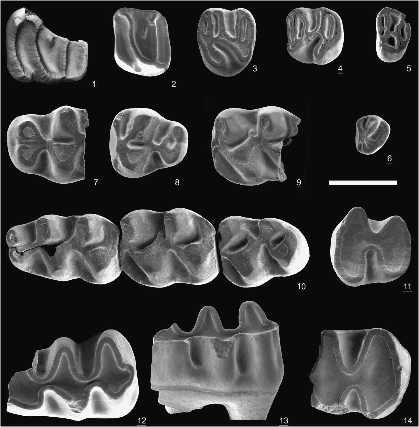
Dryomimus aff. D. eliomyoides molars: (1) M2, MAH-14 from Mahmutlar; (2) M1, KT2-16 from Kale Tepe 2. Keramidomys aff. K. ermannorum molars from Kale Tepe 2: (3) M2, KT2-14; (4) m1 or m2, KT2-15. Keramidomys aff. K. ermannorum molars from Kale Tepe 1: (5) M3, KT1-45. Cricetidae genus and species indet. molar from Kale Tepe 1: (6) M3, KT1-36. Allocricetus sp. indet. molars from Kele Tepe 1: (7) mesial part of M1, KT1-41; (8) M3, KT1-42; (9) damaged m2, KT1-43; (10) m1-m3 series, KT1-44. Pseudomeriones sp. indet. molar from Kale Tepe 1: (11) worn M2, KT1-53. Pseudomeriones sp. indet. molars from Mahmutlar: (12) damaged m1, MAH-12; (13) same specimen in labial view; (14) distal part of a worn m1, MAH-10. All molars are illustrated as left; the underlined numbers indicate reversed pictures. Scale bar=1 mm.

Occurrence
The genus Allocricetus is known with nine named species all over Eurasia from the late Miocene to early Pleistocene.
Description
M1 with anterocone divided into two equally large cusps without anterior groove; both cusps connected to the anterior arm of the protocone; precingulum present; mesial protolophule indicated by short spur issued from anterolophule; distal protolophule strong, connected backward to longitudinal crest. M3 with labial anteroloph stronger than lingual one; mesoloph short, pointed toward metacone; four roots. The m1 anteroconid bifid; labial anteroconid connected to junction of protoconid-metaconid by oblique, thick anterolophulid; mesolophid lacking; posterolophid thick, connected to distal base of entoconid. The m2 and m3 with protosinusid as deep as sinusid; labial anterolophid strong; lingual one reduced. The m2 with short mesolophid directed transversely; mesolophid stronger on m3, directed backward.
Materiel from KT1 and measurements
Fragment of left M1 (KT1-41, >1.17 mm W); left M3 (KT1-42, 1.17 mm L x 1.07 mm W); fragment of left mandibular ramus with m1−m3 (KT1-44, m1: 1.78 mm L x 1.09 mm W; m2: 1.46 mm L x 1.20 mm W; m3: 1.40 mm L x 1.07 mm W); fragment of right m2 (KT1-43, 1.17 mm W).
Remarks
Identification of late Miocene−Pleistocene cricetines of Eurasia is a debated issue due to the great similarities of their dental patterns. McKenna and Bell (Reference McKenna and Bell1997) recognized 23 genera belonging to the subfamily Cricetinae Fischer de Waldheim, Reference Fischer de Waldheim1817. De Bruijn et al. (Reference de Bruijn, Doukas, van den Hoek Ostende and Zachariasse2012, p. 73) listed 20 genera as valid for only small to medium-sized hamsters. They also noted that “The appalling number of generic names for what, judging by the figures, often seem to be animals with a similar dental morphology, suggests that a number of them may be synonymous.” Although these numbers demonstrate the great dynamics and diversity of Cricetinae during this time interval, it is true that a global systematic revision of fossil and extant cricetines is lacking. Therefore, it is difficult to identify genus and species when the material is limited.
The specimens from KT1 are clearly smaller than the molars of any species referred to the genera Cricetus Leske, Reference Leske1779, Apocricetus Freudenthal, Mein, and Martín-Suárez, Reference Freudenthal, Mein and Martín-Suárez1998, Hypsocricetus Daxner-Höck, Reference Daxner-Höck1992, Stylocricetus Topachevskii and Skorik, Reference Topachevskii and Skorik1992, Pseudocricetus Topachevskii and Skorik, Reference Topachevskii and Skorik1992, and clearly larger than those of the genera Phodopus Miller, Reference Miller1910, Cricetulus Milne-Edwards, Reference Milne-Edwards1867, Tscherskia Ognev, Reference Ognev1914, Nannocricetus Schaub, Reference Schaub1934 (except N. qiui Li et al., Reference Li, Stidham, Ni and Li2018 from Tibet), and Odessamys Topachevskii and Skorik, Reference Topachevskii and Skorik1992. The genera Cansumys Allen, Reference Allen1928, Chuanocricetus Zheng, Reference Zheng1993, and Allocricetulus Argyropoulo, Reference Argyropulo1933 are limited to central and eastern Asia, and they are poorly documented in the fossil record.
The genus Neocricetodon Schaub, Reference Schaub1934 (including Kowalskia Fahlbusch, Reference Fahlbusch1969, sensu Freudenthal et al., Reference Freudenthal, Mein and Martín-Suárez1998) is a common element of late Miocene and Pliocene faunas all over Eurasia. Sinitsa and Delinchi (Reference Sinitsa and Delinschi2016) listed 23 species referred to this genus, but recognized 18 as valid and used 15 in a phylogenetic analysis. Neocricetodon includes small to medium-sized species. Although some of them are similar in size to the cricetine from KT1, they are different in having wide anterocone and anteroconid on the first molars, the presence of well-developed mesolophs and mesolophids, and anterolophulids often double on the m1.
The KT1 molars are within the size range of Nannocricetus qiui from the early Pliocene of Tibet. However, this species is different in lacking mesolophids in all lowers, and in having an anterolophulid labially directed on the m1 and a strong lingual anterolophid on the m2.
Among the small to medium-sized cricetines, another common genus is Allocricetus, which is known all over Eurasia from late Miocene to early Pleistocene localities. Its type species is A. bursae from Fortyogoberg, a late Pliocene locality in Hungary. Eight other species have been referred to this genus (see Cuenca-Bescos, Reference Cuenca-Bescos2003). Allocricetus is characterized by its slender lower molars, anterocone and anteroconid of the first molars narrow, mesolophs and mesolophids absent or weakly developed and when they exist, their size increases from the first to the last molar. On the M1, the anterocone is anteriorly bifid, and connected to the protocone by a forked anterolophule. On the m1, the anteroconid is slightly bifid, posteriorly grooved, and connected by a single anterolophulid to the protoconid. All of these characters match the molars from KT1. Among the species referred to this genus, the size and morphology of the KT1 molars are similar to those of A. bursae, the most common Plio-Pleistocene species of the genus, and A. primitivus Wu and Flynn, Reference Wu and Flynn2017 from the early Pliocene of China. Despite these similarities in size and morphology, an identification of the KT1 cricetine at the species level would be hazardous due to the limited material.
In Turkey, small to medium-sized cricetines are known in several localities. De Bruijn et al. (Reference de Bruijn, Ünay and Hordijk2013) reported Allocricetus sp. from three localities in Turkey: the oldest Altıntaş in western Turkey correlated to MN 9, Düzyayla (Sivas Basin) correlated to MN 12, and Süleymanlı 2 (Çankiri Basin) correlated to MN 13. These records are not yet described. Allocricetus aylasevimae Ünay et al., Reference Ünay, de Bruijn and Suata-Alpaslan2006, from the late Miocene (MN 11) hominoid locality of Çorakyerler in the Çankiri Basin, is distinguished from the other species of Allocricetus in having a short and weak metalophul on the M1, whereas its size is similar to that of A. bursae. Rummel (Reference Rummel1998) referred specimens from Karaözü (Sivas Basin, MN 11) and Amasya (western Turkey, MN 13) to A. cf. A. ehiki Schaub, Reference Schaub1930, and those from Kavurca (Çankiri Basin, MN 13) to Allocricetus sp. without justifying these identifications. The size of these specimens rather match A. bursae and A. aylasevimae, whereas the connection of the metalophule on the M1 and M2 is like that in A. bursae. In addition, Rummel (Reference Rummel1998) illustrated some molars from Yeni Eskihisar (western Turkey) as cf. Allocricetus sp. This locality yielded quite a rich fauna that is correlated to the latest middle Miocene (i.e., MN 8), and these molars, although limited in number, are of great interest in documenting the ancestry and relationships of the small to medium-sized cricetines. Suata-Alpaslan et al. (Reference Suata-Alpaslan, Ünay and Ay2010) recorded A. bursae in the early Pliocene (MN 14) locality of Igdeli. Erten and Görmüs (Reference Erten and Görmüs2011) referred some molars to A. bursae from the Çameli Basin in southwestern Turkey, whereas from the same localities, van den Hoek Ostende et al. (Reference van den Hoek Ostende, Diepeveen, Tesakov, Saraç, Mayhew and Alçiçek2015a, Reference van den Hoek Ostende, Gardner, van Bennekom, Alçiçek, Murray, Frank, Wesselingh, Alçiçek and Tesakovb) reported Cricetulus cf. C. migratorius (Pallas, Reference Pallas1773) and C. sp. These localities are correlated to the Villanyian/Biharian transition, ca. 1.9 Ma. In summary, the late Miocene to Pleistocene cricetines are abundant in Turkey but a comprehensive study of their systematics is lacking at present.
Cricetidae gen. indet. sp. indet.
Description
Unique M3 with short, wide occlusal outline; labial anteroloph stronger than lingual; distal portion reduced to ridge-shaped cusp connected to protocone; one strong root forked at tip.
Materiel from Kale Tepe 1
Right M3 (KT1-16: 0.5 mm L x 0.6 mm W).
Remarks
This M3 is smaller than that of any other cricetid. The small cricetids such as Cricetulus, Allocricetus, Cricetinus Zdansky, Reference Zdansky1928, Nannocricetus, and Democricetodon Fahlbusch, Reference Fahlbusch1964 have all larger M3s of different morphologies. We also compared it with several species referred to Calomyscidae Vorontsov and Potapova, Reference Vorontsov and Potapova1979, Gerbillidae Gray, Reference Gray1825, and Eomyidae Depéret and Douxami, Reference Depéret and Douxami1902 known from Miocene and Pliocene localities of southeastern Europe and Turkey, without success. The mesial portion of this molar with two strong anterolophs and almost symmetrical protocone and paracone recalls cricetids, hence its tentative attribution to this family.
Family Gerbillidae Gray, Reference Gray1825
Subfamily Gerbillinae Gray, Reference Gray1825
Genus Pseudomeriones Schaub, Reference Schaub1934
Type species
Lophocricetus abbreviatus Teilhard de Chardin, Reference Teilhard de Chardin1926; Pliocene, China.
Pseudomeriones sp. indet.
Occurrence
The genus Pseudomeriones known by eight named species in southeastern Europe and Asia, from the late Miocene to late Pliocene.
Description
M2 with reduced distal part and transverse sinus. The m1 semihypsodont; occlusal outline elongated; without labial spur of anteroconid; protoconid situated distal to metaconid; posterolophid as distal process in moderately worn specimen.
Material from Mahmutlar
Left m1, distal part (MAH-10: 1.37 mm W; Fig. 8.14), right m1 with damaged anteroconid (MAH-12: estimated 2.20 mm L x 1.34 mm W; Fig. 8.12, 8.13), fragment of right M1 (MAH-11).
Material from Kale Tepe 1
Right M2 (KT1-53: 1.16 mm L x 1.17 mm W; Fig. 8.11).
Remarks
The lack of a labial spur on the m1 distinguishes these specimens from Pseudomeriones latidens Sen, Reference Sen2001 and P. pythagorasi Black, Kristalka, and Solounias, Reference Black, Kristalka and Solounias1980. In addition, P. pythagorasi, P. rhodius Sen, Reference Sen1977, and in particular, P. megistos Sylvestrou and Kostopoulos, Reference Sylvestrou and Kostopoulos2007 each have a clearly larger m1 than the specimens from Mahmutlar and KT1. The size and pattern of these teeth share great similarities with P. abbreviatus (Teilhard de Chardin, Reference Teilhard de Chardin1926) from King yan Fou (China) and Pul-e Charkhi (Afghanistan), P. hansi Suata-Alpaslan, Reference Suata-Alpaslan2009 from Igdeli (Turkey), and P. thaltaensis Sen, Reference Sen1977 from Çalta (Turkey). The limited number of specimens and their poor state of preservation do not allow further comparisons, hence their identification as Pseudomeriones sp. indet.
Famille Arvicolidae Gray, Reference Gray1821
Genus Mimomys Major, Reference Major1902
Type species
Arvicola pliocaenicus Major, Reference Major1889; Pliocene, Europe.
Mimomys sp. indet.
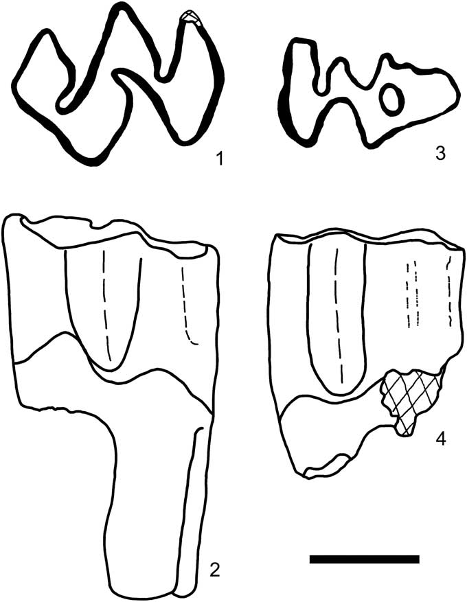
Mimomys sp. indet. molars from Mahmutlar (line drawings): (1) right M2, MAH-33; (2) same molar in lingual view; (3) left M3, MAH-34; (4) same molar in lingual view. Scale bar=1 mm.

Occurrence
The genus Mimomys with over 20 named species is known all over Eurasia from the early Pliocene to early Pleistocene.
Description
Molars rooted, devoid of cement; linea sinuosa low, slightly undulated; enamel of posterior edges of triangles unthickened. M2 with two mesial and one distal roots. M3 elongated, tapering distally; second lingual re-entrant fold transformed into enamel island; two roots. Maximum enamel 2.77 mm H (measured on fragment of m1).
Material from Mahmutlar
Right M2 (MAH-33: 1.98 mm L x 1.49 mm W; Fig. 9.1, 9.2), left M3 (MAH-34: 1.75 mm L x 1.04 mm W; Fig. 9.3, 9.4), and ~10 fragments of upper and lower molars.
Remarks
Rooted molars, lack of cement, low linea sinuosa, and short M3 are characters of primitive arvicolids such as Microtodon Miller, Reference Miller1927, Promimomys Kretzoi, Reference Kretzoi1955, Mimomys, and Propliomys Kretzoi, Reference Kretzoi1959 (see Hordijk and de Bruijn, Reference Hordijk and de Bruijn2009, p. 52−54 and van den Hoek Ostende et al., Reference van den Hoek Ostende, Gardner, van Bennekom, Alçiçek, Murray, Frank, Wesselingh, Alçiçek and Tesakov2015b, p. 314 for detailed discussion on the systematics of early arvicolids). Microtodon and Promimomys have crowns much lower than in other genera, and their M3s are short and round distally. The M3 of Promimomys is different in having the distal lingual re-entrant fold open in early stages of wear, whereas on the Mahmutlar M3, it is transformed into an enamel island. In these characters, the specimens from Mahmutlar match more closely with the early species of Mimomys, e.g., M. davakosi van de Weerd, Reference van de Weerd1979 and M. occitanus Thaler, Reference Thaler1955. Mimomys davakosi is well documented in Turkey and Greece (van de Weerd, Reference van de Weerd1979; Sen, Reference Sen1998; Hordijk and de Bruijn, Reference Hordijk and de Bruijn2009). The molars from Mahmutlar match this species in size and in having strong roots, a low crown, lack of cement, weakly differentiated enamel, and a distal enamel island on the M3. However, the limited number of specimens and the lack of the most characteristic tooth m1 prevent species identification for the Mahmutlar arvocolid.
Family Gliridae Thomas, Reference Thomas1897
Subfamily Myomiminae Daams, Reference Daams1981
Genus Dryomimus Kretzoi, Reference Kretzoi1959
Type species
Dryomimus eliomyoides Kretzoi, Reference Kretzoi1959; Csarnota-2, Hungary, late Pliocene.
Dryomimus aff. D. eliomyoides Kretzoi, Reference Kretzoi1959
Cotypes
Kretzoi (Reference Kretzoi1959, Reference Kretzoi1962) gave no specimen number or holotype, but illustrated in 1962 three upper and one lower cheek teeth from Csarnota-2 that can be considered cotypes of this species. The type material is preserved at the HGI (no catalog number).
Occurrence
Dryomimus eliomyoides is known in Hungary, the Balkans, and Turkey during the Pliocene and early Pleistocene.
Description
Upper molars with four main ridges and posterior centroloph; anteroloph separated from protoloph by wide lingual and narrow labial valleys; protoloph, metaloph, and posteroloph connected lingually to form endoloph along lingual margin.
Material from Mahmutlar
A lingual fragment of M1 (MAH-13) and a damaged M2 (MAH-14: 1.26 mm L x estimated 1.23 mm W; Fig. 8.1).
Material from KT2
Left M1 (KT2-16: 0.89 mm L x 1.07 mm W; Fig. 8.2).
Remarks
These molars are similar in size and morphology to those of Dryomimus eliomyoides from Csarnota-2 (Hungary), which is the type locality of this species. Dryomimus eliomyoides or compared forms are known in Turkey by scarce remains from the localities of Çalta, Ortalica, Hamamayagi, and Çaybasi (Sen, Reference Sen1998; Ünay and de Bruijn, Reference Ünay and de Bruijn1998; Ünay et al., Reference Ünay, Emre, Erkal and Keçer2001), and Dryomimus sp. from Develi and Tekman (Suata-Alpaslan, Reference Suata-Alpaslan2011; Mayda, Reference Mayda2014). These localities are correlated to MN 14 to MN 16.
Family Eomyidae Depéret and Douxami, Reference Depéret and Douxami1902
Genus Keramidomys Hartenberger, Reference Hartenberger1966
Type species
Keramidomys pertesunatoi Hartenberger, Reference Hartenberger1966; Can Llobateres, Spain, late Miocene.
Keramidomys aff. K. ermannorum Daxner-Höck and Höck, Reference Daxner-Höck and Höck2009
Holotype
Right m1 (NHMW RH-A/7) from Richardhof-Golfplatz, Vienna Basin, Austria
Occurrence
Keramidomys ermannorum is known in several localities in Austria, Greece (Maramena), and Turkey. Late Miocene to earliest Pliocene.
Description
M2 wider than long; lingual part narrower than labial part; sinus deep, anteriorly oblique; weak connection between protoloph and mesoloph; mesoloph straight; anterosinus short, posterosinus elongated, both closed labially to form enamel islands; three roots. M3 with half-circle occlusal outline; four transverse lophs separated by three sinuses, all closed labially.
The m1 rectangular in occlusal outline, with five transverse ridges; mesial ridge thinner than others; longitudinal ridge between mesolophid and protolophulid complete; mesolophid straight, strong, connected lingually to metaconid; anterosinusid, anterior mesosinusid, and posterosinusid closed lingually to form enamel islands; sinusid oblique, directed backward; one anterior and two posterior roots.
Material from KT1
Left M3 (KT1-45: 0.51 mm L x 0.77 mm W; Fig. 8.5).
Material from KT2
Left M2 (KT2-14: 0.86 mm L x 1.03 mm W; Fig. 8.3); right m1 or m2 (KT2-15: 0.85 mm L x 0.82 mm W; Fig. 8.4).
Remarks
The small size of these three molars and the occlusal outline of M2 and m1 with five ridges match the diagnostic features of the genus Keramidomys. The best-known species of this genus, K. thaleri Hugueney and Mein, Reference Hugueney and Mein1968 (type locality Vieux Collonges, early part of MN 5) and K. carpathicus (Schaub and Zapfe, Reference Schaub and Zapfe1953) (type locality Neudorf-Spalte, MN 6), are from early and middle Miocene localities. De Bruijn (Reference de Bruijn1995a) considered K. thaleri as well as K. octaviae Baudelot, Reference Baudelot1972 subjective junior synonyms of K. carpathicus, following the opinions of Fahlbusch (Reference Fahlbusch1975) and Bolliger (Reference Bolliger1992). Contrary to this, Prieto (Reference Prieto2010) showed that K. thaleri is a distinct species and its dentition displays morphological variations as seen in central European localities at the transition of early−middle Miocene, i.e., longitudinal crest interrupted, weak or strong in different percentages in the fossil assemblages, length of the mesoloph, and mesolophid variable. Prieto (Reference Prieto2010) also showed with dispersion diagrams great size variation inside an assemblage when it is well documented.
Studying the rich Keramidomys material from Maramena (northern Greece, late MN13), de Bruijn (Reference de Bruijn1995a, p. 100) noted that it displays, as in other late Miocene and Pliocene occurrences of this species, primitive characters compared to the type material of K. carpathicus, e.g., “the anteroloph and the protoloph of the P4 not completely fused and the longitudinal ridge of the M1-2 not interrupted in about 50% of the specimens,” or derived characters such as the reduction of third molars and of their mesoloph/mesolophid (see de Bruijn, Reference de Bruijn1995a for a detailed discussion).
Daxner-Höck and Höck (Reference Daxner-Höck and Höck2009) studied late Miocene Eomyidae from the Vienna Basin and erected the new species K. ermannorum (type locality Richardhof-Golfplatz, MN 9). This species differs from K. carpathicus and K. thaleri by its larger dimensions and in having a tendancy to develop lophodonty, deeper sinus(id), flattened occlusal surface, and wider lower molars. These authors also included the material from Maramena (Greece) and Stanjantsi (Meotian, Bulgaria) in K. ermannorum. The size and occlusal pattern of the molar from the KT2 (complete longitudinal crest, strong mesoloph(id), deep sinus(id), and flat occlusal surface) compare best to those of K. ermannorum. However, the limited material from Kale Tepe does not allow observation of morphological variation, hence its identification as K. aff. K. ermannorum.
Another European middle−late Miocene species, K. mohleri Engesser, Reference Engesser1972 (type locality Anwil, MN 8) has cheek teeth larger than those at Kale Tepe, with frequent interruption of the longitudinal crest, shorter mesoloph on M1-2, and the sinus less oblique and deep.
Order Lagomorpha Brandt, Reference Brandt1855
Family Ochotonidae Thomas, Reference Thomas1897
Genus Prolagus Pomel, Reference Pomel1853
Type species
Lagomys sansaniensis Lartet, Reference Lartet1851; Sansan, France, middle Miocene.
Prolagus sorbinii Masini, Reference Masini1989
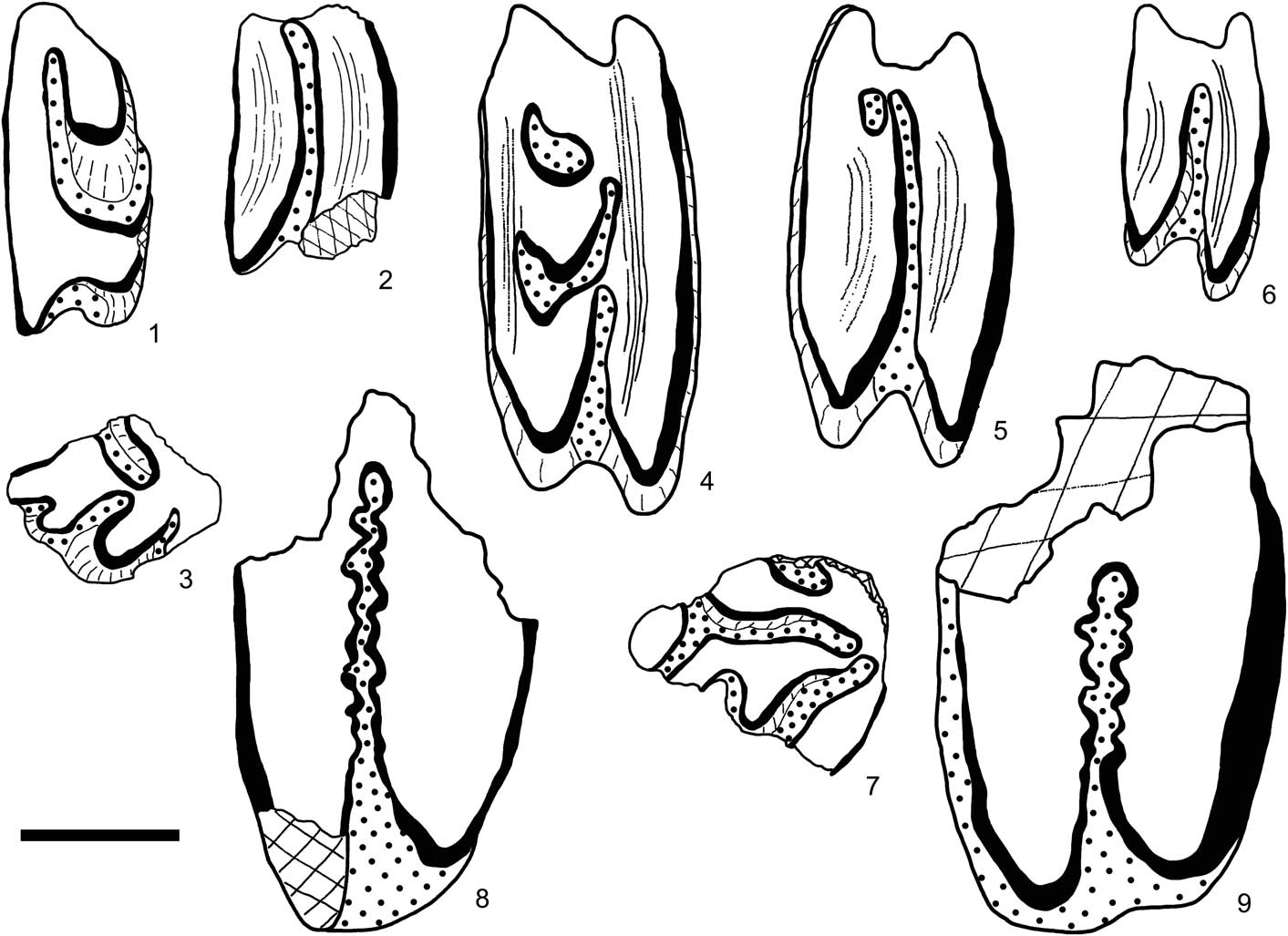
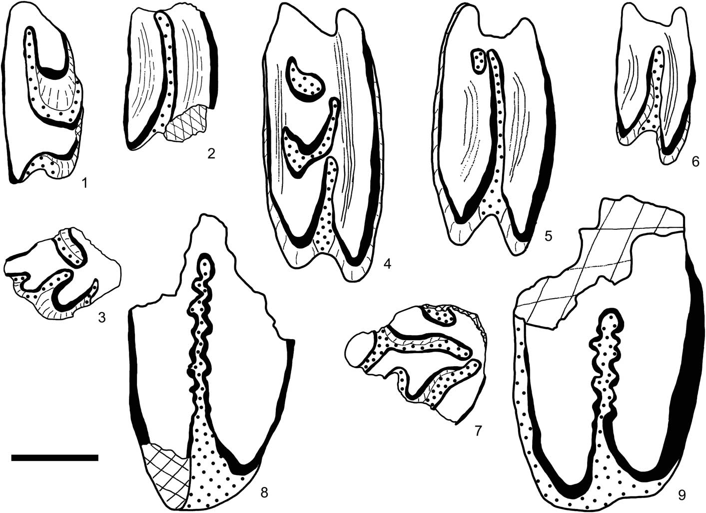
Lagomorph cheek teeth in occlusal view (line drawings) from three localities in the Çankırı-Çorum Basin. Ochotonoma sp. indet. from Mahmutlar: (1) right P3, MAH-22; (2) right P4 or M1, MAH-21; (3) fragment of left p3, MAH-30. Prolagus sorbinii from Kale Tepe 2: (4) right P4 (KT2-47); (5) right M1 (KT2-48); (6) right M2 (KT2-49); (7) left p3 (KT2-57). Leporidae indet. from Kale Tepe 1: (8, 9) two right upper molariform teeth (KT1-46 and KT1-47). Blackened lines=enamel; white=dentine; dotted=cement infill. Scale bar=1 mm.
Holotype
Articulated skeleton with skull (MNHV V.702) from Monte Castellaro (Pesaro, Italy).
Occurrence
Prolagus sorbinii is known in several localities in Italy, Greece (Maramena), and Turkey; late Miocene and Pliocene.
Description
Medium-sized cheek teeth as typical for Prolagus. P4 with short hypoflexus and short parafossette labially limited to level of mesofossette. M1 with deep hypoflexus and small mesofossette. Distinctive p3 with small, rounded anteroconid, narrow protoisthmus (protoconid-talonid connection), without crochet.
Material from Kale Tepe 2
Five fragments of upper incisors (KT2-52 to 56); right P4 (KT2-47: 1.46 mm L x 3.03 mm W; Fig. 10.4); right M1 (KT2-48: 1.36 mm L x 2.72 mm W; Fig. 10.5); right M2 (KT2-49: 0.98 mm L x 1.79 mm W; Fig. 10.6); damaged left p3 (KT2-57; Fig. 10.7), and several fragments of upper and lower cheek teeth.
Remarks
Masini (Reference Masini1989) defined Prolagus sorbinii based on an articulated skeleton from Monte Castellaro (Italy), which is dated to the latest Miocene (MN 13). According to Masini (Reference Masini1989), this is a fairly large species. On its dentition, P2 bears a well-developed anteroflexus; P4 has a short hypoflexus and M1 preserved the mesoflexus; p3 has very reduced or absent crochet, and lacks the protoisthmus. The specimens from KT2 have all of these features. Subsequently, materials from several other localities in Italy (Brisighella, Moncucco Torinese, Borro Strolla 1 and 2, Ciabot Cagna, Velona, Arcille, Solava, Casa Inferno; see Angelone, Reference Angelone2007; Abbazzi et al., Reference Abbazzi, Benvenuti, Ceci, Esu, Faranda, Rook and Tangocci2008; Angelone and Cavallo, Reference Angelone and Cavallo2010; Angelone and Rook, Reference Angelone and Rook2012), in Greece (Maramena; de Bruijn, Reference de Bruijn1995b), in Moldova (Novaya Andriyashevka and Leordoaya; see Delinschi, Reference Delinschi2014), in Ukraine (Grebeniki-2; see Tesakov and Averianov, Reference Tesakov and Averianov2002), and in Turkey (Develi; see Sen et al., Reference Sen, Jaeger, Dalfes, Mazin and Bocherens1989) were referred to or compared with P. sorbinii. We suggest that the specimen from Ptolemais 1 in northern Greece (van de Weerd, Reference van de Weerd1979) should also be referred to this species.
On the other hand, Vasileiadou et al. (Reference Vasileiadou, Koufos and Syrides2003, Reference Vasileiadou, Konidaris and Koufos2012) rightly referred the remains of a pika from the latest Miocene−early Pliocene localities of Silata and Kessani in northern Greece to Prolagus michauxi López Mártinez in López Mártinez and Thaler, Reference López Mártínez and Thaler1975. Indeed, the material from these localities is different from P. sorbinii in having a crochet, a large protoisthmus, and a curved mesoflexid on the p3. These discoveries indicate that two medium-sized Prolagus species existed during the late Miocene−early Pliocene in southern Europe from the Iberian Peninsula to Anatolia.
Genus Ochotonoma Sen, Reference Sen1998
Type species
Ochotonoma anatolica Sen, Reference Sen1998; Çalta, Turkey; early Pliocene.
Ochotonoma sp. indet.
Occurrence
Species referred to Ochotonoma recorded in Hungary, Romania, Bulgaria, Greece, Turkey, and China, in Pliocene localities.
Description
Upper incisor with anterior groove situated laterally. P3 short, large, its hypoflexus moderately deep and filled with cement, its mesial hyperloph short and not constricted. Lophs mesially concave on P4 or M1. Anteroconid triangular in outline on p3, with labial flexid filled with cement; anteroconid connected to main part of tooth by narrow, rather central isthmus (0.09 mm); protoflexid and paraflexid deep, oriented backward.
Material from Mahmutlar
Left upper incisor (MAH-37: 1.69 mm L x 1.06 mm W), right P3 (MAH-22: 0.97 mm L x 2.14 mm W; Fig. 10.1), fragment of right P3 (MAH-23), right P4 or M1 (MAH-21: 1.14 mm L x 1.82 mm W; Fig. 10.2), two fragmentary p3 (MAH-30 and 31; Fig. 10.3), several fragments of upper and lower cheek teeth.
Remarks
The dental pattern and size of these teeth match Ochotonoma anatolica, which was initially defined from the early Pliocene locality of Çalta (Kazan Basin, Ankara, Turkey; Sen, Reference Sen1998). That same year, Ünay and de Bruijn (Reference Ünay and de Bruijn1998) erected the new species O. ortalicensis from Ortalica in the Tosya Basin (Kastamonu, Turkey) and reported the occurrence of O. cf. O. ortalicensis from the locality of Tasova (Tasova-Erbaa Basin, Turkey) and O. sp. from the locality of Sürsürü (Elazig Basin, Turkey). All of these localities are correlated to MN 15. The cheek teeth from all of these localities are apparently identical with those from Çalta, although the single p3 from Sürsürü is smaller and should be referred to the genus Ochotonoma (Sen, Reference Sen2003). According to Suata-Alpaslan (Reference Suata-Alpaslan2010), O. ortalicensis also exists in the localities of Babadat and Dinar-Akçaköy in western Turkey, both correlated to late MN 14. The material from these localities is not described.
The central European occurrences of this genus are from Csarnota-2 and Beremend-26 (Hungary), Ciuperceni-2 (Romania), and Muselievo (Bulgaria). The ochotonids from all of these localities were initially referred to Ochotonoides csarnotanus that Kretzoi (Reference Kretzoi1959) briefly described as a new species from Csarnota-2. Sen (Reference Sen1998, Reference Sen2003) included this species in Ochotonoma together with other records from Rhodos Island (Greece) and Songshan-1 (China). Lastly, Čermák (Reference Čermák2007) revised the central European occurrences of O. csarnotanus and confirmed their attribution to the genus Ochotonoma. Most important is that Čermák (Reference Čermák2007) suggested the synonymy of O. anatolica and O. ortalicensis with O. csarnotana. His argument was that the variation of dental pattern in the southeastern European localities (Csarnota-2, Beremend-26, Ciuperceni-2, and Muselievo) also covers that of the ochotonids from Turkey, in particular those from Çalta and Ortalica. Consequently, Čermák (Reference Čermák2007) included all of the occurrences of this ochotonid from southeastern Europe and Turkey in O. csarnotana. However, the type specimen of O. csarnotana and the sole p3 from Csarnota-2 is larger than all p3s from Çalta (N=15) and Ortalica (N=8), and its occlusal pattern is clearly different from those of O. anatolica or O. ortalicensis (see Sen, Reference Sen1998). It is beyond the aim of the present study to provide a new investigation on the genus Ochotonoma. While awaiting a detailed comparative analysis of the specimens from different regions, the poor material from Mahmutlar is referred to Ochotonoma sp.
Family Leporidae Gray, Reference Gray1821
Leporidae genus indet. species indet.
Description
Two upper molariform teeth with moderately deep hypoflexus (>50% width); enamel thin on mesial border but rather thick on distal border of lophs; enamel equally plicated along both margins of hypoflexus.
Material from KT1
Right upper molariform (KT1-46: 2.08 mm L x 3.68 mm W; Fig. 10.8), fragment of right upper molariform (KT1-47; Fig. 10.9), trigonid of a lower molariform (KT1-55: 2.79 mm W).
Remarks
Enamel plication around the hypoflexus recalls that of Alilepus Dice, Reference Dice1931 (Angelone and Rook, Reference Angelone and Rook2011; Delinschi, Reference Delinschi2013), Trischizolagus Radulescu and Samson, Reference Radulescu and Samson1967 (Averianov and Tesakov, Reference Averianov and Tesakov1997), and Pliopentalagus Gureev and Konkova in Gureev, Reference Gureev1964 (Tomida and Jin, Reference Tomida and Jin2009; Čermák and Wagner, Reference Čermák and Wagner2013). Oryctolagus is different in having enamel plication along the anterior margin of the hypoflexus, whereas the enamel is quite flat along the posterior margin (de Marfa and Mein, Reference de Marfa and Mein2007; de Marfa, Reference de Marfa2009; Angelone and Rook, Reference Angelone and Rook2012). The lack of characteristic teeth, e.g., P2 and p3, makes it not possible to identify the genus and species to which these specimens belong.
Conclusions
As a result of this study, we can list the rodents and lagomorphs for the localities of KT1, KT2, and Mahmutlar as follows. The number of studied cheek teeth for each taxon is given in parentheses; (+) means additional fragments.
KT1: Apodemus gorafensis (13), A. gudrunae (23), Allocricetus sp. indet. (6), Pseudomeriones sp. indet. (1), Cricetidae gen. indet. sp. indet. (1), Keramidomys aff. K. ermannorum (1), and Leporidae gen. indet. sp. indet (3+). Number of studied specimens 48.
KT2: Apodemus gorafensis (13), A. gudrunae (30), Micromys sp. indet. (4), Dryomimus aff. D. eliomyoides (1), Keramidomys aff. K. ermannorum (2), and Prolagus sorbinii (4+). Number of studied specimens 54.
Mahmutlar: Apodemus dominans (10), Pseudomeriones sp. indet. (3), Mimomys sp. indet. (2+), Dryomimus aff. D. eliomyoides (2), and Ochotonoma sp. indet. (5+). Number of studied specimens 22.
These three localities are situated in the southern part of the Çankırı Basin where fluvio-lacustrine deposits of the Akkaşdağı Formation, in which these localities are included, cover large areas. The fossiliferous outcrops of Kale Tepe and Mahmutlar are fresh road cuts that were opened when the new Kırıkkale-Çorum highway was built. Consequently, the stratigraphic succession of these three localities is well controlled (Fig. 2).
To define the age of these localities, one must compare them with well-documented and well-dated sites in Turkey and nearby Greece. In Turkey, Amasya-1 and Süleymanlı-2 (MN 13), Igdeli (MN 14), and Çalta (MN 15) are the best-documented and best-dated micromammal localities. There are many other sites that have yielded small mammals, but either their faunas are poor in species, probably due to insufficient sampling, or they are known with preliminary lists without detailed study of the material to make them reliable for comparison.
In Greece, numerous localities in the Ptolemais Basin yielded rich small mammalian faunas (van de Weerd, Reference van de Weerd1979; Hordijk and de Bruijn, Reference Hordijk and de Bruijn2009). In addition, the stratigraphy of this basin is well established, and mammal localities are correlated to the magnetic polarity time scale or dated by radiometric methods (Van Vugt et al., Reference Van Vugt, Steenbrink, Langereis, Hilgen and Meulenkamp1998; Steenbrink et al., Reference Steenbrink, Van Vugt, Hilgen, Wijbrans and Meulenkamp1999, Reference Steenbrink, Hilgen, Krijgsman, Wijbrans and Meulenkamp2006). These studies evidenced a succession of small mammal localities for the 5.5−3.9 Ma interval. Consequently, the small-mammal localities of this basin became the benchmarks for correlation of other small-mammal localities close in age to the Miocene/Pliocene boundary. In Greece, other localities such as Maramena (Schmidt-Kittler, Reference Schmidt-Kittler1995), Kessani and Silata (Vasileiadoou et al., 2003, 2012) in northern Greece, and Maritsa on the island of Rhodes (de Bruijn et al., Reference de Bruijn, Dawson and Mein1970), are also important clues for documenting small mammals of latest Miocene and early Pliocene times.
The assemblages from KT1, KT2, and Mahmutlar are dominated by muroid rodents, in addition to a few glirids, eomyids, and lagomorphs. The assemblages from KT1 and KT2 are quite similar in sharing three species (Apodemus gorafensis, A. gudrunae, and Keramidomys aff. K. ermannorum), but the fauna from Mahmutlar is different because it shares only one species with KT1 (Pseudomeriones sp. indet.) and one other species with KT2 (Dryomimus aff. D. eliomyoides). The arvicolids are apparently absent at KT1 and KT2, whereas a rooted arvicolid similar to Mimomys davakosi is present at Mahmutlar.
The lack of Cricetodontini and Arvicolidae brackets the localities of KT1 and KT2 to the latest late Miocene and earliest Pliocene. These groups are well documented in Greece and Turkey, and they are reliable chronological markers for dating terrestrial deposits. In both localities, the dominant species are Apodemus gorafensis and A. gudrunae, which represent 75% and 80% of the studied specimens, respectively. These species co-occur in Turkey at Süleymanlı-2, ~70 km NNW of Kale Tepe, in the northern part of the Çankırı Basin, and also probably at Amasya (western Turkey), both correlated to the MN 13 zone. As discussed above, both species probably exist at Tomea Eksi 1 and 2 in northern Greece. The age of these localities is bracketed between 5.43 and 5.30 Ma (Hordijk and de Bruijn, Reference Hordijk and de Bruijn2009), i.e., just before the Miocene/Pliocene boundary at 5.33 Ma (Hilgen et al., Reference Hilgen, Lourens and van Dam2012). Apodemus gorafensis is also known in Greece at Maramena, Kessani, and Silata, and in over 30 localities in Spain where most localities are correlated to the second part of MN 13 zone, and a few other to MN 14, in particular its type locality Gorafe A. Apodemus gudrunae provides a similar age, because it is reported in Turkey from Süleymanlı-2 and Amasya, both correlated to the MN 13 zone. The other rodents and lagomorphs are also well-known elements across the Miocene/Pliocene transition.
Keramidomys aff. K. ermannorum from KT1 and KT2 is similar in size and morphology to K. ermannorum from Austrian localities, correlated to the Turolian, and from Maramena in northern Greece (Daxner-Höck and Höck, Reference Daxner-Höck and Höck2009). The age of the latter locality was estimated as close to the late Miocene/early Pliocene boundary (Schmidt-Kittler, Reference Schmidt-Kittler1995). Based on the dominance of Apodemus gorafensis and A. gudrunae at KT1 and KT2, and also the presence of K. aff. K. ermannorum, KT1 and KT2 are tentatively correlated to the end of the late Miocene, i.e., in term of the Mammalian Neogene zones to the upper part of MN 13.
The rodent assemblage from Mahmutlar is dominated by Apodemus dominans (59% of rodent molars). The type locality of this species (Csarnota-2 in Hungary) is correlated to MN 15. In the Ptolemais Basin in northern Greece, this species occurs at Notio-1, the age of which is calibrated by magnetostratigraphy as between 3.92 and 3.97 Ma (Hordijk and de Bruijn, Reference Hordijk and de Bruijn2009). In Turkey, A. dominans is known in several localities (Çalta, Develi, Hamamayağı, Igdeli, Ortalıca, Tozaklar, Yağmurlu), all correlated to MN 14 or MN 15 (Ünay and de Bruijn, Reference Ünay and de Bruijn1998; Saraç, Reference Saraç2003). The primitive arvicolid from Mahmutlar referred to Mimomys sp. indet. resembles M. davakosi, which is only known in localities correlated to the early part of MN 15 zone, between 3.5 and 4.0 Ma. Dryomimus cf. D. eliomyoides and Ochotonoma sp. indet. from Mahmutlar also match this time interval.
The sedimentary features of deposits at KT1, KT2, and Mahmutlar indicate more-or-less permanent lacustrine environments, ponds, and temporarily flooded lands (Karadenizli, Reference Karadenizli2011). As shown in Figure 2, all of the lower part of the Kale Tepe section is mainly composed of coal-bearing and organic-rich clays. Kayseri-Özer et al. (Reference Kayseri-Özer, Karadenizli, Akgün, Oyal, Saraç, Sen, Tunoglu and Tuncer2016) sampled this part of the section for pollen and ostracods. Their study concluded that this section recorded the late Miocene/Pliocene transition. Pollen analyses showed that the late Miocene vegetation was dominated by warm-temperate elements such as oaks, deciduous and evergreen types, bitternut hickory, hornbeam, zelkova, elm, alder, hackberries, lime-trees, and willow, in addition to grasses and aquatic herbs. Steppe elements (sage and ephedra) are only recorded in low percentages. The paleovegetation underwent a significant change from the late Miocene to early Pliocene probably due to changes in temperature and precipitation. The paleovegetation in early Pliocene deposits of the Kale Tepe section indicates cooling temperature and increasing moisture by diversity and abundance of Sapotaceae, chestnut, and chinquapin. Spore and pollen distribution of the early Pliocene consisted of abundant herbaceous and shrub elements.
Habitat preferences of rodents and lagomorphs recorded at Kale Tepe and Mahmutlar can be extrapolated in comparison with their extant relatives. Apodemus gorafensis and A. gudrunae from KT1 and KT2 can be compared to their extant relatives A. mystacinus (Danford and Alston, Reference Danford and Alston1877) and A. flavicollis (Melchior, Reference Melchior1834), respectively, both well known in Turkey and surrounding countries. Although Ruiz Bustos et al. (Reference Ruiz Bustos, Sesé, Dabrio, Peña and Padial1984) hypothesized the origination of A. mystacinus from A. gorafensis, this is not fully demonstrated because of the large time gap between two species. However, the dentition of these two species shares many common features such as large and globular cusps connected with short ridges on upper molars, cusp-shaped t12 on M1 and M2, m1 with a large tma, and thick labial cingulum, which bears several large cuspules. We hypothetise that these two species could have had similar habitats. Apodemus mystacinus, also known as the ‘rock mouse,’ inhabits open forests with rocky areas and a sparse cover of grass or shrubs (Krytufek et al., Reference Kryštufek, Vohralík and Janžekovič2009). The yellow-necked mouse, A. flavicollis, is mostly a woodland species, often living near the forest verge, but in mountainous regions, it occupies any part of the forest. It is usually found in mature deciduous woodland and scrubby areas (Krytufek et al., Reference Kryštufek, Vohralík and Janžekovič2009). Micromys sp. indet. is the third murid at KT2. The main habitat of the extant species M. minutus (Pallas, Reference Pallas1771), the harvest mouse, is open landscapes with grassy or bushy vegetation. The presence of the the dormouse, Dryomimus cf. D. eliomyoides, at KT2 is consistent with this assessment.
At Mahmutlar, the dominant species is Apodemus dominans, which is similar in size and dental morphology to the extant A. flavicollis, the yellow-necked mouse that is well established throughout Anatolia. As noted above, it mainly occupies deciduous woodland and scrubby areas. Contrary to this, the gerbil Pseudomeriones sp. indet. and the pika Ochotonoma sp. indet., inhabitants of steppic environments in Asia, might indicate that the environnement of Mahmutlar was somewhat more open and drier than at KT1 and KT2.
It is worth noting that the large-sized species of Apodemus—A. gorafensis, A. gudrunae, A. jeanteti, as well as their Quaternary relatives A. mystacinus and A. epimelas (Nehring, Reference Nehring1902)—only occur in the southern margins of Europe (Spain, southern France, Italy, Greece) and around the eastern Mediterranean. They are not known in central and eastern Europe where only small to medium-sized species—A. atavus, A. dominans, A. flavicollis, A. sylvaticus (Linnaeus, Reference Linnaeus1758), A. witherbiyi (Thomas, Reference Thomas1902), and A. alae Nesin, Reference Nesin2011—dominate in the latest Miocene and later rodent faunas.
Rodents from Kale Tepe and Mahmutlar, as well as from other latest Miocene−early Pliocene localities in Greece and Turkey, evidence great affinities with those of the southern regions of Europe. This is particularly true for Muridae, Cricetidae, and Eomyidae. This resemblance was already noted by van de Weerd (Reference van de Weerd1979), Sarica-Filoreau (Reference Sarıca-Filoreau2002), Hordijk and de Bruijn (Reference Hordijk and de Bruijn2009), and Vasileadou et al. (2003, 2012). However, the occurrence of Pseudomeriones in several localities in Greece and Turkey, and here at KT1 and Mahmutlar, also suggests some connection with Asia.
The remains of lagomorphs are scarce and fragmentary in all three localities, hence incomplete identifications at the genus and species levels. Three species are identified: Leporidae gen. indet. sp. indet. at KT1, Prolagus sorbinii at KT2, and Ochotonoma sp. indet. at Mahmutlar. Prolagus sorbinii is only known in southern European regions and in Turkey. On the contrary, Ochotonoma has Asiatic affinities although its distribution during the early Pliocene extends from Hungary to Turkey and probably to China. In summary, the rodent and lagomorph assemblages from KT1, KT2 and Mahmutlar share common elements mainly with southern and central Europe, but also to some extent with eastern Asia.
Acknowledgments
The field work in the Çankırı Basin was done in the context of a joint project between the General Directorate of Mineral Research and Exploration (MTA) in Ankara and Natural History Museum (MNHN) in Paris. The MTA provided permit and field assistance. We received financial support from the DARIUS Program (Paris) and from CR2P-CNRS for travel and field expenses. E. Albayrak, F. Arslan, Y. Büyükmeriç, O. Erdal, C. Karaca, S. Karaman, M. Koç, G. Métais, M.J. Orliac, N. Oyal, C. Öztürk, S. Peigné, V. Rommevaux, and A. Vural participated in the field work. A. Lethiers (CR2P-Sorbonne Université) redrew the map and the section for Figures 1 and 2. S. Pont (MNHN, Paris) produced the SEM photos with a Surface Electronic Microscope TESCAN Vega2 at the Muséum, and P. Loubry (CR2P-CNRS-MNHN) helped with improving them. We are grateful to all of them. We would like to thank the referees L.J. Flynn and H. de Bruijn, and also the Editor-in-Chief H.-D. Sues and J. Kastigar for their comments that greatly contributed to improve this study.