INTRODUCTION
In Argentina, the superfamily Grapsoidea comprises marine, brackish and freshwater crabs; there are five species grouped in three genera and two families (Martin & Davis, Reference Martin and Davis2001). The family Varunidae is represented by Cyrtograpsus angulatus (Dana, 1851), Cyrtograpsus altimanus (Rathbun, 1914), Cyrtograpsus affinis (Dana, 1851) and Neohelice granulata (Dana, 1851) (=Chasmagnathus granulata). The last one included in the subfamily Cyclograpsinae. Metasesarma rubripes (Rathbun, 1897) belongs to the family Sesarmidae (Boschi, Reference Boschi1964).
This study is focused on Cyrtograpsus, an endemic South Atlantic and Pacific Ocean genus (Rathbun, Reference Rathbun1918). Other previous studies on this genus were carried out on larval development (Scelzo & Litchstein de Bastida, Reference Scelzo and Lichtschein de Bastida1979; Rieger & Vieira, Reference Rieger and Vieira1997; Spivak & Cuesta, Reference Spivak and Cuesta2000), ecological and population structure parameters (Rognone, Reference Rognone1984; Rascio, Reference Rascio1986; Castiglioni & Santos, Reference Castiglioni and Santos2000), moult and growth (Spivak, Reference Spivak1988), use of habitat, mating systems and sexual selection (Gavio & Spivak, Reference Gavio and Spivak1994; Gavio, Reference Gavio2003), feeding (Capítoli & Ortega, Reference Capítoli and Ortega1996), fecundity (Luppi et al., Reference Luppi, Bas, Spivak and Anger1997), reproduction (López Greco & Rodriguez, Reference López Greco and Rodríguez2004) and morphometric and molecular comparisons (Spivak & Schubart, Reference Spivak and Schubart2003).
Cyrtograpsus species present a wide distribution: C. angulatus is distributed along the south-western Atlantic Ocean coast from Rio de Janeiro, Brazil to Puerto Deseado, Argentina and in the south-eastern Pacific Ocean from San Lorenzo Island, Perú to Talcahuano Bay, Chile; C. altimanus is distributed from Rio Grande do Sul, Brazil to Puerto Deseado, Argentina; and C. affinis is distributed from San Roque Cape, Brazil to San Matías Gulf, Argentina (Boschi, Reference Boschi1964).
Spivak (Reference Spivak1999) and Spivak & Schubart (Reference Spivak and Schubart2003) report differences in body size and habitat for these species; maximum carapace width in adult specimens of C. angulatus is three to five times larger than that of C. altimanus and C. affinis, respectively. Cyrtograpsus angulatus and C. altimanus coexist in rocky intertidal habitats (Rathbun, Reference Rathbun1918; Boschi, Reference Boschi1964; Scelzo & Lichtschein de Bastida, Reference Scelzo and Lichtschein de Bastida1979) and display diverse abilities to invade estuarine habitats (Spivak, Reference Spivak1999). These species are very different in their external morphology; whereas C. altimanus is morphologically similar to C. affinis. The latter species can be distinguished by a more squarish carapace and because it inhabits subtidal habitats. At present, C. altimanus and C. affinis are considered putative species (Spivak & Schubart, Reference Spivak and Schubart2003), but there is a great controversy about their taxonomic status.
Due to the fact that no other study regarding gastric mill was found in the literature on the genus Cyrtograpsus, it was considered relevant to undertake a descriptive and comparative morphological study of the gastric mill ossicles within the species that this genus comprises. The present study increases the knowledge on Cyrtograpsus species' morphological characteristics that allow them to be identified. The chitinous gastric mill ossicles are resistant to the digestive process because of their hardness. Thus, they are useful to recognize the species in predator stomach contents when there are no other external morphological features available.
Stomach and gastric mill studies have been carried out on many species of decapod crustaceans; those of Nauck (Reference Nauck1880), Patwardhan (Reference Patwardhan1934, Reference Patwardhan1935), Meiss & Norman (Reference Meiss and Norman1977), Dall & Mortiary (Reference Dall, Mortiary and Mantel1983), Felgenhauer & Abele (Reference Felgenhauer, Abele and Felgenhauer1989), Brösing (Reference Brösing2002), Brösing et al. (Reference Brösing, Richter and Scholtz2002) and Castro & Bond-Buckup (Reference Castro and Bond-Buckup2003) include a critical review of the current terminology in Reptantia (Macrura: Anomura: Brachyura).
Several authors (Boschi & Angelescu, Reference Boschi and Angelescu1962; Dall & Mortiary, Reference Dall, Mortiary and Mantel1983; Felgenhauer & Abele, Reference Felgenhauer, Abele and Felgenhauer1989; Huang et al., Reference Huang, Yang and Ng1998; Brösing et al., Reference Brösing, Richter and Scholtz2001; Brösing, Reference Brösing2002; Sakai et al., Reference Sakai, Türkay and Yang2006) have used the gastric mill structure as an additional characteristic to define genera, families and species. These authors have discussed the gastric mill structural complexity, stating that this structure reflects the evolutionary trends and relationships within decapods.
The gastric mill is a triturative device with several strongly chitinized ossicles of which the dorsal ones are important for the shape and stabilization of the anterior stomach chamber (Brösing, Reference Brösing2002). The anterior, posterior, ventral and dorsal side refers to the position of the gut within the body. In this study only the most conspicuous ossicles, easy to identify and considered of taxonomic value, were included (Brösing et al., Reference Brösing, Richter and Scholtz2001; Brösing, Reference Brösing2002), those supporting the stomach chamber being excluded.
Mesocardiac (I), pterocardiac (II), post-pterocardiac (IIb), zygocardiac (V), urocardiac (VII), propyloric (VI), exopyloric (IV) and pyloric (III) ossicles are placed dorsally in the cardiac chamber; the central unpaired ossicles (mesocardiac and urocardiac) can be fused according to the species. The unpaired mesocardiac ossicle is laterally hinged to the paired pterocardiac ossicle and this articulates with the paired post-pterocardiac which acts as a pivot for the zygocardiac (paired) and prepectineal (IX) (paired) ossicles, the latter is hinged anteriorly to pectineal ossicle (VIII) (paired). In some species prepectineal and pectineal ossicles are fused; both are placed in the lateral walls of cardiac stomach. Urocardiac, zygocardiac and pectineal ossicles have a dorsal median tooth, lateral and accessory teeth, respectively. All these teeth are responsible for mastication and grinding of the food that enters through the oesophagus. The accessory teeth assist in pushing the material towards the central region of the cardiac chamber.
The cardiopyloric valve (CPV) placed ventrally between the cardiac and pyloric stomach, lies under the urocardiac median dorsal tooth. This valve is conformed by the unpaired anterior (XVI) and posterior (XVII) ossicles, and the lateral ossicle (XVIII) (paired). This valve directs and regulates food material passage from the cardiac to pyloric stomach. Meiss & Norman (Reference Meiss and Norman1977), Brösing (Reference Brösing2002) and Brösing et al. (Reference Brösing, Richter and Scholtz2002) compare the cardiopyloric valve ossicles in different decapod crustacean species.
The objectives of this research were to: (i) describe C. angulatus, C. altimanus and C. affinis gastric mill; and (ii) differentiate each species by comparing the gastric mill structures. Morphology of the dorsal median tooth in urocardiac ossicle; number of ridges and molar processes (cusps and plates) in zygocardiac ossicle; number of accessory teeth in pectineal ossicle and in cardiopyloric valve development and shape of median tooth on the posterior margin, and length, density and distribution of setae on its lateral and posterior margins were considered.
MATERIALS AND METHODS
Sampling and study area
Cyrtograpsus altimanus (N = 193) and Cyrtograpsus angulatus (N = 97) specimens were manually collected during low tide in Punta Cuevas (42°46′ 50.8″ S 65°0′ 06.0″ W), April 2003 and Punta Este (42°47′ 06.6″ S 64°57′ 0.9″ W), December 2002, February and March 2004. These sampling sites are located in Golfo Nuevo (Chubut, Argentina). Cyrtograpsus affinis (N = 8), were supplied by E. Spivak, and collected at 13 m depth, in Río de la Plata estuary (36°05′ S 56°37′ W) during the field work of the fishing research ship ‘Capitán Cánepa’ (Instituto Nacional de Investigación y Desarrollo Pesquero, INIDEP).
Methodology
Crabs were transported alive to the laboratory, once there some specimens were: (i) fixed in 10% formaldehyde for a week and then they were kept in 70% ethanol; or (ii) frozen at −20ºC. Frozen animals facilitate stomach dissection and muscles removal; in these specimens appendages lost is less frequent.
In all specimens carapace length (CL) and carapace width (CW) were measured with a Vernier caliper and then the stomach was extracted by lifting up the carapace, since it is adhered to it. Ossicles were measured in a stereomicroscope and microscope with an ocular micrometer.
Stomachs were cleaned and observed under a stereomicroscope (Zeiss, Stemi SV6). Afterwards, they were opened in an antero-posterior direction through the ventral mid-line from the oesophagus up to the pleuropyloric valve and the stomach content was discarded so the gastric mill ossicles (urocardiac, zygocardiac and pectineal) and cardiopyloric valve could be observed, described and compared in order to establish interspecific and intraspecific differences in the three studied species.
Those stomachs to be observed under scanning electron microscopy (SEM) (ALUAR S.A.I.C.) were prepared as follows: (i) organic debris and tissues adhered to the stomach structures were removed with 10% sodium hydroxide, time varied from 5 to 20 minutes according to stomach size and ossicles hardness (intermoult or premoult crabs); (ii) alkali was neutralized with 5% acetic acid and then rinsed in distilled water; and (iii) each stomach was extended and mounted with a double-sided adhesive tape on a labelled slide.
Under a JEOL JSM-6460 LV microscope dry SEM samples without gold coating were viewed using the back-scattered electron (BSE) technique, at low vacuum (15–20 Pa). Digital images of gastric mills' general views and ossicles details were taken, as well as each photograph condition (magnification, plate inclination and rotation, scale, resolution, etc.).
RESULTS
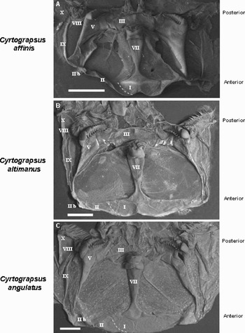
Cyrtograpsus gastric mill ossicles shape and their relative position follow the same general pattern in the studied species (Figure 1 A–C).
Gastric mill, internal dorsal view: (A) Cyrtograpsus affinis; (B) Cyrtograpsus altimanus; and (C) Cyrtograpsus angulatus. Ossicles: I, mesocardiac; II, pterocardiac; IIb, postpterocardiac; III, pyloric; V, zygocardiac; VII, urocardiac; VIII, pectineal; IX, prepectineal; X, post-pectineal. Scale bars: 1 mm.

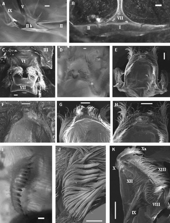
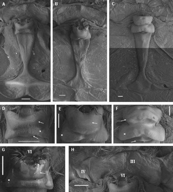
The unpaired urocardiac ossicle (VII) is an elongated centred T-shaped ossicle that narrows to its anterior region (Figure 3 A–C). There is a small protuberance at the end of this narrowing. The urocardiac ossicle articulates anteriorly with the mesocardiac ossicle and laterally is separated from the pterocardiac ossicle by an incision (Figure 2 B). The posterior end is hinged with the vertical unpaired subtriangular propyloric ossicle (VI) constituting a dorsal median tooth (Figures 2C & 3H).
(A) Cyrtograpsus altimanus ossicles: postpterocardiac (IIb), pterocardiac (II), zygocardiac (V), prepectineal (IX); (B) Cyrtograpsus affinis ossicles: mesocardiac (I), pterocardiac (II), urocardiac (VII); (C) Cyrtograpsus angulatus ossicles: propyloric (VI), urocardiac (VII), pyloric (III), cardiopyloric valve general view; (D) C. angulatus, ventral posterior view; (E) C. angulatus, dorsal view. Detail; (F) C. affinis; (G) C. altimanus; (H) C. angulatus; (I) and (J) C. angulatus pectineal ossicle, accessory teeth; and (K) C. altimanus ossicles: pectineal (VIII), prepectineal (IX), postpectineal (X) and ‘Quill’ (Xa), inferior lateral cardiac (XIII) and posterior lateral cardiac plate (XII). (A), (B), (D) and (I) stereo-microscope photographs. Scale bars: 200 µm.

Urocardiac ossicle. General view: (A) Cyrtograpsus affinis; (B) Cyrtograpsus altimanus; (C) Cyrtograpsus angulatus. Dorso-median tooth, detail: (D) C. affinis; (E) C. altimanus; (F) C. angulatus and (G) C. affinis; and (H) C. affinis: pyloric (III), exopyloric (IV) and propyloric (VI) ossicles. (Arrows show central teeth and crosslateral teeth in urocardiac dorsal median tooth.) Scale bars: 200 µm.

The unpaired pyloric ossicle (III) looks like a paired one because it is thinner in the median line (Figure 3H). This ossicle is hinged anteriorly to the propyloric ossicle and laterally contacts the subtriangular exopyloric ossicle (IV) (Figure 3H).
The mesocardiac ossicle (I) (unpaired) is an anteriorly bilobated triangular plate which is thinner in the median line looking like a paired ossicle (Figures 1 & 2B), is laterally fused to the pterocardiac (II) ossicle and is posteriorly hinged to the urocardiac one. Mesocardiac (I) and pterocardiac (II) ossicles fusion is incomplete and is pointed out by an incision (Figures 1A–C, dotted line on the left side, and 2B).
The pterocardiac ossicle (II) (paired) is subrectangular, tapering to its distal lateral end (Figures 1 & 2B).
The postpterocardiac ossicle (IIb) (paired) is a Y-shape ossicle, sometimes very difficult to observe due to its small size and location. It is hinged anteriorly to the pterocardiac ossicle and posteriorly to the paired zygocardiac and prepectineal ossicles (Figure 2A).
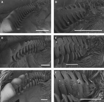
The zygocardiac ossicle (V) (paired) is a large triangular plate bearing the gastric mill lateral teeth. These heavily sclerotized teeth have dorsal ridges with upwardly curved endings and ventral molar processes with large cuspidate processes (cusps) shaping the anterior part followed by flattened ones (plates) (Figure 4A–F). This ossicle posteriorly articulates with the exopyloric (IV) one.
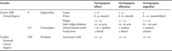
In the three Cyrtograpsus species, the number of molar processes (cusps and plates) is similar to that of the ridges in the zygocardiac ossicle (Table 1). Cusps are smooth and constitute half of the lateral teeth length; but the plate's surface differs within the species.
Number of teeth, molar processes (cusps and plates) and ridges in cardiac stomach ossicles.

The long and narrow paired prepectineal ossicle (IX) (Figure 1A–C) articulates anteriorly with the posterocardiac ossicle and posteriorly with the pectineal ossicle.
The pectineal ossicle (VIII) (paired) is a mobile oblong plate bearing the lateral accessory teeth, directed forward and posteriorly hinged to the postpectineal ossicle (X). The lateral accessory teeth (12–16) (Figures 1A–C & 2 I–J) are long, narrow and flexible setae arranged bordering the pectineal ossicle. These teeth externally surrounded with long setae, slightly project towards the centre where there is a group of long thin setae. Teeth number variation is less in small size specimens.
The postpectineal ossicles (X), ‘quill’ (Xa) of the postpectineal ossicles, posterior lateral cardiac plates (XII) and inferior lateral cardiac ossicles (XIII) are similar in the three Cyrtograpsus species (Figure 2K). Anterior lateral cardiac plates (XI) were not observed due to the way the stomach was opened.
The cardiopyloric valve (Figure 2D–H) is ventral and placed posteriorly to the urocardiac dorso-median tooth. This valve is conformed by three ossicles: (i) anterior (XVI), unpaired, forms a small middle ridge; (ii) posterior (XVII), unpaired, slightly curved with a well-developed tooth shaped median projection. Lateral to this median tooth there are eight spines usually strongly worn out (rounded and shorter or broken) (Figure 2F–H). The postero-dorsal surface of the median tooth has a smooth triangular area whose borders bear densely arranged setae (Figure 2D); and (iii) lateral (XVIII), paired, with setae of different length on the postero-median edge. These lateral ossicles have big ear-like projections on their antero-lateral border (Figure 2E). Posterior and lateral ossicles form a unit.
Table 1 shows the main structures in cardiac stomach ossicles that can be used to identify Cyrtograpsus species.
The interspecific and intraspecific variation observed in the zygocardiac (number of ridges) and pectineal (number or teeth) ossicles (Table 1) is based in data obtained from all the Cyrtograpsus altimanus (N = 193) and Cyrtograpsus angulatus (N = 97) specimens studied. Their size-range was 8.5–25.3 mm for C. altimanus and 16–47.4 mm for C. angulatus. In Cyrtograpsus affinis (N = 8) intraspecific variation could not be established because of the scanty specimens.
The differences observed in cardiac stomach ossicles in Cyrtograpsus species are the following ones:
UROCARDIAC OSSICLE (VII) (FIGURE 3A–G)
The well-developed quadrangular dorsal median tooth has two lobed projections on its anterior margin in C. angulatus (Figure 3C & F, cross) and C. affinis (Figure 3A, D & G, cross); there is another smaller tooth (Figure 3D & F, inferior arrow) anterior to this bilobate edge. The anterior small protuberance in the urocardiac ossicle is very conspicuous in C. angulatus (Figure 3F), less evident in C. affinis (Figure 3D & G) and absent in C. altimanus (Figure 3E).
ZYGOCARDIAC OSSICLE (V) (FIGURE 4A–F)
In C. angulatus (Figure 4C & F), this ossicle has four cusps and six to ten mammillated plates whose rough aspect is given by small rounded elevations (mammillas/mammelons). The dorsal ridges (11–14), spaced 62–100 µm, curve upward on their distal end. Stout setae form these ridges, in a compact arrangement; they appear on the surface as small cusps whose height increases gradually towards the ridge's anterior distal end and surpassing its edges. In the apex of the zygocardiac ossicle, there is a dense group of short and thin setae (Figure 4F).
Zygocardiac ossicle. General view: (A) Cyrtograpsus affinis; (B) Cyrtograpsus altimanus; (C) Cyrtograpsus angulatus. Lateral tooth, posterior end detail: (D) C. affinis; (E) C. altimanus; (F) C. angulatus. Scale bars: 200 µm.

In C. altimanus (Figure 4B & E) there are three to four cusps followed by six to eight smooth plates. Zygocardiac ridges (11–13) are similar to those of C. angulatus, except for the inter-ridges distance (20–60 µm) and the setae that barely surpass the ridge's edges. As in C. angulatus a dense group of setae is present at the apex of the zygocardiac ossicle (Figure 4E).
Cyrtograpsus affinis (Figure 4A & D) has five to six cusps, six to nine smooth plates and a row of 13–14 ridges spaced by 20–42 µm; the ridges are very similar to those of C. altimanus. At the apex of the zygocardiac ossicle, there is a less dense group of thin setae and stout ones (Figure 4D).
DISCUSSION
Sakai et al. (Reference Sakai, Türkay and Yang2006) found that the differences in the form of the gastric mill in the genera Helice/Chasmagnathus complex support the generic groupings within families and can be used in brachyuran systematics. The authors stated that ‘The gastric mill teeth differ significantly between genera but are relatively homogenous within a genus,’ and also that ‘The species can be sometimes distinguished by the number of the lateral comb-shaped teeth and by details of the median tooth plate. Such differences have been referred to in the species descriptions, but there is a great deal of overlap, and the structure of the teeth is not useful in distinguishing between closely related species (p.5).’
Within the family Varunidae, the ossicle structures in Cyrtograpsus differ from those shown by Sakai et al. (Reference Sakai, Türkay and Yang2006) and Covelo de Zolessi & Spiritoso (Reference Covelo de Zolessi and Spiritoso1985) for the genera Helice/Chasmagnathus complex and Neohelice granulata (=Chasmagnathus granulata), respectively.
In Cyrtograpsus the gastric mill has the same general pattern in the three studied species and there are no differences related to specimens' size or sex.
Zygocardiac and urocardiac ossicles' structures are useful to distinguish species. They can be observed under SEM, but can also be recognized using a microscope or a stereomicroscope, allowing their measurement with an ocular micrometer.
The incision that laterally separates the urocardiac ossicle from the pterocardiac ossicle is similar to that shown by Brösing (Reference Brösing2002) and Nauck (1880) in Pseudosesarma moeschi (Sesarmidae) and Cardisoma carnifex (Gecarcinidae), respectively. In the latter species, the pterocardiac and postpterocardiac ossicles are like those found in Cyrtograpsus. Yang's (Reference Yang1986) drawings of mesocardiac and pterocardiac ossicles in Uca (Ocypodidae) and Metaplax (Grapsidae) are quite similar to Cyrtograpsus (Varunidae) ones.
The urocardiac ossicle dorso-median tooth and the zygocardiac ossicle lateral teeth (ridges, cusps and plates) are useful to identify Cyrtograpsus species. Since they show interspecific differences, these structures are helpful when found intact in the stomach contents of their predators.
The urocardiac dorso-median tooth is similar in C. affinis and C. altimanus, but C. angulatus differs only by a small well-developed central tooth in front of the lateral projections.
The zygocardiac ossicle differs in the type, surface and number of molar processes (cusps and plates) in the lateral tooth. There are 5–6 cusps and 6–9 smooth plates in C. affinis, 3–4 cusps and 6–8 smooth plates in C. altimanus and 4 cusps and 6–10 mammilated plates in C. angulatus. The number of ridges (14) is similar in Cyrtograpsus species and the variability observed in C. angulatus and C. altimanus (11 to 14) is considered to be intraspecific variation. The Cyrtograpsus altimanus inter-ridge distance size-range (20–60 µm) overlaps that for C. affinis (20–42 µm); but, this distance size-range is quite different in C. angulatus (62–100 µm).
Shape and number of molar processes as well as inter-ridge distance differences in the zygocardiac ossicle could be related to the species feeding type associated to their habitat.
In Cyrtograpsus the pectineal (VIII) and prepectineal (IX) ossicles are slightly chitinized compared with urocardiac and zygocardiac ones. Therefore, it is less probable to find them complete in predators' stomach contents. In the three species studied, the pectineal ossicle is similar. The number of accessory teeth is quite constant (12–16) and the differences observed are considered as due to specimen size.
The morphology of the cardiopyloric valve is similar in all Cyrtograpsus species. According to Felgenhauer & Abele (Reference Felgenhauer, Abele and Felgenhauer1989), the morphology of the cardiopyloric valve provides an idea of the species feeding type, as this valve limits the size of the food that enters the pyloric chamber. Therefore, a valve with a prominent median tooth (posterior ossicle) indicates that together with zygocardiac lateral teeth it contributes to food grinding. In Cyrtograpsus species, cardiopyloric valve similarities suggest filtering of same size food particles. The well-developed median tooth in the posterior valve's ossicles (XVII) implies large food intake (macrophagy).
Gastric mill comparisons in Cyrtograpsus species have shown very few differences. Gastric mill morphology in C. altimanus and C. affinis are similar but they differ from C. angulatus in zygocardiac and urocardiac ossicles. Huang et al. (Reference Huang, Yang and Ng1998) have also found that these ossicles are different in two very closely related Ocypodidae species (Ocypode sinensis and O. cordimanus).
The few internal morphological differences found in the three Cyrtograpsus species are consistent with larval morphology studies (Spivak & Cuesta, Reference Spivak and Cuesta2000). The differences stated in zoeal and megalopae morphology descriptions were based on laboratory-reared larvae (Scelzo & Lichtschein de Bastida, Reference Scelzo and Lichtschein de Bastida1979; Rieger & Vieira, Reference Rieger and Vieira1997; Spivak & Cuesta, Reference Spivak and Cuesta2000), finding minimum differences in morphology and zoeae (I–IV) size. Megalopae sizes are similar in C. altimanus and C. affinis, but they are larger in C. angulatus. The morphology of this larval stage is similar in the three species and the main differences among the megalopae are in the morphology of antennule and the mandible (Spivak & Cuesta, Reference Spivak and Cuesta2000).
Spivak & Schubart's (Reference Spivak and Schubart2003) comparisons of morphometrics and 16S mtDNA did not find diagnostic differences between C. affinis and C. altimanus and these putative species are considered closely related species. These authors stated that C. affinis and C. altimanus differences found in molecular 16S mtDNA comparisons, external morphology and size-range could be considered interpopulational differences related to life in different habitats. Cyrtograpsus altimanus is distributed in subtidal and intertidal habitats but C. affinis is restricted to subtidal habitats.
Spivak & Schubart (Reference Spivak and Schubart2003) did not find diagnostic differences between these two putative species considering that both represent different morphs of a single species. These results are confirmed by Lezcano et al. (Reference Lezcano, Dellatorre and Gónzales-José2006) in a geometric morphometric study carried out in Puerto Madryn (Chubut, Argentina). The authors' findings showed the same continuous gradient in subtidal habitats and a strong alometric effect. They also consider that C. affinis and C. altimanus represent morphs of a single species. Spivak & Schubart (Reference Spivak and Schubart2003) attribute morphological variability to a high phenotypic and ecological plasticity. A similar case of closely related species is that of Brachynotus sexdentatus and B. gemmellari (Varunidae). Their 16S mtDNA are identical and have overlapping morphometries, differing only in habitat preferences (Cuesta et al., Reference Cuesta, Schubart and Rodríguez2000; Schubart et al., Reference Schubart, Cuesta and Rodríguez2001). A 16S mtDNA molecular study should be performed in C. angulatus to establish if there are molecular differences with C. affinis and C. altimanus.
Our findings demonstrate enough evidence to conclude that C. altimanus and C. affinis should belong to a single species as mentioned by Spivak & Schubart (Reference Spivak and Schubart2003) and Lezcano et al. (Reference Lezcano, Dellatorre and Gónzales-José2006). These new data validate the existence of only two species, C. affinis and C. angulatus, and allow us to recognize these species in the stomach content of their predators.
ACKNOWLEDGEMENTS
The authors thank ALUAR SAIC for SEM. We are grateful to Rodolfo Acuña Laje for providing SEM authorization, Jaime Groizard for operating the scanning microscope and his generous technical assistance during this work. Thanks to Mariana Bulgarella, Tamara Rubilar and Eva Fitz for their suggestions and critical reading of the manuscript, and Eduardo Spivak for generously providing Cyrtograpsus affinis specimens.