The aetiology, natural progression, and prognosis of aortic aneurysm in children remain unknown because of the rarity of presentation. Although aortic aneurysm is rare in children, it can be easily diagnosed by imaging studies such as computed tomographic scan or ultrasound, but delineation of the anatomy is best obtained with a computed tomographic angiogram or magnetic resonance imaging. They can be asymptomatic or present dramatically with rupture and cardiovascular collapse.Reference Roy, Azakiea, Moon-Grady and Blurton 1 Here we present the case of a 7-year-old boy diagnosed with ascending thoracic aortic aneurysm. Analytical determinations including karyotyping and genetic mapping were all normal and a diagnosis of idiopathic aneurysm was made. To our knowledge, idiopathic aneurysm of the ascending aorta in children is very rare.
Case report
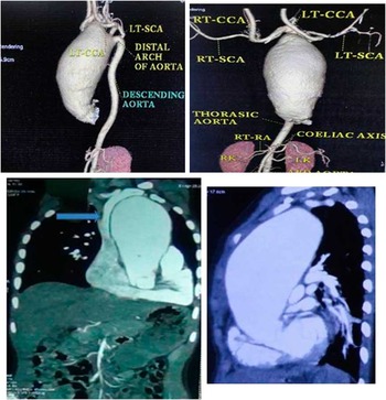
A 7-year-old boy weighing 19 kg was referred to us for mild difficulty in breathing. He was diagnosed to have ascending aorta aneurysm causing mild bronchial obstruction. Echocardiogram revealed a large dilatation of the ascending aorta, trivial aortic regurgitation, and normal annulus. Magnetic resonance angiogram showed aneurysmal dilatation of the ascending aorta and the proximal arch (60×67 mm), no evidence of dissection, or thrombosis, aortic root not dilated, right brachiocephalic trunk and left common carotid artery originating from the aneurysmal segment of the aortic arch, left subclavian artery originating from the normal segment of the aortic arch, and normal descending thoracic and abdominal aorta (Fig 1). The left main bronchus was mildly compressed by the aneurysmal segment of the aorta (Fig 1). The left radial and right dorsalis pedis artery was used for invasive arterial pressure monitoring. Monitoring included electrocardiogram, pulse oximeter, central venous pressure, bispectral index, temperature, and urine output. Anaesthesia was induced with fentanyl 50 mcg, midazolam 1 mg, etomidate 8 mg, vecuronium 3 mg, and intubated with 5.5 mm cuffed endotracheal tube. Anaesthesia was maintained with oxygen, nitrous oxide, and sevoflurane.
Computed tomographic angiogram in the coronal and sagittal section showing the innominate artery and left carotid artery coming from the aneurysmal sac and the left subclavian artery arising from the normal arch. The blue arrow mark shows the mildly compressed bronchus.

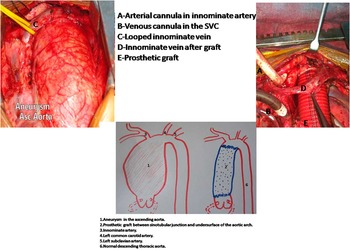
After median sternotomy and full heparinisation, cardiopulmonary bypass was established with an arterial cannula in the right subclavian artery through a 6 mm gortex tube graft and bicaval cannulations (Fig 2). Total circulatory arrest was achieved for a short period with low-flow antegrade cerebral perfusion. The aneurysmal sac was opened and was seen extending from the sinotubular junction to the mid-arch of the aorta. The graft (26 mm collagen-coated woven polyster – INTER GUARD, MAQUET) was anastomosed proximally from the sinotubular junction and distally to the undersurface of the arch preserving the native lie of all the three arch branches (Fig 2). The right brachiocephalic trunk and left common artery was seen arising from the normal segment and not from the aneurysmal segment as mentioned in the magnetic resonance angiogram report (Fig 2). After complete rewarming, the child was weaned off pump with Inj. Dobutamine 5 mcg/kg/minute and Inj. Adrenaline 0.05 mcg/kg/minute. The total duration of cardiopulmonary bypass time was 120 minutes, aortic cross-clamp time was 54 minutes, and total circulatory arrest was 12 minutes. The resected aortic aneurysmal sac was sent for histopathology report, which showed degenerative fibromyxoid changes of the wall of the aorta. The duration of ventilation was 6 hours. The duration of intensive care unit stay was 3 days and the child was discharged on the seventh post-operative day.
Intra-operative diagram showing the aneurysm, looped innominate vein, and the cannula placement. Schematic representation of the aneurysm, arch vessels, and the graft placement. SVC=superior vena cava.

Discussion
Ascending aortic aneurysm is uncommon in the paediatric population. The majority occur in the descending thoracic aorta and post-coarction repair aneurysms are the most frequent type.Reference Sterpetti, Hunter and Schultz 2 Arterial aneurysms in childhood may be due to arterial infection secondary to staphylococcus aureus and salmonella, giant cell aortoarteritis, or autoimmune vasculitis due to Kawasaki disease, degeneration of the media layer as in Ehlers–Danlos and Marfan syndromes, disruption of the arterial layers as in pseudoaneurysm, congenital as in congenital defect in type III collagen and may be idiopathic.Reference Marın-Manzano, Gonzalez-de-Olano, Haurie-Girelli and Herraiz-Sarachaga 3 The genetic test included fibrillin gene (FBN 1) for Marfan syndrome, collagen type V (COL 5 A 1 and COL 5 A 2) for classical type of Ehlers–Danlos, transforming growth factor receptor β1 and 2 (TGF B R1/TGF B R2) for Loeys–Dietz syndrome and myosin heavy chain 11 (MYH 11) for familial thoracic aortic aneurysms and dissections. Clinically, the patient did not have marfanoid habitus or cleft palate andbifid uvula as in Loeys–Dietz syndrome. In addition, none of the first-degree family member had a similar disease. All the karyotypic and genetic work-up done to find the aetiology were also normal. None of the first-degree family members had a similar disease. Owing to the fact that our case did not present with any of the above-mentioned characteristics, it should be classified among idiopathic aneurysms.
The association of aneurysm of the aorta in childhood is mainly linked to aortic valvular stenosis, coarctation of the aorta, abnormality of the sinus of Valsalva, congenital abnormalities of the aorta, or congenital connective tissue disorders. Coarctation of the aorta and aortic valvular stenosis are the most common congenital cardiac abnormalities associated with aortic aneurysm, and when both coexisted aneurysms were more likely to develop. Associated factors such as hypertension, infection, trauma, and cystic medial necrosis share in the pathogenesis of the paediatric aneurysm.Reference Fricker, Park, Neches and Mathews 4 In our case, none of the above associations was present.
The paediatric aneurysm literature is limited to case reports and small case series, precluding a meaningful evaluation of outcomes or conclusions regarding the ideal repair technique. The most important fact to be considered during infancy is the permanent growth of the patient. Options vary between resection, lateral repair, pulmonary homograft patch, venous patch, end-to-end anastomosis, endovascular repair, and autologous vein or dacron patch.Reference Marın-Manzano, Gonzalez-de-Olano, Haurie-Girelli and Herraiz-Sarachaga 3 Although avoidance of prosthetic material seems diligent in the setting of infection, multiple studies have demonstrated the safety of prosthetic conduits if actively infected tissue is absent or can be fully debrided.Reference Ting, Cheng, Ho, Poon and Tsu 5 Our patient had no evidence of active aortic infection, minimising concern over placement of prosthetic material. The oversized interposition graft used should accommodate future growth and obviate the need for additional surgery.Reference Andersen, Bhattacharya, Williams and McCann 6 Although endovascular techniques have diminished the morbidity of thoracic aortic repair, such as thoracotomy, full heparinisation, aortic clamping, and the risks of distal, medullary, and visceral ischaemia, its application in paediatric population is very limited. Despite covered stents being frequently used to repair coarctations of the aorta, its application in paediatric aortic aneurysm is yet to be seen.Reference Butera, Piazza and Chessa 7
The surgical technique has to be individualised. Cannulation of the right subclavian artery allowed good bypass flows in this child and the proximal clamp could be placed in a way to allow flow in the left carotid artery. It is equally important to continue appropriate anticoagulation and avoid dehydration as it may precipitate graft thrombosis.Reference Mendeloff, Stallion, Hutton and Goldstone 8 The main problem with the use of artificial blood vessels in children is that they may become relatively narrow compared with normal blood vessels as children mature, and that may result in limb ischaemia or affect growth and development.Reference Ye, Yin, Lin and Zhou 9 Of course, the long-term effects can only be determined with long-term follow-up, and re-operation may be required should stenosis occur.
Conclusion
Idiopathic ascending aortic aneurysm is a very rare condition in children. As can be noted from this case report, the causes of ascending aortic aneurysm in children are varied, with inherited syndromes being the predominant causes of aortic disease in this age group. Owing to the frequent association of aortic aneurysms with inherited connective tissue disorders, as well as cardiac and extracardiac malformations, chromosomal, genetic, and pedigree analysis should be performed. Our experience indicates that good outcomes can be obtained with accurate diagnosis and surgical management with artificial grafts. This case report may assist physicians in diagnosing and treating similar patients.
Acknowledgement
The author likes to acknowledge the help of Dr Sivamuthukumar, Dr Rajeshkmar Kodali, Dr Kamalakannan, and Dr Sushma.
Financial Support
This research received no specific grant from any funding agency, commercial or not-for-profit sectors.
Conflicts of Interest
None.