Introduction
Total aortic cusp replacement such as the Ozaki procedure is one of the techniques of aortic valve repair. Reference Ozaki, Kawase, Yamashita, Uchida, Takatoh and Kiyohara1 While some studies have described the short-term results of the Ozaki procedure in paediatric patients, Reference Wiggins, Mimic and Issitt2–Reference Baird, Cooney, Chávez, Sleeper, Marx and del Nido4 long-term results have rarely been reported. Herein, we report the 13-year follow-up data of a patient undergoing total aortic cusp replacement.
Case
A boy diagnosed with critical aortic valve stenosis at birth underwent balloon valvuloplasty at the age of 6 days. The procedure resulted in an improved aortic valve peak pressure gradient (from 60 mmHg to 20 mmHg) with trivial aortic valve regurgitation; however, the regurgitation worsened gradually during follow-up. At 9 years of age (weight = 28.2 kg; height = 133 cm), aortic valve regurgitation worsened to severe with a peak pressure gradient of 35 mmHg (Figure 1a and b). Cardiac MRI showed a regurgitant fraction of 30.2 %. Because the pulmonary valve regurgitation was mild, the Ross procedure was not recommended, and we selected aortic valve repair.
Transthoracic echocardiogram. Preoperative echocardiogram in the diastolic phase (( a ): short axis, ( b ): long axis) showing severe aortic valve regurgitation. Postoperative echocardiogram in the diastolic phase (( c ): short axis, ( d ): long axis) showing no aortic valve regurgitation. Diastolic phase echocardiogram from the last follow-up (( e ): short axis, ( f ): long axis) showing mild aortic valve regurgitation. Systolic phase echocardiogram from the last follow-up showing slight thickening of the leaflets ( g ). Change in the aortic annular diameter, diastolic left ventricular diameter, and body weight since the operation until the last follow-up ( h ). The annular diameter and left ventricular diameter remained around 100 % of normal, while the body weight increased steadily. LVDd, diastolic left ventricular diameter.

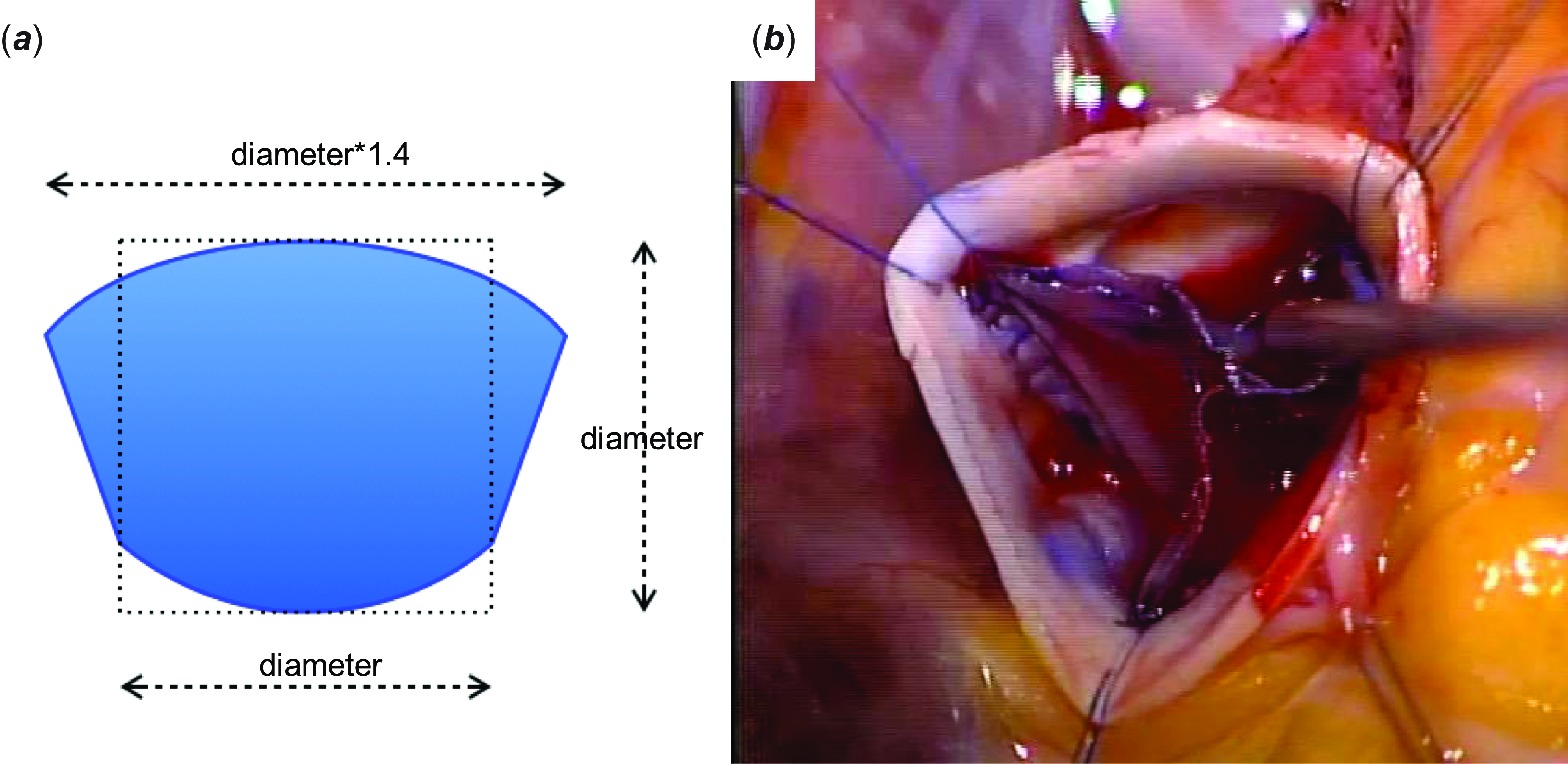
Through a standard median full sternotomy, the autologous pericardium was harvested and treated with 0.6% glutaraldehyde for 3 minutes. A cardiopulmonary bypass was established with ascending aortic and right atrial cannulation. After cardioplegic arrest, the ascending aorta was transected approximately 1 cm above the sinotubular junction, and the aortic valve was exposed. All cusps appeared thickened with fused right and noncoronary cusps. Since an aortic valve repair could not be performed without any materials, and we decided to perform a total cusp replacement. All cusps were excised. The aortic annular diameter was 20 mm with intraoperative measurement by Hegar dilator and 20.5 mm (132% of normal) with preoperative transesophageal echocardiography at the cusp hinge point. Mainly according to the intraoperative measurement, we trimmed the glutaraldehyde-treated autologous pericardium with a cusp template of 20 mm as the annulus (Figure 2a). Each cusp was sutured by continuous suture with 7-0 Pronova (Johnson & Johnson Services, Inc., New Brunswick, NJ, USA). The commissure was made at the same position as the native valve a little lower of the sinotubular junction (Figure 2b).
Template of the aortic valve cusp and operative images. Our template for the leaflet ( a ); diameter is the aortic annular diameter. Operative image after suturing each leaflet ( b ).

The coaptation test was performed by negative pressure created using a venting tube, which revealed adequate coaptation. Intraoperative transesophageal echocardiography showed trivial aortic valve regurgitation without stenosis. The cardiopulmonary bypass and cardiac arrest times were 163 and 134 minutes, respectively. The postoperative course was uneventful. At discharge, the peak pressure gradient was 8 mmHg with trivial aortic valve regurgitation (Figure 1c and d). Oral aspirin was induced for antiplatelet therapy.
Thirteen years postoperatively, he is doing well without any physical limitations. The peak pressure gradient at the last follow-up was 22 mmHg with mild aortic valve regurgitation (Figure 1e–g, and Supplementary Figure S1), and cardiac MRI showed a regurgitant fraction of 12 %. The annulus and the left ventricle remained 100 % of normal, indicating that the annulus and the left ventricle also expanded with somatic growth (Figure 1h). Oral aspirin was continued.
Discussion
So far, the results for paediatric aortic valve repair without any materials have been good; Reference Vergnat, Asfour and Arenz5 nevertheless, additional materials, such as the autologous pericardium, may be used in some cases. A previous study reported using the autologous pericardium for cusp extension; however, the results were unsatisfactory probably because of the difficulty in reconstructing appropriate valve morphology or difference in characteristics of the native valve and pericardium. Reference d’Udekem, Siddiqui and Seaman6 Alternatively, the Ross procedure may be selected if there is no problem with pulmonary valve function. Valve replacement is another option, but lifelong anticoagulation is required for mechanical valves, and early degeneration is a major limitation of bioprosthetic valves in paediatric patients. Reference Kaza and Pigula7 Finally, in cases where valve repair is impossible without any materials, total aortic cusp replacement, such as the Ozaki procedure, can be performed for both adult and paediatric patients. Reference Ozaki, Kawase, Yamashita, Uchida, Takatoh and Kiyohara1–Reference Baird, Cooney, Chávez, Sleeper, Marx and del Nido4 Since 2005, we have been performing aortic cusp replacement (including one- or two-cusp replacement) with our original template Reference Ishidou, Ikai and Sakamoto8 for paediatric patients before long-term results of the Ozaki procedure were reported in adult patients. The present case report contains the longest follow-up and includes a mostly grown-up patient for whom no reintervention was required.
At our institute, this technique is also used for small children including neonates. Reference Ishidou, Ikai and Sakamoto8,Reference Ishidou, Hirose, Ikai and Sakamoto9 However, in such patients, it is not easy to measure the intercommissural width; therefore, our template was designed based on the annular diameter. Our patient’s aortic annular diameter was dilated to 132 % of normal. We created the aortic cusps according to this annular diameter, therefore a little larger for his body size. Moreover, our template had a higher geometric height than the native cusp. Although the created cusps did not grow, the coaptation depth could be maintained with only slight doming along the somatic growth and annular growth.
The peak blood flow velocity through the aortic valve repaired using the Ozaki procedure tends to be faster than that for the Ross procedure. Reference Polito, Albanese and Cetrano3,Reference Secinaro, Milano and Ciancarella10 Using a cusp bigger than required is inappropriate because it may encourage this trend. In this regard, selected size was appropriate, and our cusp design is not inferior to that of the Ozaki procedure. Furthermore, in the case of reoperation, thickening of the pericardium owing to adhesions may increase the peak velocity through the valve. In our patient, we performed total aortic cusp replacement as the first operation; thus, good quality of the autologous pericardium was available.
This operation is not standardised like the Ozaki procedure, which is one of the problems to be solved.
In conclusion, total aortic cusp replacement is a viable option for school-aged patients with aortic valve disease in whom aortic valve repair without materials cannot be performed as the first operation. Our study provides substantial evidence supporting its effectiveness over a 13-year follow-up period.
Supplementary material
The supplementary material for this article can be found at https://doi.org/10.1017/S1047951125101480
Acknowledgements
None.
Financial support
This research received no specific grant from any funding agency, commercial, or not-for-profit sectors.
Competing interests
None.
Ethical standard
The authors assert that all procedures contributing to this work comply with the ethical standards of the relevant national guidelines on human experimentation and with the Helsinki Declaration of 1975, as revised in 2008, and has been approved by the institutional committees (Mt. Fuji Shizuoka Children’s Hospital).