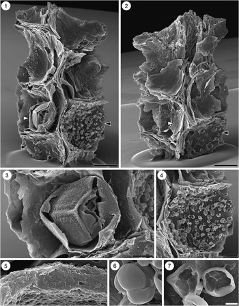
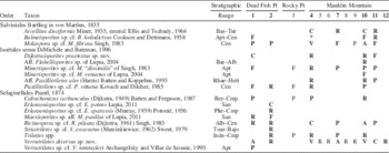
Introduction
Fossil megaspores record the diversity and distribution of heterosporous lycopsids and ferns through time and offer evidence for the origin and diversification of the latter during the later Mesozoic and Cenozoic eras (e.g., Collinson, Reference Collinson1991; Kovach and Batten, Reference Kovach and Batten1993; Collinson et al., Reference Collinson, Smith, Van Konijnenburg-van Cittert, Batten, Van der Burgh, Barke and Marone2013), concurrent with the radiation of flowering plants (e.g., Lidgard and Crane, Reference Lidgard and Crane1988; Heimhofer et al., Reference Heimhofer, Hochuli, Burla and Weissert2007; Friis et al., Reference Friis, Crane and Pedersen2011) and other leptosporangiate ferns (e.g., Schneider et al., Reference Schneider, Schuettpelz, Pryer, Cranfill, Magallón and Lupia2004; Schuettpelz and Pryer, Reference Schuettpelz and Pryer2009). Although of botanical interest, megaspores have not been used widely for biostratigraphic purposes (but see, e.g., Fulgewicz, Reference Fulgewicz1973, Reference Fulgewicz1979, Reference Fulgewicz1980, and Marcinkiewicz, Reference Marcinkiewicz1971, Reference Marcinkiewicz1976, Reference Marcinkiewicz1992, for Triassic and Jurassic usage), in part because of the large volume of sediment required to adequately characterize diversity as compared to much smaller pollen and spores. Nevertheless, Tschudy (Reference Tschudy1976) championed the potential utility of megaspores for biostratigraphic zonation in combination with palynomorphs, if only widespread, routine sampling would be conducted. This is the third in a series of contributions (Lupia, Reference Lupia2004, Reference Lupia2011) aimed at documenting megaspore floras through time along the East and Gulf coasts of North America.
Complete Albian megaspore floras from western and central North America have been described by Singh (Reference Singh1964, Reference Singh1971) from Alberta, Brideaux et al. (Reference Brideaux, Clowser, Copeland, Jeletzky, Norford, Norris, Pedder, Sweet, Thorsteinsson, Uyeno and Wall1976) from the Yukon Territories, Hopkins and Sweet (Reference Hopkins and Sweet1976) from Ontario, and Sweet (Reference Sweet1979) from the Canadian Arctic. Complete Cenomanian megaspore floras from western and central North America have been described by Schemel (Reference Schemel1950) and Hall (Reference Hall1963) from Iowa, Hall and Peake (Reference Hall and Peake1968) from Minnesota, Sweet (Reference Sweet1979) from the Canadian Arctic, Singh (Reference Singh1983) from Alberta, and Kovach and Dilcher (Reference Kovach and Dilcher1988) from Kansas. However, no one has described similarly complete Albian and Cenomanian megaspore floras from the East Coast. Isolated megaspores from the Albian and Cenomanian have been identified based mainly on specimens appearing in palynological preparations from the East and Gulf coasts by Kimyai (Reference Kimyai1966), Paden Phillips and Felix (Reference Paden Phillips and Felix1971), Wolfe and Pakiser (Reference Wolfe and Pakiser1971), and Tschudy (Reference Tschudy1975); and from elsewhere in North America by Potter (Reference Potter1963), Ellis and Tschudy (Reference Ellis and Tschudy1964), Hedlund (Reference Hedlund1966), Agasie (Reference Agasie1969), Playford (Reference Playford1971), Bergad (Reference Bergad1973), Romans (Reference Romans1975), Tschudy (Reference Tschudy1976), Wingate (Reference Wingate1980), Zippi and Bajc (Reference Zippi and Bajc1990), Ravn (Reference Ravn1995), Ravn and Witzke (Reference Ravn and Witzke1995), Scott and Stea (Reference Scott and Stea2002), and Hu et al. (Reference Hu, Dilcher, Jarzen and Taylor2008).
In this paper, I describe a new megaspore flora from 12 samples from three sites in the Potomac Group of Maryland’s Elk Neck Peninsula. Angiosperm remains have been described previously from the Mauldin Mountain locality and include flowers (Mauldinia mirabilis Drinnan, Crane, Friis, and Pedersen, Reference Drinnan, Crane, Friis and Pedersen1990; Spanomera mauldinesis Drinnan, Crane, Friis, and Pedersen, Reference Drinnan, Crane, Friis and Pedersen1991); fruits, seeds, and stamens (Couperites mauldinensis Pedersen, Crane, Drinnan, and Friis, Reference Pedersen, Crane, Drinnan and Friis1991; unnamed taxa illustrated in Crane and Herendeen, Reference Crane and Herendeen1996); and wood (Paraphyllanthoxylon marylandense Herendeen, Reference Herendeen1991). The megaspore flora contains 18 species of megaspores in 14 genera, including two new species. The range of variation in form within the sample population is illustrated to delineate differences in form from established species as appropriate. In addition, a single fragmentary strobilus of selaginellalean affinity containing a megaspore and microspores found among the mesofossils is described.
Materials and Methods
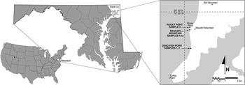
Megaspores were recovered from three sites on the Elk Neck Peninsula in Cecil County, northern Maryland (Fig. 1). Two samples were collected ~1.5 km south of Mauldin Mountain (“Dead Fish Point,” Earleville 7.5’ Quadrangle, 39°28.40’ N latitude, 75°59.67’ W longitude)—Sample #1 from a gray carbonaceous clay layer exposed at low tide, and Sample #2 from a gray carbonaceous clay layer exposed ~1 m above the high tide mark. Sample #3 was collected from a gray carbonaceous clay layer exposed in a slump ~100 m north of Mauldin Mountain, and ~1.2 m above the bay, on Rocky Point Bluff (Earleville 7.5’ Quadrangle, 39°29.32’ N latitude, 75°59.72’ W longitude).
Map of localities on Elk Neck Peninsula in Cecil County, Maryland, USA.

Samples #4–#12 were collected from a thick (~1.5 m), gray, carbonaceous clay layer exposed in a west-facing cliff face, ~20 m above the Chesapeake Bay on Mauldin Mountain, on the west shore of the Elk Neck Peninsula (Earleville 7.5’ Quadrangle, 39°29.24’ N latitude, 75°59.72’ W longitude). Mauldin samples were collected in three stratigraphic sets (4–6, 7–9, 10–12), with each sample separated 20–30 cm vertically, and each set separated laterally 2–5 m across the lens. Additional locality and sample information is available in Supplemental Data 1.
Geologists have designated sediments overlying the crystalline basement in Maryland and Virginia as the Potomac Group and divided it into three formations. The Patuxent Formation is the lowest stratigraphically, followed in succession by the Arundel and Patapsco formations (Wolfe and Pakiser, Reference Wolfe and Pakiser1971; Higgins and Conant, Reference Higgins and Conant1990), although a fourth, higher formation called the Raritan, by correlation with the Raritan Formation in New Jersey, was included in the original description by Clark and Bibbins (Reference Clark and Bibbins1897). In Cecil and neighboring Harford counties, recognition of the Arundel Formation is difficult, and the Patapsco and Raritan formations (as originally defined) show no lithological break, leading to the entire succession being mapped only as the Potomac Group (Owens, Reference Owens1969; Higgins and Conant, Reference Higgins and Conant1990). Potomac Group sediments represent extensive non-marine deposition in channels, floodplains, and swamps along the western margins of the North Atlantic Ocean (Higgins and Conant, Reference Higgins and Conant1990).
Given the non-marine origin of the Potomac Group, most recent work on age determination has relied on palynology. Beginning with Groot and Penny (Reference Groot and Penny1960) and Brenner (Reference Brenner1963), and extended by many others (e.g., Doyle, Reference Doyle1969, Reference Doyle1973, Reference Doyle1992; Wolfe and Pakiser, Reference Wolfe and Pakiser1971; Doyle and Hickey, Reference Doyle and Hickey1976; Doyle and Robbins, Reference Doyle and Robbins1977), these investigations led to a pollen zonation for the Potomac Group. Based on these studies, current zonation and age equivalents are: the Patuxent is lower Zone I (Aptian; Doyle, Reference Doyle1992), the Arundel is upper Zone I (early Albian; Hochuli et al., Reference Hochuli, Heimhofer and Weissert2006; Doyle and Upchurch, Reference Doyle and Upchurch2014), and the Patapsco is Zone II-A to II-B (middle Albian, and middle to late Albian, respectively; Doyle and Upchurch, Reference Doyle and Upchurch2014, contra Hochuli et al., Reference Hochuli, Heimhofer and Weissert2006). Sediments formerly referred to as the Raritan Formation at the top of the Potomac Group in Maryland contain a palynological assemblage distinctly different—notably lacking Normapolles Group pollen (e.g., Wolfe and Pakiser, Reference Wolfe and Pakiser1971; Christopher, Reference Christopher1977)—than that of the type Raritan Formation in New Jersey (Zones IV and V, dated as middle Cenomanian and Turonian, respectively; Christopher, Reference Christopher1977). These Maryland sediments are assigned to Zones II-C and III (late Albian and early Cenomanian, respectively; Doyle and Robbins, Reference Doyle and Robbins1977; Doyle and Upchurch, Reference Doyle and Upchurch2014) and have been treated informally as the “Elk Neck Beds” of the Potomac Group (Wolfe and Pakiser, Reference Wolfe and Pakiser1971). Previously, Owens (Reference Owens1969) described several samples from the Elk Neck Peninsula, including samples NE-1 (near North East, at the north end of the peninsula) and ST-1 (Turkey Point, at the southern tip of the peninsula). NE-1 was assigned to Zone II-B, whereas ST-1 was suggested to be stratigraphically lower. Wolfe and Pakiser (Reference Wolfe and Pakiser1971) described a palynological sample (#11069) containing an angiosperm-dominated flora with polycolpate pollen types from a black clay on Mauldin Mountain in the middle of the peninsula and considered the sample to be early Cenomanian. Doyle (personal communication, cited in Drinnan et al., Reference Drinnan, Crane, Friis and Pedersen1990) assigned a different Mauldin Mountain sample, from which Mauldinia mirabilis was isolated, to lower Zone III (early Cenomanian).
Reserved sediment available for Dead Fish Point Sample #1 was processed using standard techniques (Global Geolabs, Ltd; Alberta, Canada), and examined for palynomorphs. Sample #1 contained rare specimens of Rugubivesiculites reductus Pierce, Reference Pierce1961 and Taurocusporites spackmani Brenner, Reference Brenner1963 that together are diagnostic of upper Zone II-B or younger sediments (Brenner, Reference Brenner1963). Common reticulate monosulcates (e.g., Clavatipollenites minutus Brenner, Reference Brenner1963 and Stellatopollis barghoorni Doyle in Doyle et al., Reference Doyle, Van Campo and Lugardon1975), and abundant, small (mostly 10–15 µm polar diameter), reticulate tricolpates and/or tricolporoidate variants, including Tricolpites albiensis Kemp, Reference Kemp1968 and Tricolpites micromunus (Groot and Penny, Reference Groot and Penny1960) Singh, Reference Singh1971, as well as Striatopollis vermimura (Brenner, Reference Brenner1963) Srivastava, Reference Srivastava1977, are consistent with assignment to upper Zone II-B. Sample #1 did not contain any grains of Rugubivesiculites rugosus Pierce, Reference Pierce1961 nor any psilate and/or triangular tricolpor-(oid)ates that would clearly indicate Zone II-C and/or Zone III. Sample #1 is assigned to upper Zone II-B (middle to late Albian) based on the observed palynoflora.
Reserved sediment available for Mauldin Mountain Sample #4 was also processed (Global Geolabs, Ltd; Alberta, Canada) and examined for palynomorphs. Among other pollen types, Sample #4 contained Rugubivesiculites rugosus Pierce, Reference Pierce1961, Retitrescolpites azemae Ward, Reference Ward1986, and cf. Tricolporopollenites sp. B of Doyle and Robbins (Reference Doyle and Robbins1977), as well as Tricolporopollenites sp. B of Doyle and Robbins (Reference Doyle and Robbins1977), and Tricolporopollenites sp. E of Doyle and Robbins (Reference Doyle and Robbins1977) that are diagnostic of Zone III as defined by Doyle and Robbins (Reference Doyle and Robbins1977). Sample #4 also yielded Striatopollis paranea (Norris, Reference Norris1967) Singh, Reference Singh1971 that is not reported above lower Zone III, and the large polycolpate Stephanocolpites tectorius Hedlund, Reference Hedlund1966 that first appears in upper Zone III (Doyle and Robbins, Reference Doyle and Robbins1977). Thus, Sample #4 from Mauldin Mountain and Samples #5–#12 from the same bed are securely assigned to the Zone III (early Cenomanian) based on the observed pollen flora.
No reserved sediment was available for Dead Fish Point Sample #2. At only ~1 m above Sample #1, its age is most likely to be upper Zone II-B, but could range between upper Zone II-B to Zone III (middle Albian-lower Cenomanian), constrained by the samples as discussed above. Likewise, no reserved sediment was available from the Rocky Point locality. Its age is inferred to be Zone II-B to Zone III (middle Albian-lower Cenomanian) only by geographic and stratigraphic proximity to the other samples.
Bulk samples of 5–15 kg of unconsolidated clays and silts were allowed to dry and then soaked in detergent solution to disaggregate the matrix. Samples #1 and #2 from Dead Fish Point and Samples #4, #7, and #10–12 from Mauldin Mountain were washed through 500 µm and 125 µm sieves; the remainder through 500 µm sieves only. Remaining sediment was removed by dissolution in HCl and HF acids. Organic debris, preserved as charcoal or lignitized material, was rinsed in water and allowed to air-dry. Residues were sorted and megaspores isolated under a dissection microscope at 20–40×magnification. Specimens for further study were mounted on stubs, coated with gold and/or gold-palladium, and examined and photographed using a LEO 1430VP and/or 1450VP scanning electron microscope (SEM). Specimens were measured using the scale from the SEM image; means in parentheses are reported with ranges.
Systematic paleontology
Specimens from Dead Fish Point samples are deposited in the Paleobotany Collection (OPC), Sam Noble Museum, Oklahoma. Specimens from Mauldin Mountain and Rocky Point samples are deposited in the Paleobotany Collection (PP), Department of Geology, The Field Museum, Chicago.
Class Polypodiopsida sensu Smith et al., Reference Smith, Pryer, Schultzpeltz, Schneider, Korall and Wolf2006
Order Salviniales Bartling in von Martius, Reference von Martius1835
Genus Arcellites Miner, Reference Miner1935, emend. Ellis and Tschudy, Reference Ellis and Tschudy1964
Arcellites disciformis Miner, Reference Miner1935, emend. Ellis and Tschudy, Reference Ellis and Tschudy1964
1935 Arcellites disciformis Miner, p. 600, pl. 20, figs. 64–66.
1964 Arcellites disciformis Miner, Reference Miner1935, emend. Ellis and Tschudy, p. 75, pl. 1, figs. 1–12, text-fig. 1.
Description
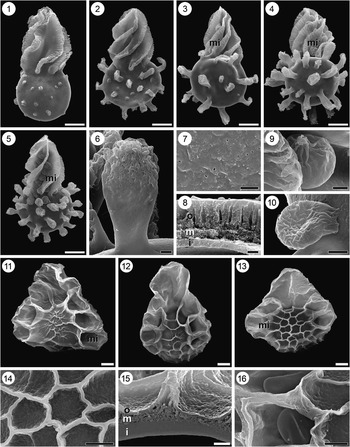
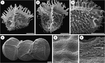
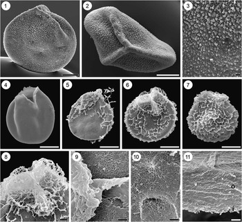
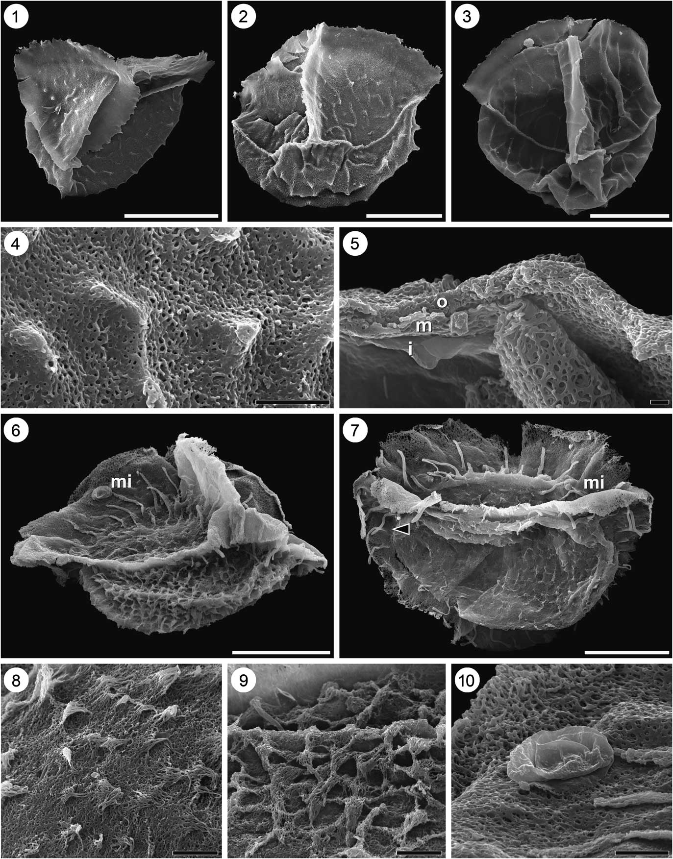
Megaspores are trilete, 360–481 (610) µm long, including acrolamella, with a spherical body that is 160–320 (260) µm (N=42) in diameter (Fig. 2.1–2.5). The body is ornamented with short to long (130 µm maximum) protuberances ranging from small mounds to spatulate appendages with foveolate to reticulate sculpture at tips (Fig. 2.1–2.6). The number of protuberances is 9–28 (20) (N=42). The acrolamella is 200–341 (277) µm long (N=42) and usually composed of six “leaves” that are twisted into a spiral above laesurae and bear distinct crenulations along their edges (Fig. 2.1–2.5). The surface is sparsely punctate (Fig. 2.7). The wall is composed of three layers (Fig. 2.8). The inner layer is ~1.8 µm thick (N=2), densely granulate to nearly homogenous; the middle layer is ~3.7 µm thick (N=2), loosely granulate, expanding and becoming an anastomosing mesh beneath and in hollow appendages and around base of acrolamella; and the outer layer is ~4.9 µm thick (N=2), gradational with underlying middle layer as granules become denser, then homogenous. The outer layer is strongly channeled, with channels appearing as punctae on surface (Fig. 2.8). Microspores found within the leaves of the acrolamella or on the surface of the megaspore are 25.2–41.5 (36.7) µm in diameter (N=15 specimens), with finely granulate (Fig. 2.9) or foveo-rugulate (Fig. 2.10) sculpture distally and acrolamella-like structure proximally.
Arcellites and Balmeisporites megaspores and microspores. (1–10), Arcellites disciformis Miner, Reference Miner1935, emend. Ellis and Tschudy, Reference Ellis and Tschudy1964: (1) megaspore in equatorial view illustrating bumps and underdeveloped appendages, PP55499; (2) megaspore in equatorial view, PP55500; (3) megaspore in equatorial view illustrating mixed bumps and sparse appendages, with microspores (mi) in acrolamella, PP55501; (4) megaspore in equatorial view bearing microspores (mi) in acrolamella, PP55502; (5) megaspore in equatorial view illustrating dense appendages, with microspores (mi) in acrolamella, PP55503; (6) close-up of an appendage illustrating distal perforations, PP55502; (7) close-up of megaspore surface illustrating sparse punctae, PP55502; (8) cross-section through megaspore wall illustrating inner (i), middle (m), and outer layers (o), PP55504; (9) close-up of a microspore in acrolamella illustrating finely granulate surface and twisted acrolamella, PP55505; (10) close-up of a microspore in acrolamella illustrating foveo-rugulate surface and untwisted acrolamella, PP55506; (11–16) Balmeisporites sp. cf. B .holodictyus Cookson and Dettmann, Reference Cookson and Dettmann1958: (11) megaspore in equatorial view illustrating low, incomplete reticulum, with microspore (mi) in wing-like extension, PP55507; (12) megaspore in equatorial view, OPC2054-14833; (13) megaspore in equatorial view illustrating complete reticulum, with microspore (mi) in wing-like extension, PP55508; (14) close-up of megaspore surface illustrating fine perforations within lumina, PP55508; (15) cross-section through megaspore wall illustrating inner (i), middle (m), and outer layers (o), OPC2054-14831; (16) close-up of monolete microspores, PP55508. (1–5) Scale bar=100 µm; (6, 9, 10, 14) scale bar=10 µm; (7, 8, 15) scale bar=2 µm; (11–13) scale bar=20 µm; (16) scale bar=5 µm.

Remarks
These specimens conform in all aspects to the emended diagnosis of Ellis and Tschudy (Reference Ellis and Tschudy1964), and the cross-section of the spore wall as observed with SEM is consistent with the description of A. disciformis given by Hueber (Reference Hueber1982) and Batten et al. (Reference Batten, Dutta and Collinson1996).
The consistent segregation of Arcellites hexapartitus and Arcellites disciformis has proved difficult. The degree of crenulations on the acrolamella (more pronounced in A. disciformis), the size of lumina on spatulate protuberances (irregularly reticulate in A. disciformis), presence/absence of swellings in place of protuberances (few or absent in A. disciformis), and the number of protuberances (fewer in A. disciformis) have been used to distinguish taxa (Batten et al., Reference Batten, Dutta and Collinson1996). Within the Mauldin Mountain population, Arcellites specimens show consistently strong crenulation, but otherwise show variation in development of protuberances, including presence of swellings, and of sizes of lumina on protuberances (Fig. 2.1–2.6). Both the mean number of protuberances and the range of variation observed here, and by Hueber (Reference Hueber1982; 11–26 [16]), for specimens assigned to Arcellites disciformis are lower than those observed from specimens assigned to A. hexapartitus (0–63 [26]) from North East Plaza (Lupia, Reference Lupia2004).
Approximately 62% (N=42) of all A. disciformis megaspores from Mauldin Mountain bear microspores in the folds of their acrolamellae. If found dispersed, the microspores would be assigned to the dispersed genus Crybelosporites Dettmann, Reference Dettmann1963 in agreement with previous reports of Arcellites (e.g., Cookson and Dettmann, Reference Cookson and Dettmann1958; Hueber, Reference Hueber1982; Li and Batten, Reference Li and Batten1986; Lupia, Reference Lupia2004). Unlike previous studies, two forms of the microspores are found on recovered megaspores: a finely granulate form (Fig. 2.9) and a distally foveo-rugulate form (Fig. 2.10). The finely granulate microspores match closely those figured by Hueber (Reference Hueber1982) on Aptian specimens of A. disciformis, whereas the foveo-rugulate microspores are similar to those observed on A. hexapartitus from North East Plaza (Lupia, Reference Lupia2004). Both forms were observed on some specimens, but a gradation in ornament could not be documented. Nevertheless, the finely granulate to foveo-rugulate ornament of A. disciformis microspores distinguishes them from the strongly reticulate microspores within broken specimens of Molaspora sp. cf. M. fibrosa (below) that are also assignable to Crybelosporites.
Genus Balmeisporites Cookson and Dettmann, Reference Cookson and Dettmann1958, emend. Dettmann, Reference Dettmann1995
Balmeisporites sp. cf. B. holodictyus Cookson and Dettmann, Reference Cookson and Dettmann1958
1958 Balmeisporites holodictyus Cookson and Dettmann, p. 42, pl. 2, figs. 1–6.
Description
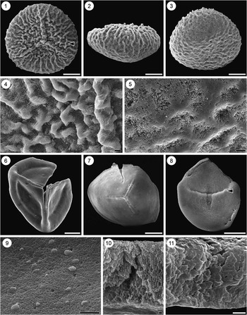
Megaspores trilete, triangular in equatorial outline (Fig. 2.11–2.13), ~164.4–183.0 (172.0) µm tall (N=6), including trifolium, and 108.2–154.4 (138.9) µm (N=3) wide, including “wing-like extensions” sensu Dettmann (Reference Dettmann1995). The trifolium is 51.8–84.9 (67.8) µm (N=3) tall and is composed of three blades fused along its entire height. The megaspore surface is irregularly (Fig. 2.11) to strongly reticulate (Fig. 2.13), with lumina generally larger at the distal pole and near wing-like extensions. The muri vary in height from 2.5 to 8.0 (4.9) µm (N=17), with irregularly reticulate specimens having shorter muri and strongly reticulate specimens having taller muri. Complete muri vary from 0.7 to 1.4 (1.1) µm wide independent of reticulum development (Fig. 2.14). Within the lumina, the megaspore surface is perforate, especially near the bases of the muri (Fig. 2.15). The wing-like extensions are tallest at the equator and confluent with the reticulum (Fig. 2.11–2.13). The wall of the megaspore is composed of three layers (Fig. 2.15). The inner layer is 1.56–1.81 µm thick (N=2), homogenous, appearing granular at high magnification. The middle layer is 0.78–0.93 µm thick (N=2) and composed of anastomosing fibers; beneath muri and the trifolium, this layer thickens (Fig. 2.15). The outer layer is 0.31–0.52 µm thick (N=2) and forms the surface of the grain. Microspores found in the lumina of the wing-like extensions are monolete and psilate to finely granular (Fig. 2.16).
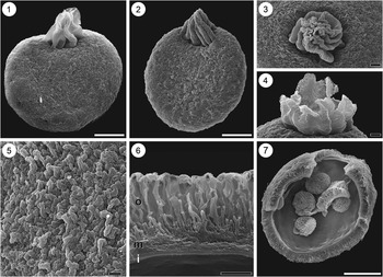
Molaspora sp. cf. M. fibrosa Singh, Reference Singh1983. (1) megaspore in equatorial view illustrating rugulate “knotty” surface, PP55509; (2) megaspore in equatorial view illustrating eight-fold acrolamella, PP55512; (3) megaspore in proximal polar view illustrating twisted, seven-fold acrolamella, PP55510; (4) open, untwisted acrolamella illustrating triangular lobes, PP55511; (5) close-up of megaspore surface illustrating rugulate “knotty” surface without conical projections, PP55512; (6) cross-section of megaspore wall illustrating inner (i) and middle (m) layers, and outer layer (o) without conical projections, PP55513; (7) fractured megaspore containing four reticulate microspores, PP55513. (1, 2, 7) Scale bar=100 µm; (3, 4) scale bar=20 µm; (5, 6) scale bar=10 µm.

Remarks
Attribution of all of the specimens from Mauldin Mountain and Dead Fish Point samples to Balmeisporites sp. cf. B. holodictyus is based on thickness of the megaspore wall, large size of lumina, and tall muri in the strongly reticulate specimens, which are comparable to Cookson and Dettmann’s (Reference Cookson and Dettmann1958) original diagnosis. The specimens described here differ strictly from the Balmeisporites holodictyus in possessing thinner muri compared to original material and SEM illustrations presented in Dettmann (Reference Dettmann1995). Most specimens from Mauldin Mountain and Dead Fish Point that were studied under SEM are strongly reticulate forms (Fig. 2.13).
Genus Molaspora Schemel, Reference Schemel1950, emend. Hall, Reference Hall1963
Molaspora sp. cf. M. fibrosa Singh, Reference Singh1983
1983 Molaspora fibrosa Singh, p. 95, pl. 26, figs. 1–7.
Description
Megaspores trilete, spheroidal (Fig. 3.1, 3.2), and 154–401 (300) µm in diameter (N=14). Acrolamellae are 73.3–144 (115) µm tall and 67.7–157 (114) µm wide (N=14), and composed of six to eight triangular lobes that are preserved pleated and twisted (Fig. 3.1–3.4) or open (Fig. 4.5). The surface of megaspores appears rugulate or “knotted” (Fig. 3.5), without prominent papillae or cones. The megaspore wall is composed of three layers (Fig. 3.6). The inner layer is 0.98–1.66 (1.26) µm thick (N=3) and homogenous. The middle layer (P1 of Batten et al., Reference Batten, Zavattieri and Collinson2011b) is densely fibrous, 5.1–5.9 (5.5) µm thick (N=3), and the outer layer (P2 of Batten et al., Reference Batten, Zavattieri and Collinson2011b) is composed of radially disposed, flattened elements that are 19.9–26.1 (22.2) µm tall. Associated microspores (Fig. 3.7) possess an acrolamella, bear rugulate-reticulate surface sculpture, and are 42.2–71.3 (54.9) µm in diameter (N=8).
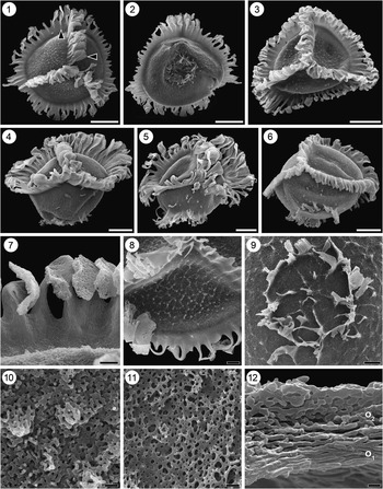
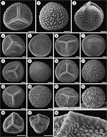
Dijkstraisporites praetextus n. sp. (1) Megaspore in proximal polar view illustrating strong reticulum and lateral expansion of elements of the margo (lower arrowhead) and of the zona (upper arrowhead), OPC2054-14861; (2) same megaspore as in Figure 4.1 in distal polar view illustrating flanges encircling distal pole and edge of auricula (arrow), OPC2054-14861; (3) megaspore in proximal polar view, OPC2054-14859; (4) megaspore in equatorial view illustrating weak reticulum on proximal hemisphere and isolate coni/capilli on distal hemisphere, holotype, OPC2054-14857; (5) megaspore in equatorial view, PP55514; (6) megaspore in equatorial view illustrating isolated coni on proximal hemisphere and short flat elements of zona, PP55515; (7) close-up of elements formed by dissection of the zona, OPC2054-14859; (8) close-up of proximal hemisphere illustrating coni forming reticulum, OPC2054-14858; (9) close-up of distal pole illustrating nearly complete circlet of flanges, OPC2054-14860; (10) close-up of coni on proximal surface of megaspore illustrated in 4.8; (11) close-up of distal surface of holotype megaspore illustrated in 4.4; (12) cross-section of megaspore wall illustrating inner laminar zone (o1) and outer fibrous zone (o2) of outer layer, PP55516. (1–6) Scale bar=100 µm; (7) scale bar=10 µm; (8, 9) scale bar=20 µm; (10–12) scale bar=2 µm.

Remarks
The Elk Neck specimens are similar to Molaspora lobata, but lack the prominent conical/papillate projections of the outermost perine seen in most published images of M. lobata (e.g., Hall, Reference Hall1963; Kovach and Dilcher, Reference Kovach and Dilcher1988; Lupia et al., Reference Lupia, Schneider, Moeser, Pryer and Crane2000; Batten et al., Reference Batten, Zavattieri and Collinson2011b). Instead, specimens appear to possess a dominantly and consistently rugulate outer perine (Fig. 3.1–3.3, 3.5). This sculpture is congruent with the text description of the surface of Molaspora rugosa Schemel, Reference Schemel1950, but the type for Schemel’s species was placed in synonymy with M. lobata by Hall (Reference Hall1963). An attempt to locate and reexamine the type material of M. rugosa reposited at the University of Michigan was unsuccessful. Singh (Reference Singh1983, p. 95) erected the species Molaspora fibrosa for Molaspora specimens from the Cenomanian of Alberta, Canada, with a “finely fibrous perine; sculpture composed of dense fibrous matting.” These Elk Neck specimens are somewhat smaller in all dimensions than Singh’s, but otherwise agree with his circumscription. Because the absence of conical/papillate projections is unlikely to be due to abrasion—conical projections are hollow and abrasion would be expected to show as abundant holes on surface (e.g., Lupia et al., Reference Lupia, Schneider, Moeser, Pryer and Crane2000, fig. 3.20)—or simple compression, as implied by Batten et al. (Reference Batten, Zavattieri and Collinson2011b) for many of their specimens, the greater affinity is considered to fall to Molaspora fibrosa.
Multiple microspores assignable to Crybelosporites were found in two broken specimens of Molaspora sp. cf. M. fibrosa from two Mauldin Mountain samples (Fig. 3.7). If merely a chance association, they could have collected in any one of many dispersed iso-/microspores known from the dispersed palynoflora. However, both specimens show an identical, strongly rugulate-reticulate form with an acrolamella and contain four or more microspores of the same form. This form matches the known microspores of Molaspora lobata established by co-occurrence in sporocarps (Lupia et al., Reference Lupia, Schneider, Moeser, Pryer and Crane2000) and on dispersed megaspores (Batten et al., Reference Batten, Collinson and Brain2011a, Reference Batten, Zavattieri and Collinsonb).
Class Lycopsida sensu DiMichele and Bateman, Reference DiMichele and Bateman1996
Order Isoëtales sensu DiMichele and Bateman, Reference DiMichele and Bateman1996
Genus Dijkstraisporites Potonié, Reference Potonié1956, emend. Batten and Koppelhus, Reference Batten and Koppelhus1993
Remarks
The genera Dijkstraisporites and Tenellisporites (Dijkstra, Reference Dijkstra1949) Potonié, Reference Potonié1956 are very similar in form based on the revised diagnoses of Batten and Koppelhus (Reference Batten and Koppelhus1993, pp. 36–38, quoted below). In equatorial outline, the spore bodies are “circular to convexly triangular” (Dijkstraisporites) or “circular to rounded triangular” (Tenellisporites). The surface may be “smooth-scabrate but usually sculptured with granules, verrucae, or a reticulum” (Dijkstraisporites) or “granules, coni, spines, or a reticulum” (Tenellisporites). The equator of the spore bodies bear “a zona or corona which may be undulose or ribbed,” and “the outer part of the corona may comprise appendages of varying length that are often connected by threads of sporopollenin” (Dijkstraisporites), or “a corona comprising a narrow flange subtended by discrete appendages” (Tenellisporites). The equatorial appendages along the zona/corona of both genera share their morphology with their respective margos. These revised diagnoses do not explicitly capture the discussion by Batten and Koppelhus (Reference Batten and Koppelhus1993, p. 25), in which they distinguish between genera on the basis of the width of the zona/corona relative to the spore body it surrounds, namely that Dijkstraisporites has “a variably developed corona or a zona that is about as wide as, or wider than the radius of the spore.” The informal distinction based on zona/corona width:body radius was used by Batten et al. (Reference Batten, Colin and Néraudeau2010, p. 162) to attribute specimens to genus level. Batten (Reference Batten1995, p. 412) further noted that the zona/corona of Tenellisporites “has moderately to strongly developed, finger-like processes extending from it whereas that surrounding Dijkstraisporites is more irregular in form with processes often slightly expanded at the their tips, and sometimes partly fused to adjacent elements.”
Dijkstraisporites praetextus new species
Figure 4.1–4.12
Types
Holotype: OPC2054-14861 (Fig. 4.1, 4.2), Sample #1, Dead Fish Point (39°28.40’ N, 75°59.67’ W), Elk Neck Peninsula, Cecil County, Maryland, United States, Potomac Group undifferentiated, Pollen Zone II-B (middle-late Albian). Paratypes: OPC2054-14857 (Fig. 4.4, 4.11), OPC2054-14859 (Fig. 4.3, 4.7), OPC2054-14858 (Fig. 4.8, 4.10); OPC2054-14860 (Fig. 4.9), FMNH PP55514 (Fig. 4.5), PP55515 (Fig. 4.6), PP55516 (Fig. 4.12).
Diagnosis
Megaspore trilete, zonate, circular to convexly triangular in polar view; margo above laesura basally continuous, ribbed, becoming dissected at various heights into discrete processes that are expanded perpendicular to margo; zona composed of continuous flange, weakly to strongly ribbed, becoming dissected at varying widths to form discrete processes which may or may not expand towards tips; width of zona greatest where margo and zona meet; proximal hemisphere unornamented or ornamented with scattered coni, dense coni expanded and/or aligned to form reticulum, or reticulum; distal hemisphere ornamented with scattered coni and/or isolated capilli or narrow flanges and/or reticulum; distal pole bears numerous, closely spaced membranous processes, often expanded basally and fused to form continuous or nearly continuous ring.
Occurrence
Elk Neck Peninsula, Cecil County, Maryland, United States, Potomac Group undifferentiated, Zone II-B (middle-late Albian) and Zone III (early Cenomanian).
Description
Megaspore is trilete and the spore body (exclusive of zona) is 183–366 (259) µm wide (N=14) and 209–309 (284) µm tall (N=5), with a circular (Fig. 4.1) to convexly triangular (Fig. 4.3) polar amb. The margo above the laesura is variable in height, 84.8–143 (108) µm tall (N=9), ribbed, entire basally becoming dissected at varying heights into individual elements above the ribs. The elements are expanded perpendicular to margo (Fig. 4.1, bottom arrow), becoming 34.2–82.2 (52.3) µm wide (N=30) and facing the proximal pole (Fig. 4.1, 4.3, 4.4). The margo reaches the equator where it joins the zona (Fig. 4.1, 4.3–4.6). The zona is 31.7–98.9 (71.5) µm wide (N=24) interradially, and 70.2–151 (93.9) µm wide (N=12) radially. The zona is continuous at its base, becoming dissected into separate elements at varying widths (Fig. 4.1–4.3, 4.7, 4.8). The elements of the zona are slightly to greatly expanded at their tips, often perpendicular to zona, especially near apices of laesura (Fig. 4.1, top arrow). Zona is smooth, undulate, or ribbed. Auriculae are not defined. Numerous flanges are concentrated around the distal pole (Fig. 4.2, 4.4–4.6), usually forming a ring of discrete elements or a (nearly) continuous circlet (Fig. 4.9). The surface of the megaspore is unornamented, or ornamented with a weak to strong reticulum (Fig. 4.1–4.4, 4.8), variably connected flanges, and/or isolated coni; some of the coni are elongated as scattered capilli on the proximal and/or distal hemispheres (4.4–4.6, 4.10). The surface of the megaspore is perforate (Fig. 4.9, 4.10). The megaspore wall is composed of two layers (Fig. 4.12). The inner layer (not figured) is thin, homogenous, and detached from the wall or lost in broken specimens. The outer layer is 5.5–6.7 µm thick (N=2) and has two zones. The inner zone appears densely laminar in cross-section transitioning to an outer zone that appears as an open, spongy network.
Etymology
from Latin praetextus, decorated, trimmed. For the dense flanges, often in form of a ring, occupying the distal pole of the megaspore.
Remarks
Specimens assigned to this morphotype resemble both Dijkstraisporites and Tenellisporites in possessing a surface ornamentation composed of a reticulum and/or coni, and a margo and zona dissected at varying depths yielding individuated elements. Above the separation of those elements from each other, no threads connect them. The ratio of the width of the zona (at auriculae) relative to the spore body varies from 0.26–0.44 (0.36) (N=14). Thus, the zona is never as wide as or wider than the spore body, arguing for placement within Tenellisporites. However, this ratio is not part of the emended diagnoses provided by Batten and Koppelhus (Reference Batten and Koppelhus1993). Most specimens show that the margo/zona is undulate or ribbed, and the elements of the margo/zona are expanded at their tips, which are more characteristic of Dijkstraisporites (Batten, Reference Batten1995). In view of all of these features and the current generic diagnoses, the Elk Neck specimens are assigned to Dijkstraisporites.
Elongated flanges concentrated at the distal pole are otherwise known among Cretaceous megaspores only from Paxillitriletes dakotaensis Hall, Reference Hall1963. Specimens herein designated Dijkstraisporites praetextus cannot be assigned to Paxillitriletes Hall and Nicolson, Reference Hall and Nicolson1973 because of the absence of capilli adjacent to the margo (Batten and Koppelhus, Reference Batten and Koppelhus1993). Therefore, the distal polar cluster or ring distinguishes this taxon from the diagnosis of all other named species within Dijkstraisporites (or Tenellisporites).
Examination of the individual populations from Dead Fish Point and Mauldin Mountain reveals morphological differences between them. All Dead Fish Point specimens have proximal hemispheres that are ornamented with a strongly (Fig. 4.1) to weakly (Fig. 4.4) developed reticulate pattern. In contrast, most Mauldin Mountain specimens are unornamented or ornamented with only scattered coni or capilli proximally (Fig. 4.5, 4.6). However, there are specimens from Mauldin Mountain that exhibit an obvious reticulum. Likewise, all observed Dead Fish Point specimens and most Mauldin Mountain specimens possess a ring of discrete or continuous flanges distally (without or with scattered extras, Fig. 4.9), but some Mauldin Mountain specimens bear a distal cluster that does not have the appearance of a ring. Furthermore, the zona in most Dead Fish Point specimens is undulate or ribbed (Fig. 4.1–4.3), whereas it is smooth/flat in many Mauldin Mountain specimens. Thus, there is some evidence to suggest a different taxon in each assemblage. Nevertheless, there is overlap in all features, and determining whether these differences have stratigraphic importance and/or represent different taxa will require larger sample sizes and additional sample sites; for now, D. praetextus is diagnosed to include all variants.
Dijkstraisporites praetextus is extremely similar to?Paxillitriletes sp. of Batten et al. (Reference Batten, Colin and Néraudeau2010, plate VII, figs. 9–11) from the Cenomanian of France, although their specimens have a stronger reticulum on both hemispheres. Tenellisporites brasinoviensis Knobloch, Reference Knobloch1984 is also similar to members of these populations with only coni, and although no distal ring is mentioned in his description, one of the figured specimens (plate IX, fig. 4) from the Senonian of South Bohemia appears to have distal processes. Scott and Stea (Reference Scott and Stea2002) presented one image of a specimen from the Aptian–Albian of Nova Scotia identified as Paxillitriletes in distal polar view that resembles Dijkstraisporites praetextus (their fig. 6a; I note that the megaspore in their fig. 6b is Arcellites sp., which is a salvinialean fern, not Bohemisporites sp., a selaginellalean lycopsid).
Genus Flabellisporites Marcinkiewicz, Reference Marcinkiewicz1978
aff. Flabellisporites sp. of Lupia, Reference Lupia2004
2004 aff. Flabellisporites sp. Lupia, p. 653,655, fig. 4.
Description
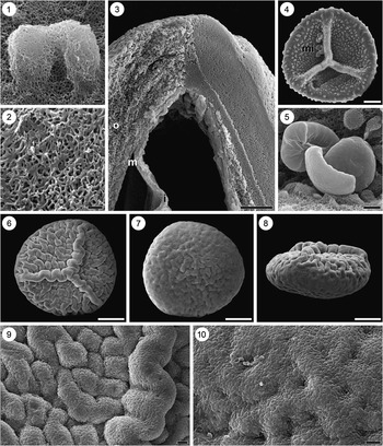
Megaspores are trilete, azonate, auriculate, with a subtriangular outline in polar view (Fig. 5.1, 5.2), 394–436 µm in diameter (N=2). The laesura is bordered by membranous margo, continuous at base but becoming dissected into elongate, flat processes, and up to 125 µm tall including processes (Fig. 5.1, 5.2). The margo over the laesura reaches the equator contacting auriculae that are 92.5–121 (110) µm long and 44.5–95.2 (67.7) µm wide (N=3). Auriculae do not merge laterally with a zona, but instead gradually disappear (Fig. 5.3). Proximal and distal hemispheres are ornamented with a weak reticulum (Fig. 5.1–5.3). Capilli emerging from intersections of muri are tubular with slightly expanded tips (Fig. 5.3). The capilli are longer adjacent to the margo (up to 103 µm long) than interradially or on distal hemisphere (Fig. 5.1). Megaspore surface is perforate between muri.
Aff. Flabellisporites and Minerisporites megaspores. (1–3) Aff. Flabellisporites sp. of Lupia, Reference Lupia2004: (1) megaspore in equatorial view, PP55517; (2) megaspore in oblique proximal polar view, PP55517; (3) close-up of auricula illustrating lateral terminal without formation of a zona, PP55518; (4–6) Minerisporites sp. cf. M. “dissimilis” of Singh, Reference Singh1964: (4) cluster of three megaspores, PP55519; (5) close-up of proximal hemisphere illustrating reticulum formed from low ridges and perforate surface, PP55519, scale bar=10 µm; (6) cross-section of megaspore wall illustrating laminar layer (i) and fibrous layer (o), PP55520, scale bar=2 µm. (1–4) scale bar=100 µm.

Remarks
As noted previously (Lupia, Reference Lupia2004), the absence of a zona excludes this morphotype from common zonate Cretaceous genera, and the restriction of the longest capilli to the area adjacent to the margo preclude it from the genus Flabellisporites Marcinkiewicz, Reference Marcinkiewicz1978. Aff. Flabellisporites sp. of Lupia (Reference Lupia2004) has a wide distribution, being documented from Barremian and/or Aptian sediments in Asia (Li et al., Reference Li, Batten, Zhang and Zhang1987), Europe (Schultz and Noll, Reference Schultz and Noll1987; Nye et al., Reference Nye, Feist-Burkhardt, Horne, Ross and Whittaker2008), and North America (Sweet, Reference Sweet1979; Lupia, Reference Lupia2004), and now from the Cenomanian of North America, but is never so abundant in any sample as to support designation of a new genus.
Genus Minerisporites Potonié, Reference Potonié1956
Minerisporites sp. cf. M. “dissimilis” of Singh, Reference Singh1983
1976 Minerisporites dissimilis Tschudy, p. E6, pl. 2, figs. 1–6 (not).
1983 Minerisporites dissimilis; Singh, p. 107, pl. 35, figs. 4–6.
Description
The megaspores are trilete and zonate (Fig. 5.4), with a round outline in polar view, 479–570 (519) µm in diameter (N=6). The margo is tallest, reaching up to 66.4 µm tall, away from the proximal pole, but without forming a prominent, deep notch at pole (Fig. 5.4). The margo is thicker, 27.4–30.3 µm (N=2), near its base thinning to its apex, 4.4–13.3 (5.3) µm (N=3). The margo reaches equator meeting the zona to form slight auriculae 23.5–48.0 (37.3) µm long (N=5) and 49.9–76.8 (60.8) µm wide (N=3). The zona is 10.2–20.0 (14.6) µm wide (N=3) interradially and 5.7–13.1 (11.1) µm thick (N=4). Proximal and distal hemispheres are ornamented with an obvious, if locally weak, reticulum (Fig. 5.4) that extends onto margo and onto zona. The megaspore surface is perforate at high magnification (Fig. 5.5). The wall is composed of three layers, including a homogenous inner layer (not figured) less than 1 µm thick that is frequently missing in incomplete specimens or separated from the wall in fractured specimens (Fig. 5.6). The middle and outer layers together are ~10.5 µm thick between muri, and ~15.6 µm thick including muri. The middle layer is strongly laminated parallel to the spore surface, grading to a spongier outer layer beneath the surface.
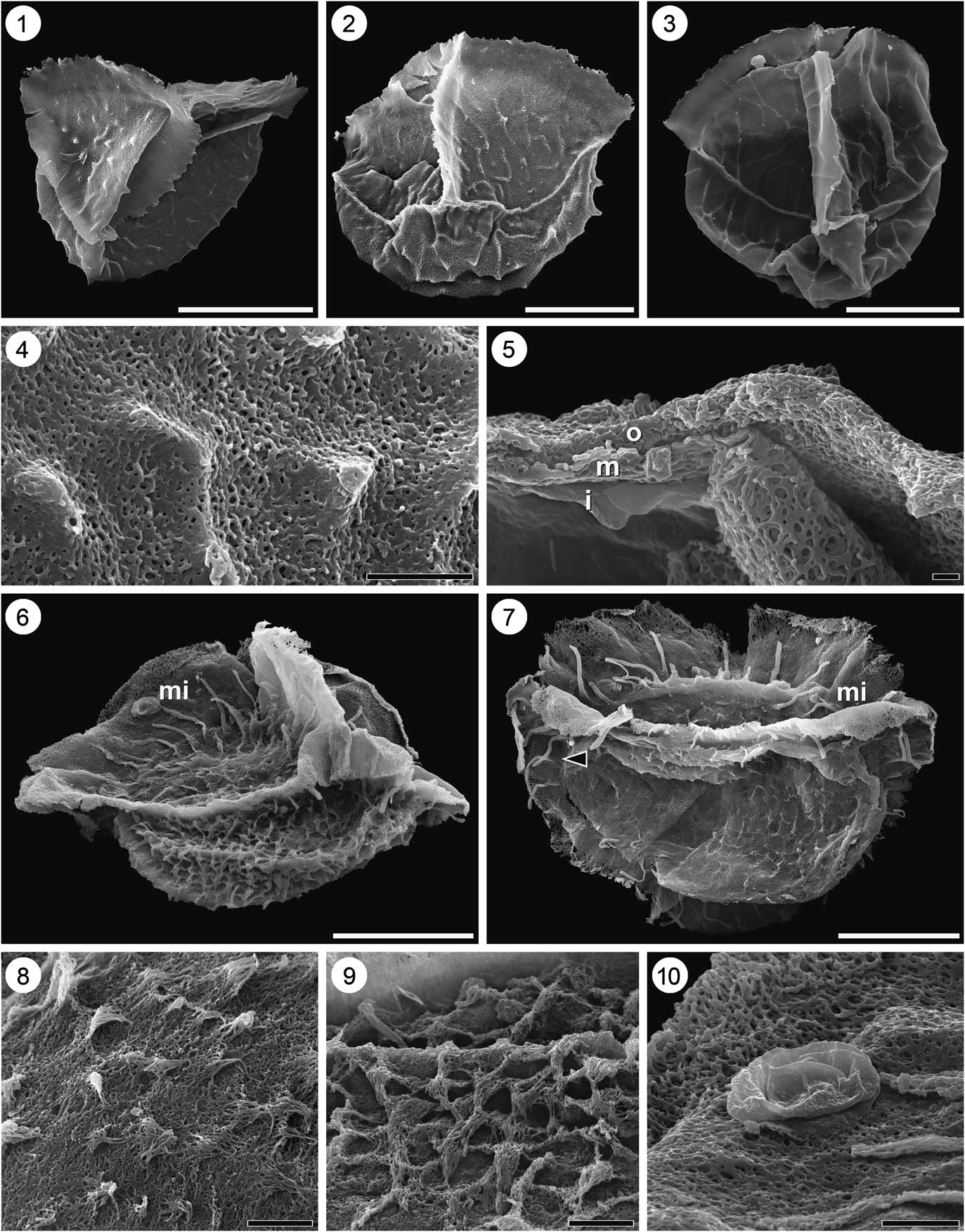
Minerisporites and Paxillitriletes megaspores. (1–5) Minerisporites sp. cf. M. venustus of Lupia, Reference Lupia2004: (1) megaspore in oblique polar view illustrating isolated coni/flanges proximally and weak reticulum distally, PP55521; (2) megaspore in equatorial view illustrating weak incomplete reticulum with prominent coni developed at some junctions, PP55522; (3) megaspore in equatorial view illustrating weak reticulum without prominent junctions, PP55523; (4) close-up of distal hemisphere of megaspore illustrated in 6.1 showing ornament and perforate surface, PP55521; (5) cross-section of megaspore wall illustrating solid inner (i), laminar middle (m), and fibrous outer (o) layers, PP55524. (6–10) Aff. Paxillitriletes ales (Harris, Reference Harris1935) Batten and Koppelhus, Reference Batten and Koppelhus1993: (6) megaspore in oblique polar view illustrating distally perforate, ragged margo, capilli that increase in length toward proximal pole, and microspore (mi), PP55527; (7) megaspore in equatorial view illustrating capillus on distal side of zona (at arrow), and microspores (mi), PP55525, (8) close-up of proximal hemisphere illustrating perforate coni/flanges, PP55526; (9) close-up of distal hemisphere illustrating reticulum with elongated junctions, PP55527; (10) close-up of granulate microspore on megaspore in 6.6. (1–3, 6, 7) scale bar=100 µm; (4, 5, 8–10) scale bar=10 µm.
Remarks
The absence of a deep notch in the margo at the pole and lack of compound reticulum exclude the Elk Neck specimens from Minerisporites dissimilis Tschudy, Reference Tschudy1976 with which they otherwise are very similar, and of similar age. These megaspores most closely resemble Minerisporites “dissimilis” from the Cenomanian illustrated by Singh (Reference Singh1983, pl. 35, figs. 4–6). The Elk Neck specimens are similar in size (although with a thinner wall), with similar uniform low reticulum; likewise, both forms lack a deep polar notch and bear the continuation of the reticulum onto the margo. Singh (Reference Singh1983, p. 107) specifically attributes the absence of compound reticulum to abrasion. Specimens similar to M. “dissimilis” of Singh (Reference Singh1983) were reported as Minerisporites borealis by Gunther and Hills (Reference Gunther and Hills1972, pl. 4, fig. 19) and as Minerisporites sp. 2 by Batten et al. (Reference Batten, Colin and Néraudeau2010, pl. II, figs. 9–11, pl. III, figs. 1, 3). The absence of a deep polar notch and the presence of a low, thin margo exclude the Elk Neck specimens from Minerisporites mirabilissimus (Dijkstra, Reference Dijkstra1961) Potonié Reference Potonié1966. Likewise, the uniform low reticulum and/or relatively thick, narrow zona without strong auriculae exclude the present specimens from M. alius Batten, Reference Batten1969; M. borealis (Miner, Reference Miner1932) Potonié, Reference Potonié1956; M. marginatus (Dijkstra, Reference Dijkstra1951) Potonié, Reference Potonié1956; M. mirabilis (Miner, Reference Miner1935) Potonié, Reference Potonié1956; M. pseudorichardsonii Gunther and Hills, Reference Gunther and Hills1972; and M. venustus Singh, Reference Singh1983, which are of generally similar form or from sediments close in age.
Minerisporites sp. cf. M. venustus Lupia, Reference Lupia2004
2004 Minerisporites sp. cf. M. venustus Lupia, p. 661, fig. 7.
Description
Megaspores are trilete and zonate, with a convexly triangular amb in polar view (Fig. 6.1–6.3) and 207–255 (242) µm in diameter (N=6). The margo is membranous, 22.9–37.0 (29.3) µm tall (N=5), with a ragged edge and extends to the equator where acutely triangular auriculae 24.4–33.9 (29.3) µm long (N=5) and 34.3–49.3 µm wide (N=2) are apparent where it meets the zona (Fig. 6.1–6.3). There is no notch in the margo at its apex (Fig. 6.2). The zona is membranous and smooth to slightly ragged, 5.4–10.1 (7.8) µm wide (N=4) interradially (Fig. 6.1–6.3). The proximal surface may be ornamented with short echinae that are isolated or expanded to form short ridges or with an incomplete to complete weak reticulum with large lumina and slight projections at the intersections of the muri (Fig. 6.1–6.4). The reticulum may extend onto the base of the margo in some specimens (Fig. 6.3). The distal surface may be ornamented with elongate ridges that join to form an incomplete reticulum or with a complete weak reticulum with large lumina and slight projections at intersections of the muri (Fig. 6.1–6.3). The megaspore surface is perforate at high magnification (Fig. 6.4). The wall is composed of three layers (Fig. 6.5), including a homogenous inner layer less than 1 µm thick that is frequently separated from the wall in fractured specimens. The middle and outer layers together are ~3.4 µm thick (N=1). The middle layer is strongly laminated parallel to the spore surface, grading to a more spongy outer layer beneath the surface.
Remarks
The end-members of this morphotype have different ornaments and could be placed in separate genera—Henrisporites for the form characterized by dominantly echinate ornament (Fig. 6.1) and Minerisporites for that characterized by dominantly reticulate ornament (Fig. 6.3). However, intermediate forms combining few echinae with a more or less complete reticulum exist (Fig. 6.2). These intermediate forms with echinae on the proximal hemisphere and reticulum on the distal hemisphere bear some resemblance to Minerisporites absonus Li and Batten in Li et al., Reference Li, Batten, Zhang and Zhang1987, from which they differ because the distal reticulum is not as strong.
Minerisporites sp. aff. M. venustus is very similar both to some specimens of Henrisporites sp. aff. H. angustus of Lupia (Reference Lupia2004, fig. 5d, e) and of Minerisporites sp. cf. M. venustus of Lupia (Reference Lupia2004, fig. 7a, b) from the Aptian of the Elk Neck Peninsula. Lupia (Reference Lupia2004) noted difficulty in separating these two forms with light microscopy, but noted no intermediate forms in that sample population. Because of the sampled intermediate forms, I recognize only one taxon due to the presence of a reticulum, however weak, on most specimens. Nevertheless, given the variation in this population, I consider even an “affinity” to M. venustus unsatisfactory. These Elk Neck specimens are smaller than M. venustus as described by Singh (Reference Singh1983), and Lupia (Reference Lupia2004). Minerisporites sp. aff. M. venustus is easily distinguished from co-occurring M. “dissimilis” of Singh (Reference Singh1983) by its much weaker reticulum with narrower muri and much larger lumina, and by its ragged, membranous margo and zona.
Genus Paxillitriletes
aff. Paxillitriletes ales (Harris, Reference Harris1935) Batten and Koppelhus, Reference Batten and Koppelhus1993
1935 Minerisporites ales Harris, p. 163, pl. 25, figs. 2, 8, 9, 11.
1993 Paxillitriletes ales (Harris, Reference Harris1935) Batten and Koppelhus, p. 38.
Description
The megaspore is trilete with a round to subtriangular polar amb (Fig. 6.6, 6.7). The spore body is 224–271 (251) µm in diameter (N=4). The margo above laesura is tall, up to 83 µm, and unornamented, becoming thinner and perforate/reticulate along margin and slightly ragged (Fig. 6.6, 6.7). Margo reaches equator where it joins zona to form auriculae (Fig. 6.6) that are 68.8–86.0 (75.1) µm long (N=4) and 89.6–168 (132) µm wide (N=4). The zona is slightly ragged and thinnest interradially, 22.4–39.8 (31.8) µm (N=4), becoming gradually wider towards contact with margo. On the proximal hemisphere, some of the ornamentation are echinae, whereas others are elongated in one dimension giving a “fimbriate” appearance (Fig. 6.8). Capilli are always present adjacent to the margo and longest at the proximal pole, up to 76 µm tall and 5.7 µm wide (Fig. 6.6, 6.7). On distal hemisphere, a strong (Fig. 6.6) to weak (Fig. 6.7) reticulum is present. In specimens with a strong reticulum, the intersections of the muri may be elongated into echinae (Fig. 6.9). Capilli are also present on the distal hemisphere immediately adjacent to and on the zona (Fig. 6.7, arrow). The microspores found on the megaspore surface (Fig. 6.10) are monolete with psilate to granulate ornament, and 20.1–23.9 (21.3) µm long (N=5) and 11.8–14.1 (13.1) µm wide (N=5).
Remarks
This morphotype possesses characteristics intermediate between Minerisporites Potonié, Reference Potonié1956, emend. Batten and Koppelhus, Reference Batten and Koppelhus1993 and Paxillitriletes Hall and Nicolson, Reference Hall and Nicolson1973 emend. Batten and Koppelhus, Reference Batten and Koppelhus1993. These specimens fall within the emendation of Minerisporites in possessing an unsegmented margo, a slightly ragged zona wider radially than interradially, auriculae, and reticulate ornamentation distally, but differ in having a echinate/fimbriate sculpture without a reticulum proximally. Capilli adjacent to the margo are permitted within Minerisporites (Batten and Koppelhus, Reference Batten and Koppelhus1993, p. 37–38). Nevertheless, these specimens conform to, and are tentatively considered to have their closest affinity to, Paxillitriletes as emended by Batten and Koppelhus (Reference Batten and Koppelhus1993), primarily based on a margo subtended by hair-like processes longest at the proximal pole, and on the presence of auriculae and reticulate ornamentation distally. A ragged to poorly dissected margo is not excluded from Paxillitriletes (Batten and Koppelhus, Reference Batten and Koppelhus1993, p. 38), and is found in several species (e.g., P. ales (Harris, Reference Harris1935) Batten and Koppelhus, Reference Batten and Koppelhus1993; P. lindstroemi Huckriede, Reference Huckriede1982; P. phyllicus (Murray, Reference Murray1939) Hall and Nicolson, Reference Hall and Nicolson1973 and P. rintoulensis Tosolini in McLoughlin et al., Reference McLoughlin, Tosolini, Nagalingum and Drinnan2002).
The Elk Neck specimens resemble the Jurassic species Paxillitriletes ales in the size of the spore body, albeit at the smaller end of the range given by Harris (Reference Harris1935), in the height of the margo, length of auriculae, and length and width of capilli. Likewise, they both possess a margo that is not deeply incised and characterized by fine perforations, and fine echinae on the distal hemisphere (at least in some specimens). The zona of the Elk Neck specimens are, however, wider than in Harris’ Paxillitriletes ales. The Elk Neck specimens differs from P. ales, and all other species of Paxillitriletes, in bearing long capilli on the distal hemisphere that are restricted to a position immediately adjacent to the zona, and therefore may belong to a new taxon.
Paxillitriletes sp. aff. P. vittatus Kovach and Dilcher, Reference Kovach and Dilcher1985
1985 Paxillitriletes vittatus Kovach and Dilcher, p. 86, 88, pl. 1, figs. 1–8, pl. 2, figs. 1, 2, 4, 5.
Description
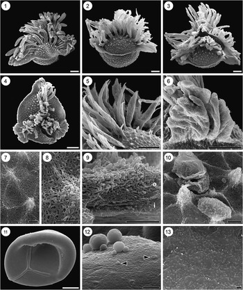
Megaspore is trilete, 314–498 (423) µm in diameter (N=4) exclusive of auriculae, with a round to subtriangular polar view (Fig. 7.1–7.4). The margo above the laesura is tall, 219–287 (252) µm (N=4), unornamented, entire basally becoming dissected into individual narrow to wide, flat to round elements (Fig. 7.1–7.3). The margo is bordered by capilli that are 208–465 (275) µm tall (N=8) and 30.3–74.1 (57.8) µm wide (N=9), and round to flattened in section (Fig. 7.1–7.5). Numerous capilli appear on each contact face, although usually seven or eight are especially prominent (Fig. 7.1, 7.2). The capilli are frequently narrower at the base expanding in diameter upward; some bifurcate, often unequally, ending bluntly or tapering (Fig. 7.5). The margo reaches equator where it joins the zona to form auriculae (Fig. 7.1–7.4, 7.6). The zona is thinnest interradially, 17.7–28.3 (21.9) µm wide (N=4), becoming wider only very close to auriculae. The auriculae are therefore sharply defined at apices of margo, 129–255 (214) µm long (N=6) and 173–208 (192) µm wide (N=9). The auriculae are lobate, with at least three lobes and frequently more (Fig. 7.6). The surface of the megaspore is ornamented on both hemispheres with a well-developed polygonal reticulum (Fig. 7.1–7.4). On both hemispheres, the intersections of the muri are elaborated as short spines. The spines become somewhat larger towards the proximal pole (Fig. 7.6), but show no trend in size on the distal hemisphere. The surfaces of the megaspore within the reticulum and of the spines and capilli are perforate (Fig. 7.7, 7.8). The megaspore wall is composed of three layers. The inner layer (not figured) is less than 1 µm thick, homogenous, and pulled away from the rest of the wall in broken specimens. The middle layer is ~3 µm thick and appears loosely laminar in cross-section, transitioning to an outer layer ~10 µm thick, which appears as a spongy network (Fig. 7.9). Microspores adherent to megaspore among capilli are monolete, 22.8–26.7 (24.3) µm long and 11.8–16.4 (14.6) µm wide (N=4), with rugulate sculpture (Fig. 7.10).
Paxillitriletes and Cabochonicus megaspores. (1–10) Paxillitriletes sp. cf. P. vittatus Kovach and Dilcher, Reference Kovach and Dilcher1988: (1) megaspore in oblique equatorial view illustrating large auriculae, OPC2054-14862; (2) megaspore in oblique equatorial view, PP55528; (3) same megaspore as in 7.2 in oblique proximal polar view; (4) megaspore in proximal polar view illustrating clearly lobate auriculae, PP55529; (5) close-up of proximal hemisphere illustrating robust, sometimes branching (arrow) capilli, PP55530; (6) close-up of lobate auricula, OPC2054-14862; (7) close-up of proximal hemisphere of megaspore in 7.3 illustrating conical elongation of junctions; (8) extreme close-up of surface in 7.7, illustrating perforate surface; (9) cross-section of megaspore wall illustrating inner laminar (i) and outer fibrous (o) layers, PP55530; (10) rugulate monolete microspores adherent to megaspore, OPC2054-14862; (11–13) Cabochonicus carbunculus (Dijkstra, Reference Dijkstra1951) Batten and Ferguson, Reference Batten and Ferguson1987: (11) megaspore in proximal polar view bearing few large gemmae, PP55531; (12) close-up of surface illustrating large gemmae and small Übisch(?) bodies (at arrows), PP55531; (13) close-up of proximal hemisphere illustrating surface with small perforations, PP55531, scale bar=2 µm. (1–6, 10, 11) Scale bar=100 µm; (7–9, 12) scale bar=10 µm.

Remarks
Within the genus Paxillitriletes, to which these Elk Neck specimens may be assigned due to the capilli adjacent to the dissected margo, they are similar, but not identical to, Paxillitriletes vittatus Kovach and Dilcher, Reference Kovach and Dilcher1985. The Elk Neck specimens are slightly smaller, but overlap the range of P. vittatus in diameter of spore body, length of auriculae, and length of capilli, and are slightly larger in width of auriculae and width of zona given by Kovach and Dilcher (Reference Kovach and Dilcher1985). The Elk Neck specimens are similar to P. vittatus in their lobate shape of auriculae, density, and long length of the capilli compared to all other species, and in the weak muri of the distal reticulum giving prominence to short coni where the muri intersect, and differ from P. vittatus in the more prominent proximal reticulum and branching capilli.
Paxillitriletes sp. cf. vittatus is distinct from other Paxillitriletes species found in Cretaceous deposits on the East Coast of North America. It differs from Paxillitriletes sp. of Hueber (Reference Hueber1982) and P. sp. aff. P. fairlightensis (Batten, Reference Batten1969) Hall and Nicolson, Reference Hall and Nicolson1973 in Lupia (Reference Lupia2004) in possessing sharply defined auriculae, weakly serrate zona, thick capilli, and short projections from distal muri. It differs from P. dakotaensis (Hall, Reference Hall1963) Hall and Nicolson, Reference Hall and Nicolson1973 in Tschudy (Reference Tschudy1975) in the absence of distal capilli. It differs from P. alatus (Batten, Reference Batten1969) Hall and Nicolson, Reference Hall and Nicolson1973 in Lupia (Reference Lupia2011) in possessing lobate auriculae relatively narrowly joined to zona, and thick capilli that frequently branch. One specimen from Upatoi Creek (Lupia, Reference Lupia2011, fig. 6.7) had similarly thick capilli but has broadly attached auriculae and longer distal spines.
Order Selaginellales Prantl, 1874
Genus Cabochonicus Batten and Ferguson, Reference Batten and Ferguson1987
Cabochonicus carbunculus (Dijkstra, Reference Dijkstra1949) Batten and Ferguson, Reference Batten and Ferguson1987
1949 Triletes carbunculus Dijkstra, p. 22, pl. 1, fig. 12.
1987 Cabochonicus carbunculus; Batten and Ferguson, p. 72. pl. 1, figs. 1–4, 7–9, pl. 2, figs. 1–8, pl. 3, figs. 1–7, pl. 4, figs. 1–6.
Description
Megaspore is trilete and round in polar view, 393–834 µm diameter (N=2) (Fig. 7.11). The margo is about three-fourths of the radius (Fig. 7.11). Interspersed small to large gemmae (up to 47.1 µm tall and 68.2 µm wide) cover the equator and extend over distal hemisphere; the contact area and distal pole are unornamented (Fig. 7.11, 7.12). Scars and/or undeveloped gemmae appear on the megaspore surface (Fig. 7.12, arrows). The surface appears psilate, but at high magnification is observed to be very finely perforate (Fig. 7.13). The megaspore wall is two-layered, with a very thin, homogenous inner layer, and a thick outer layer that is uniformly composed of dense fibers.
Remarks
The presence of gemmae that have a resinous appearance under reflected light unambiguously identify this taxon. Only one complete, albeit unusually small specimen with small gemmae (Fig. 7.1–7.13), one abraded specimen, and three fragments were recovered.
Genus Erlansonisporites Potonié, Reference Potonié1956
Erlansonisporites sp. cf. E. potens Lupia, Reference Lupia2011
2011 Erlansonisporites potens Lupia, p. 11, fig. 8.
Description
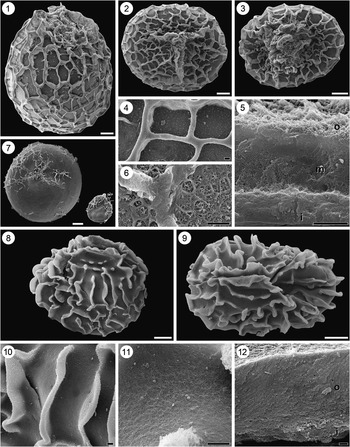
Megaspore is trilete, ovate in equatorial view, and circular to ovate in polar view (Fig. 8.1–8.3). The dimensions are 606–842 (749) µm (long axis, N=8) and 475–706 (607) µm (short axis, N=8) in specimens proximo-distally compressed. The specimens measure 628–753 µm tall (N=2) when equatorially compressed. The margo extends to approximately two-thirds of the radius (Fig. 8.2). In some specimens, the trilete margo is obscured to a lesser (Fig. 8.1) or greater degree (Fig. 8.3) by a dense, spongy network of threads that occupy the area between the arms. The entire surface is ornamented with a reticulum that is complete, except near margo where muri frequently terminate blindly in lumina (Fig. 8.1–8.3). The muri are 2.9–54.0 (23.6) µm tall (N=20) and 2.9–27.0 (11.6) µm wide (N=17). The muri are wider at their top then at their base, smooth, with a blunt, rounded apex (Fig. 8.4). Within the lumina, the surface bears granules or fine echinae (Fig. 8.4). The megaspore wall is composed of three layers (Fig. 8.5). The inner layer is composed of dense, aligned granules ~12 µm thick, transitioning to the middle layer composed of less dense, aligned granules ~15 µm thick; the outer zone forming the surface is composed of an open mesh ~2 µm thick. The muri are composed of dense granules without apparent alignment (Fig. 8.6).
Erlansonisporites megaspores. (1–7) Erlansonisporites sp. cf. E. potens Lupia, Reference Lupia2011: (1) megaspore in equatorial view illustrating margo obscured by fibrous mesh, OPC2101-14844; (2) megaspore in proximal polar view illustrating obvious margo, OPC2101-14840; (3) megaspore in proximal polar view illustrating margo obscured by fibrous mesh, OPC2101-14837; (4) close-up of reticulum on proximal hemisphere illustrating granular ornament in lumina, OPC2101-14841; (5) cross-section of megaspore wall illustrating inner fibrous layer (i), middle layer (m) of aligned granules, and outer mesh (o), OPC2101-14839; (6) close-up of broken muri illustrating granular construction, OPC2101-14838; (7) comparison at same scale of specimens of Erlansonisporites sp. cf. E. potens (OPC2101-14843) and Ricinospora sp. cf. R. pileata (Dijkstra, Reference Dijkstra1961) Singh, Reference Singh1983 (PP55533), both of which have lost their reticulum; (8–12)Erlansonisporites sp. cf. E. sparassis (Murray, Reference Murray1939) Potonié, Reference Potonié1956, specimen OPC2101-14845: (8) megaspore in equatorial view; (9) megaspore in proximal polar view; (10) close-up of ridges on proximal hemisphere illustrating granular edge; (11) close-up of surface between ridges illustrating fine mesh; (12) cross-section of megaspore wall illustrating inner fibrous layer (i) and outer layer (o) of aligned granules. (1–3, 7–9) Scale bar=100 µm; (4–6, 10, 11) scale bar=10 µm; (12) scale bar=2 µm.

Remarks
In almost every feature of ornament, surface, and wall construction, most Elk Neck specimens closely resemble the type material from the Upatoi Creek locality (Lupia, Reference Lupia2011). The Elk Neck population differs from type E. potens primarily in those specimens that have the spongiose network of sporopollenin threads that obscure the trilete form of the margo proper (Fig. 8.1, 8.3). These spongiose threads obscuring the margo (but not necessarily above it) bring those specimens close to the circumscription of the genus Ricinospora Bergad, Reference Bergad1978, emend. Li and Batten in Li et al., Reference Li, Batten, Zhang and Zhang1987. However, the other diagnostic feature of Ricinospora—an easily detachable reticulum—is not observed in most specimens. On most specimens where damage and loss of the muri is evident, a wide, pronounced scar in the former position of the muri are visible (Fig 8.6). Likewise, although not quantified, the margo and spongiose material are as likely to be damaged and/or missing from the same specimen, suggesting that the ornament is not so easily detached. A few Elk Neck specimens do show a secondarily lost reticulum indicated by narrow, faint scars, but the margo and spongiose material are missing as well (Fig. 8.7), whereas the apical ornamentation is the least detachable component of Ricinospora (Li et al., Reference Li, Batten, Zhang and Zhang1987).
Ricinospora pileata (Dijkstra, Reference Dijkstra1961) Singh, Reference Singh1983 is recorded from the Elk Neck samples (see below), but with a diameter over three times smaller (Fig. 8.7). Moreover, even ignoring size, the Elk Neck specimens with missing muri are not conspecific with R. pileata because the former have smooth and blunt muri while the latter have ragged, perforate muri. With relatively few specimens bearing fully obscured margos, and of those, few with missing muri, all of these specimens are assigned to Erlansonisporites sp. cf. E. potens.
Erlansonisporites sp. cf. E. sparassis (Murray, Reference Murray1939) Potonié, Reference Potonié1956
1939 Triletes sparassis Murray, p. 480, 482, text-figs. 3, 4.
1956 Erlansonisporites sparassis (Murray, Reference Murray1939) Potonié, p. 47.
Description
Megaspore is trilete, 599 µm tall, 682 µm wide along long axis and 442 µm (N=1) along short axis of equatorially compressed specimen (Fig. 8.8, 8.9). The margo is ~66 µm tall at proximal pole and extends one-half to two-thirds of the radius (Fig. 8.8). The surface is ornamented with ridges of varying lengths, 15.5–40.0 (27.8) µm tall and 6.7–13.0 (10.4) µm thick, that may join to form a complete lumen (Fig. 8.8, 8.9). The apices of the ridges are ornaments with granules (Fig. 8.10). The surface between the ridges is a finely perforate mesh (Fig. 10.11). The megaspore wall is composed of two layers (Fig. 8.12). The inner layer is composed of dense fibers and is 4–5 µm thick, transitioning abruptly to the outer layer that is ~15–17 µm thick and composed of less dense, aligned granules.
Remarks
The Elk Neck specimens are larger, with a thicker wall, and with a perhaps more distinct margo, but otherwise conform to the circumscription of the Erlansonisporites sparassis. Specimens of E. sparassis can be distinguished from Erlansonisporites sp. cf. E. potens by their taller margo, thicker muri, and rarely complete reticulum.
Genus Maexisporites Potonié, Reference Potonié1956
Maexisporites sp. aff. M. pusillus Lupia, Reference Lupia2011
1988 Maexisporites soldanellus Kovach and Dilcher, p. 92, pl. 1, fig. 1 (part)
2011 Maexisporites pusillus Lupia, p. 15, figs. 4–8.
Description
Megaspore is trilete, azonate, with pyramidal proximal hemisphere and rounded distal hemisphere in equatorial view, ~414 µm tall, and a convexly triangular outline in polar view, ~321 µm in diameter (Fig. 9.1, 9.2). The margo is prominent, ~52 µm tall at proximal pole and ~30 µm wide (Fig. 9.1, 9.2). The margo and surface of both hemispheres are ornamented by verrucae that are more or less isodiametric except where fused. The granules on the proximal hemisphere are wider, 2.9–17.6 (6.0) µm (N=18), and taller on average than on the distal hemisphere, 1.2–7.2 (3.2) µm wide (N=18). At higher magnification, the verrucae are seen to be composed of “piles” of submicron granules, and the surface is a mesh (Fig. 9.3).
Maexisporites and Ricinospora megaspores. (1–3) Maexisporites sp. cf. M. pusillus of Lupia, Reference Lupia2011, specimen OPC2101-14835: (1) megaspore in equatorial view illustrating size difference in granules/verrucae on proximal and distal hemispheres; (2) megaspore in proximal polar view; (3) close-up of surface illustrating change in ornament size across equator. (4–11) Ricinospora sp. cf. R. pileata (Dijkstra, Reference Dijkstra1961) Singh, Reference Singh1983: (4) megaspore in equatorial view illustrating faint scars of completely lost reticulum, PP55532; (5) megaspore in equatorial view illustrating partially lost reticulum, PP55533; (6) megaspore in equatorial view illustrating mostly intact reticulum, PP55534; (7) megaspore in equatorial view illustrating completely intact reticulum, PP55535; (8) close-up of fibrous mesh obscuring margo, PP55534; (9) close-up of reticulum illustrating perforate upper edge, PP55534; (10) close-up of megaspore surface mesh, PP55534; (11) cross-section of megaspore wall illustrating inner fibrous layer (i) and outer layer (o) of aligned granules, PP55536. (1, 2, 4–7) Scale bar=100 µm; (3, 8) scale bar=10 µm; (9–11) scale bar=2 µm.

Remarks
The Elk Neck specimen is larger than the similar form from the Santonian Allon locality (Lupia, Reference Lupia2011), but otherwise matches in possessing granulate verrucae that are larger on the proximal hemisphere than on the distal hemisphere.
Genus Ricinospora Bergad Reference Bergad1978
Ricinospora sp. cf. R. pileata (Dijkstra, Reference Dijkstra1961) Singh, Reference Singh1983
1961 Triletes pileatus Dijkstra, p. 13, pl. 1, figs. 16–19.
1983 Ricinospora pileata; Singh, p. 105, pl. 35, fig. 1.
Description
Megaspore is trilete; most specimens are laterally compressed, yielding an ovate to round equatorial view (Fig. 9.4–9.7). The polar axis is 177–271 (230) µm long (N=8) and the equatorial axis is 152–274 (227) µm long (N=8). The margo is short, about half or less of the radius, and elevated above body of megaspore (Fig. 9.4, 9.6). In most specimens, the margo is covered by spongiose network of fibrous threads (Fig. 9.5–9.8). The surface is covered by a reticulum composed of muri that are solid and smooth basally, becoming perforate and ragged apically (Fig. 9.9). The muri are 7.33–13.2 (10.6) µm tall (N=21) and 0.6–1.4 (1.0) µm thick (N=11). The reticulum is partially missing from most specimens (Fig. 9.5, 9.6) and only occasionally is completely lost (Fig. 9.4) or completely present (Fig. 9.7). At high magnification, the surface is finely perforate (Fig. 9.10). The megaspore wall has two layers (Fig. 9.11). The inner layer is ~2 µm thick and composed of fibers loosely parallel to the spore surface. The outer layer is ~8 µm thick and composed of aligned granules.
Remarks
The Elk Neck specimens conform to the circumscription of Ricinospora pileata given by Dijkstra (Reference Dijkstra1961) and the material illustrated by Singh (Reference Singh1983) except that they are smaller in all dimensions. Specimens of R. pileata can be distinguished from other Elk Neck specimens assignable to Erlansonisporites sp. cf. E. potens and to E. sparassis by their smaller size, and variably complete reticulum of thin, low muri with perforate upper edges.
Genus Striatriletes van der Hammen, 1955 ex Potonié, Reference Potonié1956
Striatriletes sp. cf. S. excavatus (Marcinkiewicz, Reference Marcinkiewicz1962) Sweet, Reference Sweet1979
1962 Erlansonisporites excavatus Marcinkiewicz, p. 476, pl. 11, figs. 3–6.
1979 Striatriletes excavatus; Sweet, p. 14, pl. 1, figs. 5, 6.
Description
Megaspore is trilete, circular in polar view (Fig. 10.1), ~426 µm in diameter. The margo appears sinuous, is ~32 µm tall and 19 µm wide, and extends about two-thirds of the radius of the spore (Fig. 10.1). The proximal hemisphere is ornamented with discrete verrucae and rugulate elements near the proximal pole that are up to 20 µm tall and up to 12 µm wide (Fig. 10.1, 10.2, 10.4). The rugulae are radially aligned starting approximately at the diameter of the arms of the margo and continue across the equator (Fig. 10.1, 10.2). The transition to radial alignment is marked by an incomplete ridge circling the proximal pole (Fig. 10.1, arrow). On the distal hemisphere, the ridges form a tight, regular reticulum with low muri (Fig. 10.3, 10.5). The individual elements on both hemispheres thin from base to apex, but end bluntly, without a sharp edge (Fig. 10.4, 10.5).
Striatriletes and Trileites megaspores. (1–5) Striatriletes sp. cf. S. excavatus (Marcinkiewicz, Reference Marcinkiewicz1962) Sweet, 1978, specimen OPC2101-14848: (1) megaspore in proximal polar view illustrating incomplete transverse ridge (arrowhead); (2) megaspore in equatorial view; (3) megaspore in oblique distal polar view; (4) close-up of proximal hemisphere illustrating rugulae (left) and sinuous margo (right); (5) close-up of distal hemisphere illustrating low reticulate ridges. (6–11) Trileites spp.: (6) megaspore in proximal polar view illustrating sunken contact facets, PP55537; (7) megaspore in proximal polar view, PP55538; (8) megaspore in proximal polar view illustrating equatorial ridge (arrowhead), PP55539; (9) close-up of distal hemisphere of megaspore in 10.7 illustrating finely perforate surface with Übisch bodies; (10) cross-section of wall of megaspore in 10.6 illustrating fibrous construction; (11) cross-section of wall of megaspore in 10.7 illustrating fibrous construction. (1, 2, 6–8) Scale bar=100 µm; (3–5, 9) scale bar=10 µm; (10, 11) scale bar=1 µm.

Remarks
The radially aligned ridges on the proximal hemisphere place this morphotype in Striatriletes, but the fact that the ridges do not extend between the arms of the margo and/or form a reticulum on the proximal hemisphere preclude assignment to Striatriletes areolatus Villar de Seoane and Archangelsky, Reference Villar de Seoane and Archangelsky2008, S. sulcatus (Dijkstra, Reference Dijkstra1961) Potonié, Reference Potonié1956, S. radialis (Gunther and Hills, Reference Gunther and Hills1972) Sweet, Reference Sweet1979, or S. minimus (Dijkstra, Reference Dijkstra1949) Batten, Reference Batten1988 from the Cretaceous. Although Striatriletes excavatus is only known from the Jurassic, the Elk Neck specimens (only two were found) closely approach the form of S. excavatus due to the robust verrucate and rugulate elements between the arms of the margo, well-delimited contact facets, and the incomplete ridge circling the proximal pole.
Striatriletes imperfectus Tosolini in McLoughlin et al., Reference McLoughlin, Tosolini, Nagalingum and Drinnan2002 is similar in possessing proximal verrucae and a delimited contact face, but its ridges and muri are poorly developed, its margo shorter, and its distal pole psilate. Striatriletes sulcatus of Tosolini et al. (Reference Tosolini, McLoughlin and Drinnan2002, fig. 8F, I), of Lupia (Reference Lupia2004, fig. 11.2, 11.3), and of Nye et al. (Reference Nye, Feist-Burkhardt, Horne, Ross and Whittaker2008); Striatriletes n. sp. of Huckriede (Reference Huckriede1982); and S. cf. sulcatus of Batten (Reference Batten1995) are also similar to Elk Neck specimens, but differ in the absence of incomplete ridge delimiting contact face. There are fewer radial ridges in the specimens of Huckriede (Reference Huckriede1982), Lupia (Reference Lupia2004), and Nye et al. (Reference Nye, Feist-Burkhardt, Horne, Ross and Whittaker2008), and the ridges thin to a fine delicate edge in Lupia’s (Reference Lupia2004) specimens.
Genus Trileites Erdtman, Reference Erdtman1947 ex Potonié, Reference Potonié1956
Trileites spp.
Description
Various trilete, psilate, circular to convexly triangular megaspores were encountered (Fig. 10.6–10.8). The diameters vary between 297 and 870 µm (N=5). The margos are low and rounded, extend about one-half to two-thirds of the radius and are set apart from the rest of the proximal hemisphere by a narrow groove all around (Fig. 10.6, 10.7). On some, the contact facets are sunken (Fig. 10.6), on some, undistinguished, and others, delimited by an equatorial ridge (Fig. 10.8, arrow). The surface appears psilate at low magnification, but at higher magnification, the surface has widely spaced to uniform perforations (Fig. 10.9). On some megaspores, scattered solid bodies, less than 5 µm wide, are seen on the distal hemisphere (Fig. 10.9). The walls are of two types, not consistently associated with presence of either equatorial ridge or scattered solid bodies. The thick walls are ~15 µm thick, and composed of narrow diameter fibers to the inside grading to larger diameter fibers on the outside (Fig. 10.10). The thin walls are ~5 µm thick and are composed of uniform fibers (Fig. 10.11).
Remarks
With no prominent ornamentation, differentiation of species assignable to Trileites relies on diameter, length of the margo, presence or absence of rims delimiting the contact facets, and wall thickness/structure (e.g., Kempf, Reference Kempf1972; Batten, Reference Batten1988). The combinations of characters seen here do not accord with any published species. A margo bounded by a groove and contact facets set off by a ridge are characters of Trileites murrayi Harris, Reference Harris1961, but the walls of Elk Neck specimens are always far thinner than 30–50 µm described originally (Harris, Reference Harris1961). Likewise, a margo extending to two-thirds of the radius without contact facets set off by a ridge are characters of Trileites persimilis Harris, Reference Harris1935, but such specimens among the Elk Neck samples are smaller than 600 µm, and have a thinner wall than the 15–20 µm described originally (Harris, Reference Harris1935). Those specimens with scattered solid bodies are reminiscent of Cabochonicus carbunculus (above), but the bodies never have a resinous appearance and never approach the size of gemmae that are normal for C. carbunculus.
Genus Verrutriletes (Dijkstra, Reference Dijkstra1951) Potonié, Reference Potonié1956
Verrutriletes diversus new species
Figures 11, 12.1–12.5
Types
Holotype: FMNH PP55540 (Fig. 11.1, 11.2, 11.18, 12.1, 12.2). Sample #7, Mauldin Mountain (39°29.24’ N, 75°59.72’ W), Elk Neck Peninsula, Cecil County, Maryland, United States, Potomac Group undifferentiated, Pollen Zone III (early Cenomanian). Paratypes: FMNH PP55541 (Fig. 11.3), PP55542 (Fig. 11.4, 11.5, 12.3), PP55543 (Fig. 11.6, 11.7), PP55544 (Fig. 11.8, 11.9), PP55545 (Fig. 11.10, 11.11), PP55546 (Fig. 11.12, 11.13), PP55547 (Fig. 11.14, 11.15), PP55548 (Fig. 11.16, 11.17), PP55549 (Fig. 12.4, 12.5).
Verrutriletes diversus n. sp. (1) Megaspore in proximal polar view, holotype, PP55540; (2) megaspore in distal polar view, holotype, PP55540; (3) megaspore in equatorial view, PP55541; (4) megaspore in proximal polar view, PP55542; (5) megaspore in distal polar view, PP55542; (6) megaspore in proximal polar view, PP55543; (7) megaspore in distal polar view, PP55543; (8) megaspore in proximal polar view, PP55544; (9) megaspore in distal polar view, PP55544; (10) megaspore in proximal polar view, PP55545; (11) megaspore in distal polar view, PP55545; (12) megaspore in proximal polar view, PP55546; (13) megaspore in distal polar view, PP55546; (14) megaspore in proximal polar view, PP55547; (15) megaspore in distal polar view, PP55547; (16) megaspore in proximal polar view, PP55549; (17) megaspore in distal polar view, PP55549; (18) close-up of ornamented margo, holotype, PP55540. (1–17) Scale bar=100 µm; (18) scale bar=10 µm.

Verrutriletes megaspores. (1–5) Verrutriletes diversus n. sp.: (1) close-up of verrucate element illustrating fibrous mesh construction, holotype, PP55540; (2) close-up of fibrous mesh forming megaspore surface, holotype, PP55540; (3) cross-section of megaspore wall illustrating thin inner homogenous (i), thin middle laminar (m) and outer unaligned granular (o) layers, noting the difference in apparent density of outer layer between torn (left) and knife-sectioned sides (right), PP55542; (4) megaspore in proximal polar view with adherent microspores (mi), PP55549; (5) close-up of megaspore in 12.4 illustrating psilate/punctate microspores, PP55549. (6–10) Verrutriletes sp. cf. V. tomtaylori Archangelsky and Villar de Seoane, Reference Archangelsky and Villar de Seoane1990, specimen OPC2101-14846: (6) megaspore in proximal polar view; (7) megaspore in distal polar view; (8) megaspore in equatorial view; (9) close-up of proximal hemisphere illustrating rugulae (left) and sinuous margo (right); (10) close-up of distal hemisphere illustrating low ridges. (1, 2) Scale bar=2 µm; (3, 5, 9, 10) scale bar=10 µm; (4, 6–8) scale bar=100 µm.

Diagnosis
Megaspore trilete, azonate, convexly triangular to circular in polar view; margo above laesura prominent, reaching equator or nearly so, always ornamented at least basally; proximal hemisphere ornamented with low to tall flat-topped verrucae (rarely baculae), often connected to form rugulae or reticula with interspersed verrucae; distal hemisphere bears same ornament as proximal hemisphere but the elements, which may not be as tall, are conspicuously less dense and show greater width or degree of connectedness.
Occurrence
Elk Neck Peninsula, Cecil County, Maryland, United States, Potomac Group undifferentiated, Zone II-B (middle-late Albian) and Zone III (early Cenomanian).
Description
Megaspore is trilete, azonate, convexly triangular to circular in polar view (Fig. 11.1, 11.2, 11.4–11.17), 238–708 (472) µm in diameter (N=22), pyramidal proximal hemisphere and rounded distal hemisphere in equatorial view when not compressed (Fig. 11.3, 11.17). The margo is prominent, 44.8–109 (74.4) µm (N=17) tall and 26.5–46.7 (37.2) µm wide (N=14), reaching equator or nearly so. The proximal hemisphere is ornamented with elements that range from low broad bumps (Fig. 11.4) to isolated verrucae (Fig. 11.6) to wide, broad verrucae or rarely baculae (Fig. 11.6, 11.10) to rugulae (Fig. 11.12) to reticula (Fig. 11.14), with interspersed verrucae. The elements are typically flat-topped (Figs. 11.18, 12.1), and verrucae are found on the margo and closer to proximal pole, whereas rugulae or reticula increase equatorially. Ornament on the distal hemisphere is similar except the elements are less densely arranged and are typically wider or more connected relative to proximal hemisphere (Fig. 11.4–11.15). The proximal ornament is sometimes taller than the distal ornament (Fig. 11.4–11.7), but always less broad/rugulate. The margo always bears ornament, sometimes only at its base (Fig. 11.4), that is similar in form but generally smaller in size than elements on the proximal hemisphere (Fig. 11.18). The verrucae are composed of anastomosing fibers (Fig. 12.1) more dense than the surrounding megaspore surface composed of a perforate mesh (Fig. 12.2). The megaspore wall is composed of three layers (Fig. 12.3). The innermost layer is homogenous, 0.37–0.78 (0.59) µm thick (N=11), while the middle layer is composed of fibers appearing aligned diagonally to spore surface, 0.91–1.69 (1.11) µm thick (N=10). The majority of the wall is formed by the outer layer that is composed of spongy fibers somewhat aligned parallel to the surface, 15.3–27 (23.2) µm thick. Microspores found on specimens (Fig. 12.4) are trilete, triangular, 31.3–36.4 (33.2) µm (N=7), with a psilate to finely pitted surface (Fig. 12.5).
Etymology
from Latin diversus, varied, diverse, several. For the wide variation in observed shape and connectedness of the elements of the ornament.
Remarks
Verrutriletes diversus is the most abundant megaspore type in all but one sample of Mauldin Mountain and Dead Fish Point. This abundance permits the characterization of the wide variance in morphology with the sampled populations. No obvious or consistent differences were observed between the two floras. The holotype (Figs. 11.1, 11.2, 11.18, 12.1, 12.2) represents the form closest to the subjective median morphology—ornament strongly developed but variable, with verrucae and short rugulae relatively dense on proximal hemisphere and wider verrucae and longer rugulae more dispersed over the distal hemisphere.
Verrutriletes diversus resembles V. composipunctatus (the type for genus) in the prominence of the margo, the presence of verrucae and rugulae, and the differentiation of the contact facets from the rest of the spore in terms of the ornament. However, the ornament on the contact facets of Verrutriletes composipunctatus is much reduced in all dimensions relative to rest of the spore, whereas in V. diversus the difference is primarily in spacing and connectedness. In addition, although size may not be reliable in separating species, the mean diameter of Verrutriletes diversus is about 170% of the mean of V. composipunctatus according to dimensions given by Batten (Reference Batten1988). In possessing (mostly) verrucae proximally and rugulae distally, Verrutriletes diversus resembles V. dubius (Dijkstra, Reference Dijkstra1949) Potonié, Reference Potonié1956, V. dasyplocoides Schultz and Noll, Reference Schultz and Noll1987, and Selaginella nehdenensis Huckriede, Reference Huckriede1982, but differs in that the density of the ornament of the latter three are not noted to vary between hemispheres, nor is the margo as prominent in S. nehdenensis. In possessing a verrucate ornament that is more dense proximally than distally, Verrutriletes diversus resembles V. maloninensis Knobloch, Reference Knobloch1984, but differs from the less prominent margo, the absence of rugulae, and the generally coarser/larger verrucae on the latter (Knobloch, Reference Knobloch1984; Batten, Reference Batten2012). Some of the specimens of V. diversus without strong ornament distally (Fig. 11.4–11.7) resemble Hughesisporites Potonié, Reference Potonié1956, but the continuity of form (specifically that there is some ornament distally and it is more elongated/rugulate) argues against recognizing a separate taxon. Hughesisporites sp. of Kovach and Dilcher (Reference Kovach and Dilcher1988) is very similar in size and ornament and may belong to this species. ?Maexisporites sp. of Li and Batten in Li et al. (Reference Li, Batten, Zhang and Zhang1987) is smaller in every dimension, but is similar in ornament.
Verrutriletes sp. cf. V. tomtaylori Archangelsky and Villar de Seoane, Reference Archangelsky and Villar de Seoane1990
1990 Verrutriletes tomtaylori Archangelsky and Villar de Seoane, p. 421, pl. 1, figs. 1–9; pl. 2, figs. 1–5; pl. 3, figs. 1–3.
Description
Megaspore is trilete, convexly triangular in polar view (Fig. 12.6, 12.7), with a diameter of 385–426 µm (N=2). The margo is sinuous, reaching the equator (Fig. 12.6), up to 39.5 µm wide and tallest, 35.5–48.1 µm (N=2), at the proximal pole (Fig. 12.8). The proximal hemisphere is ornamented with very broad, rounded verrucae to rugulae (Fig. 12.6, 12.9), ~20 µm tall and 25 µm wide, appearing somewhat disposed to radial alignment. The distal hemisphere is weakly undulate to dimpled (Fig. 12.7, 12.10). At high magnification, the surface is composed of a perforated fibrous network (Fig. 12.9, 12.10).
Remarks
The two recovered Elk Neck specimens are most similar to Verrutriletes tomtaylori in size and ornament, including vague alignment of elements (see Archangelsky and Villar de Seoane, Reference Archangelsky and Villar de Seoane1990, pl. 1, fig. 3), differing only in possessing elements that include rugulae. In giving the appearance of radial alignment of elements near the equator, these specimens approach the circumscription of Striatriletes, but elongate ridges never develop from the discontinuous elements. Verrutriletes depressus Tosolini et al., Reference Tosolini, McLoughlin and Drinnan2002 is similar, but differs in weaker ornament and in being diagnosed with sunken contact facets. In view of some specimens of Verrutriletes diversus (e.g., Fig. 11.16, 11.17), I do not regard sunken contact facets as a species character, but a preservational state, and suggest that Verrutriletes depressus should be considered a preservational state of and synonymous with V. tomtaylori unless the ultrastructure is shown to be different. Hughesisporites dettmanniae Tosolini et al., Reference Tosolini, McLoughlin and Drinnan2002 is also similar, but differs in possessing a shorter margo and a psilate, scabrate, or verrucate distal hemisphere.
Selaginellalean strobilus
Figure 13.1–13.7
Description
The fragment of the strobilus was 1.77 mm tall and 1.04 mm wide. Strobilus was compressed with two columns of 2–4 incomplete sporangia on each side (Fig. 13.1, 13.2). The fragment did not preserve either the base or apex of the strobilus. A fractured in-situ megaspore (Fig. 13.3) and in-situ microspores (Fig. 13.4) indicated that the strobilus was not unisexual. The position of the nearly complete megaspore in the same column above a microsporangium indicated that megasporangia were not exclusively basal, and that at least one column bore mixed sporangia (Fig. 13.1). A fragment of another megaspore wall in another sporangium (Fig. 13.2, arrow) indicated that at least two columns bore megasporangia. The megaspore was trilete, verrucate (Fig. 13.1), and ~350 µm in diameter. The outer megaspore wall was ~5 µm thick and composed of dense fibers (Fig. 13.5). The microspores were trilete, psilate to granulate (Fig. 13.4, 13.6, 13.7), and 25.5–30.4 (27.6) µm (N=16) in diameter.
Selaginellalean strobilus, specimen PP55550. (1) Side of strobilus illustrating two columns of fractured sporangia, one with a megaspore (white arrowhead) and two with microspores (black arrowheads); (2) reverse side of strobilus illustrating two columns of fractured sporangia, one with a megaspore fragment (white arrowhead) and one with microspores (black arrowhead); (3) close-up of verrucate megaspore; (4) close-up of fractured microsporangium; (5) cross-section of the wall of megaspore illustrated in 13.3; (6) psilate microspore tetrad; (7) close-up of fractured microspores illustrating trilete laesurae. (1, 2) Scale bar=250 µm; (3, 4) scale bar=100 µm; (5) scale bar=1 µm; (6, 7) scale bar=10 µm.

Remarks
This specimen was accidentally shattered while manipulating an adjacent specimen during study after only preliminary imaging was completed. With only fragments remaining and no additional specimens, it is not appropriate to erect a new taxon. The surface morphology of in-situ megaspore (Fig. 13.5) is similar to the dispersed Verrutriletes diversus, but the outer fibrous layer of the wall is thinner than the similar layer of V. diversus (Fig. 12.3). A smooth inner body is preserved inside the megaspore wall; its thick homogenous wall is likewise unlike the inner wall of V. diversus (Fig. 12.3). The in-situ microspores (Fig. 13.4, 13.6, 13.7) differ from those preserved on the dispersed V. diversus (Fig. 12.4, 12.5) in being smaller and mostly psilate, occasionally granulate, never punctate. Whether the differences in spore morphologies reflect different taxa, or just the effects of incomplete or aborted development is unknown. The strobilus was recovered from Mauldin Mountain Sample #10 (early Cenomanian).
Discussion
Age
The majority of the species to which the recovered megaspores are referred are solidly Cretaceous, and largely middle Cretaceous in age (Aptian through Cenomanian), notwithstanding those with morphological affinities to long-ranging or Jurassic forms (Table 1). However, resolution to middle-to-late Albian for Dead Fish Point samples, or to the early Cenomanian for Mauldin Mountain samples, as permitted by the dispersed pollen and microspore assemblages, is not strongly supported.
Stratigraphic ranges and abundance of megaspore taxa by sample at three sites. Stratigraphic ranges from Batten and Kovach (Reference Batten and Kovach1990), with updates exclusive of this study, assuming taxonomic assignment is certain (i.e., ignoring cf./aff.). Samples in bold indicate sampling with 125 µm and 500 µm sieves; otherwise, only 500 µm was used. Abundance based on minimum number of individuals in samples with fragments.

Abbreviations: for Triassic, Indu=Induan; Rhae=Rhaetian; for Jurassic, Hett=Hettangian; Plie=Pliensbachian; Toar=Toarcian; Bajo=Bajocian; for Cretaceous, Ber=Berriasian; Bar=Barremian; Apt=Aptian; Alb=Albian; Cen=Cenomanian; Tur=Turonian; San=Santonian, Cmp=Campanian.
P(resent)=1; R(are)=2-5; F(requent)=6-20; C(ommon)=21-50; A(bundant)=51-100; V(ery abundant)=more than 100
* Present only in palynological preparation
In the Dead Fish Point floras, no taxa were recovered that are uniquely Albian in age. Only four species (B. holodictyus and C. carbunculus in Sample #1, Erlansonisporites cf. E. sparassis in Sample #2, Ricinospora cf. R. pileata in both) are known from and/or range through the Albian globally (Table 1; Batten and Kovach, Reference Batten and Kovach1990). Verrutriletes cf. V. tomtaylori from Sample #1 is otherwise known only from the Aptian of Argentina (Archangelsky and Villar de Seoane, Reference Archangelsky and Villar de Seoane1990; Cesári et al., Reference Césari, Limarino, Llorens, Passalia, Loinaze and Vera2011). Sample #1 shares Minerisporites sp. cf. M. “dissimilis” of Singh, Reference Singh1983 with the Cenomanian of Alberta, Canada (Singh, Reference Singh1983), and both samples share Paxillitriletes cf. P. vittatus with the early Cenomanian of Kansas (Kovach and Dilcher, Reference Kovach and Dilcher1985), and Molaspora fibrosa with the middle Cenomanian (Singh, Reference Singh1983). Sample #2 shares Erlansonisporites cf. E. potens, and both samples share Maexisporites cf. M. pusillus of Lupia, Reference Lupia2011 with younger but geographically closer Santonian floras from Eutaw-equivalent sediments in Georgia (Lupia, Reference Lupia2011). Neither sample from Dead Fish Point produced specimens of Arcellites, although Arcellites is present and abundant in Aptian (Hueber, Reference Hueber1982; Lupia, Reference Lupia2004) and Cenomanian (herein) sediments in the Potomac Group. Overall, there is no clear corroboration of the middle–late Albian age determination for Sample #1 from palynomorphs. Likewise, Sample #2 shares exclusively one taxon with Sample #1 and one taxon with Mauldin Mountain samples; thus, megaspores offer no further resolution to its age.
In contrast, eight of the 13 species from Mauldin Mountain have stratigraphic ranges that include the Cenomanian. Minerisporites sp. cf. M. “dissimilis” of Singh, Reference Singh1983; Paxillitriletes cf. P. vittatus; and Molaspora fibrosa; previously known only from the Cenomanian, are also found at the middle–late Albian Dead Fish Point. The Mauldin Mountain flora shares Arcellites, Cabochonicus, Flabellisporites, and Ricinospora with older Aptian floras from the Potomac Group, but lacks Bohemisporites (Hueber, Reference Hueber1982; Lupia, Reference Lupia2004). Reworking could possibly explain the exclusive occurrence of older taxa at Mauldin Mountain, but the megaspores are for the most part well preserved. The Rocky Point flora contains only four taxa—all of which occur in both Dead Fish Point and Mauldin Mountain floras. Therefore, its age, assigned to a middle Albian–early Cenomanian range based on proximity and position relative to the other two sites, cannot be improved further by comparisons based on megaspores.
These floras contain the mix of taxa one might expect of middle–late Albian and early Cenomanian floras in this region, given previous sampling of Aptian and Santonian sediments. Absent from these Maryland floras are specimens assignable to the genus Ariadnaesporites Potonié, Reference Potonié1956 emend. Tschudy, Reference Tschudy1966, which was reported from the Woodbury Clay Member of the Raritan Formation (middle Cenomanian, Christopher, 1979) of New Jersey by Kimyai (Reference Kimyai1966). Species of Ariadnaesporites are common elements of Cenomanian and younger megaspore floras throughout North America (Batten and Kovach, Reference Batten and Kovach1990), but also have been reported from late Albian to early Cenomanian sediments from Canada (Sweet, Reference Sweet1979; Singh, Reference Singh1983). Ariadnaesporites may therefore be a good marker for post-Zone III sediments in the Potomac Group. Sweet (Reference Sweet1979, p. 14) suggested that the number of appendages and the relative coarseness or fineness of perforations of the surface of Arcellites disciformis may be correlative with age. Sweet’s review of Singh (Reference Singh1964, Reference Singh1971) and his sampling of Aptian through Cenomanian strata suggested that numerous appendages and fine perforations characterize Aptian through early Albian samples, whereas fewer appendages and coarser perforations characterize middle to late Albian and Cenomanian samples. Allowing that Arcellites disciformis and A. hexapartitus may be conflated, then A. hexapartitus from the Aptian of Maryland (Lupia, Reference Lupia2004) and A. disciformis from the Cenomanian of Maryland (above) follow this pattern, but A. disciformis in Hueber (Reference Hueber1982) from the Aptian of Virginia does not (i.e., fewer appendages).
Paleoecology and taphonomy
Extant heterosporous lycopsids (Isoëtales sensu DiMichele and Bateman, Reference DiMichele and Bateman1996 and Selaginellales Prantl, 1805) and heterosporous ferns (Salviniales Bartling in von Martius, Reference von Martius1835) are aquatic to semi-aquatic plants (Tryon and Tryon, Reference Tryon and Tryon1982), although great ecological diversity is observed among extant Selaginella Palisot de Beauvois, Reference Palisot de Beauvois1805 including epiphytism (Jermy, Reference Jermy1990) and the iterative evolution of xerophytism (Korall and Kenrick, Reference Korall and Kenrick2002). The presence of both clades is consistent with at least seasonal availability of standing water at Mauldin Mountain and Dead Fish Point, but detailed information on the sedimentology and depositional environment for these sites was not collected due to very limited outcrop exposure. The absence of Arcellites (with affinities to extant Marsileaceae Mirbel, 1803: Cookson and Dettmann, Reference Cookson and Dettmann1958; Ellis and Tschudy, Reference Ellis and Tschudy1964) and the presence of Erlansonisporites (with affinities to extant Selaginella: Korall and Taylor, Reference Korall and Taylor2006)—in Dead Fish Point Sample #2 contrast with the presence of the former and absence of the latter in Mauldin Mountain samples, and might suggest a more subtle difference in moisture availability between sites.
Lycopsids are more diverse than heterosporous ferns at all localities and in each individual sample except Sample #6 (Table 1). The greater diversity of lycopsids relative to ferns in these floras is consistent with global diversity patterns of heterosporous plants for the Albian–Cenomanian interval (Kovach and Batten, Reference Kovach and Batten1993). Lycopsids are also more abundant than ferns in all but Sample #1 from Mauldin Mountain (Table 1). Sweet (Reference Sweet1979) proposed that samples containing well-preserved megaspores dominated by a few species are local, but that highly diverse samples are the product of the mixing of extralocal taxa into the assemblage. Therefore, diversity and abundance may reflect taphonomic as much as ecological processes. The diversity and abundance of megaspores at Mauldin Mountain in Table 1 illustrate variation among closely spaced samples. However, the consistency in composition and their ranked abundance among Mauldin Mountain samples (those sieved with 125 µm seive) even as overall diversity and abundance decreases (Table 1) is an argument for an underlying ecological signal about the regional pool, even if absolute numbers vary. The one exception, Minerisporites cf. M. “dissimilis” of Singh, Reference Singh1983, in which abundance class increases from “present” in Sample #10 to “rare” in #4 as diversity drops, is due to only a single specimen difference. In contrast, at Dead Fish Point, the differences in composition and abundance between Samples #1 and #2, separated vertically by ~1 m, would argue for deposition in different events or from different sources (or possibly age). Thus, heterogeneity in recovered floras is observed across very short horizontal and vertical distances, attesting to the imperative for close sampling in large volumes to document megaspore diversity.
Conclusions
Sieving of middle–late Albian to early Cenomanian sediments along the Atlantic Coastal Plain of North America has yielded abundant and diverse megaspore floras containing 14 genera and 18 species. These floras show a markedly greater diversity of lycopsid megaspores relative to ferns, consistent with both previous local studies for the Aptian and Santonian (Hueber, Reference Hueber1982; Lupia, Reference Lupia2004, Reference Lupia2011) and globally (Kovach and Batten, Reference Kovach and Batten1993). However, the relative abundance of lycopsid and fern megaspores is not always congruent with their diversities (Hueber, Reference Hueber1982; Lupia, Reference Lupia2004, Reference Lupia2011). These Elk Neck samples help fill a gap between previously documented Aptian (Hueber, Reference Hueber1982; Lupia, Reference Lupia2004) and Santonian (Lupia, Reference Lupia2011) samples. However, current sampling remains insufficient to provide a robust biostratigraphic schema for the Cretaceous of the Atlantic Coastal Plain of North America. Developing a workable biostratigraphic schema employing megaspores will require considerable sampling effort to overcome natural, taphonomic, and spatial heterogeneity.
Acknowledgments
I thank P. R. Crane, P. S. Herendeen, and S. Magallón for assistance in collecting the samples. Reviews by S. Hu and an anonymous reviewer are greatly appreciated. This work was supported by NSF grants DEB-008908 and DBI-010156.
Accessibility of Supplemental Data
Supplemental data deposited in Dryad data package: http://dx.doi.org/10.5061/dryad.3n216