There has been considerable interest in facilitating electron transfer between electrodes and microorganisms to create bioelectrochemical systems. Such electron transfer can be used to break down organic matter in streams such as waste water to generate power,[Reference Logan, Wallack, Kim, He, Feng and Saikaly1,Reference Venkata Mohan, Velvizhi, Annie Modestra and Srikanth2] to synthesize biologic molecules from inorganic carbon,[Reference Tremblay and Zhang3] and to transmit signals between microorganisms and electrodes, both for sensing and for triggering cell responses.[Reference Fraiwan and Choi4,Reference Tschirhart, Kim, McKay, Ueda, Wu, Pottash, Zargar, Negrete, Shiloach, Payne and Bentley5] While some organisms have intrinsic electron transfer capabilities, many organisms have only limited capabilities and, in general, intrinsic electron transfer rates are slow, which have led to employing material science and chemistry approaches to alter either microorganisms or electrode properties to enhance this transfer. Chemical approaches include the addition of membrane-permeabilizing stains[Reference Jahnke, Bazan and Sumner6,Reference Du, Catania and Bazan7] and the use of soluble redox mediators.[Reference Sund, McMasters, Crittenden, Harrell and Sumner8] The electrode can also be modified to promote charge transfer, either by attaching functional groups[Reference Popov, Kim, Dinsdale, Esteves, Guwy and Premier9] or by structuring it through the immobilization of, for example, nanoparticles on its surface.[Reference Fan, Xu, Schaller, Jiao, Chaplen and Liu10,Reference Xu, Liu, Fan, Schaller, Jiao and Chaplen11] Nanoparticles can also be attached to the microbes rather than the electrode.[Reference Chen, Zhou, Liu, Zeng, Zhang, Ye and Zhou12,Reference Jiang, Hu, Lieber, Jackan, Biffinger, Fitzgerald, Ringeisen and Lieber13] High enough densities of nanoparticles may provide conduction paths for cells distant from the electrodes, but even lower densities of nanoparticles can serve to permeabilize the cell membrane, and the release of additional redox active molecules promotes charge transfer.[Reference Catania, Ajo-Franklin and Bazan14]
Nanoparticle interactions can be controlled in many ways. Many nanoparticle functionalizations exist to have either non-specific (e.g., electrostatic)[Reference Jahnke, Cornejo, Sumner, Schuler, Atanassov and Ista15] or specific interactions (e.g., ligand binding).[Reference Saha, Agasti, Kim, Li and Rotello16] Alternatively, the microbe can be engineered to controllably display moieties to bind the nanoparticles. A number of intrinsic mechanisms can be modified to control adhesion, including membrane-embedded proteins, appendages (e.g., fimbriae and flagella), and extracellular polysaccharide scaffolds.[Reference Petrova and Sauer17] A large number of material-binding peptides are known from nature, peptide libraries, and rational design, and have been shown to have affinities for many types of nanoparticles.[Reference Naik, Stringer, Agarwal, Jones and Stone18] By engineering the microbe to bind nanoparticles, it is possible to take advantage of the sensing and genetic logic that can be programmed into microbes, and the combination with bioelectrochemical systems could provide new ways to interface biology and devices.
Recently, we have shown that the membrane protein eCPX could be engineered to display peptides to bind gold nanoparticles,[Reference Dong, Sarkes, Rice, Hurley, Fu and Stratis-Cullum19] and here, we show that Escherichia coli that have bound nanoparticles through peptides displayed via the enhanced circularly permuted OmpX (eCPX) scaffold have improved electron transfer in microbial fuel cells (MFCs), a common bioelectrochemical system that oxidizes organic matter to generate electrical power. To attach the gold nanoparticles to the bacteria, we examine two peptides. The first was one of the best gold-binding peptides from our previous study and had six methionines and nine glycines (M6G9; MMMGGGMGGGMGGGM). The second peptide is an extension of this peptide, having more methiones (9) to increase binding and more glycines to maintain chain flexibility (18); it is referred to as M9G18 (MMMGGGMGGGMGGGMGGGMGGGMGGGM). When 5 nm gold particles are added to cells displaying either a gold-binding peptide, an enhanced current is observed, but no enhancement is observed when gold nanoparticles are added to cells not displaying a peptide, or if larger (20 nm) nanoparticles are used. SEM images show little difference in cell attachment to the electrodes before and after the run. TEM images show nanoparticles bound to cells under similar conditions as the MFC experiments and that M9G18 binds gold nanoparticles more effectively than M6G9, consistent with the larger MFC current enhancement with the M9G18.
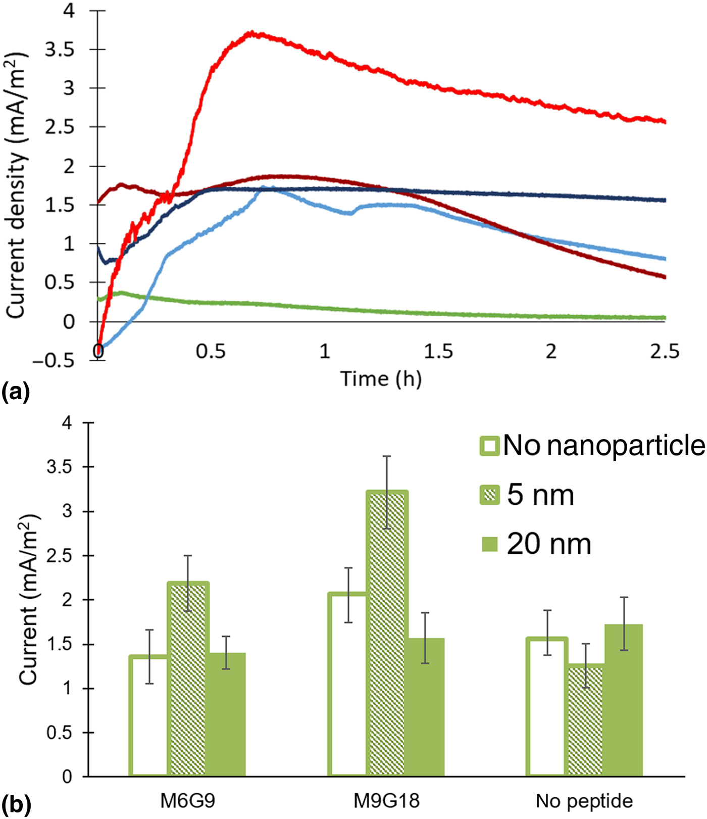
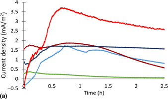
Cells displaying two methionine-based gold-binding peptides were characterized into MFCs to understand whether gold-binding peptides can enhance MFC currents by directing gold nanoparticles to the cell surface. The MFC design is based on a common literature design,[Reference Hou, Chen, Thomas, Catania, Kirchhofer, Garner, Han and Bazan20,Reference Hou, Li, Cho, de Figueiredo and Han21] using two 120 µL chambers separated by a Nafion membrane; the anode chamber was filled with a high OD culture while the cathode was filled with a potassium ferricyanide solution. Both peptides were under inducible control (induced by l-arabinose) and furthermore expressed at similar levels on the cell surface of E. coli upon induction for 1 h under shaking at 250 RPM at 37 °C (Supplementary Fig. S1). After induction, nanoparticles were added to the culture to achieve a similar mass of nanoparticles added across different sizes; this resulted in a 10,000:1 nanoparticle to cell ratio for 5 nm particles and a 150:1 nanoparticle to cell ratio for 20 nm particles. The effects of combining nanoparticles with gold-binding peptide expression on the currents in MFCs are shown in Fig. 1. Figure 1(a) shows representative traces from uninduced cells with and without 5 nm gold nanoparticles and cells displaying the M6G9 peptide with and without 5 nm gold nanoparticles. A run with heat-killed cells is also included to show the abiotic response of the system. Within 1–2 h, the fuel cells reach a maximum current and then slowly decline in current because of the high initial cell concentration and small (120 µL) MFC compartment volumes. While the addition of nanoparticles produced a higher current when the cells are displaying a gold-binding peptide, no effect is observed when no peptide is displayed.
(a) Representative traces of MFCs running with E. coli displaying the M9G18 (red) and with no peptide displayed (blue). A run with heat-killed cells is shown in green. Darker shades show runs without nanoparticles added, while lighter shades show runs with 5 nm particles added. (b) A comparison between the peak currents of fuel cells run with different peptides and nanoparticle conditions. The error bars show the standard error (N > 4). Five nanometer particles enhance peak currents when a gold-binding peptide is displayed, with a larger enhancement occurring with the M9G18 peptide.
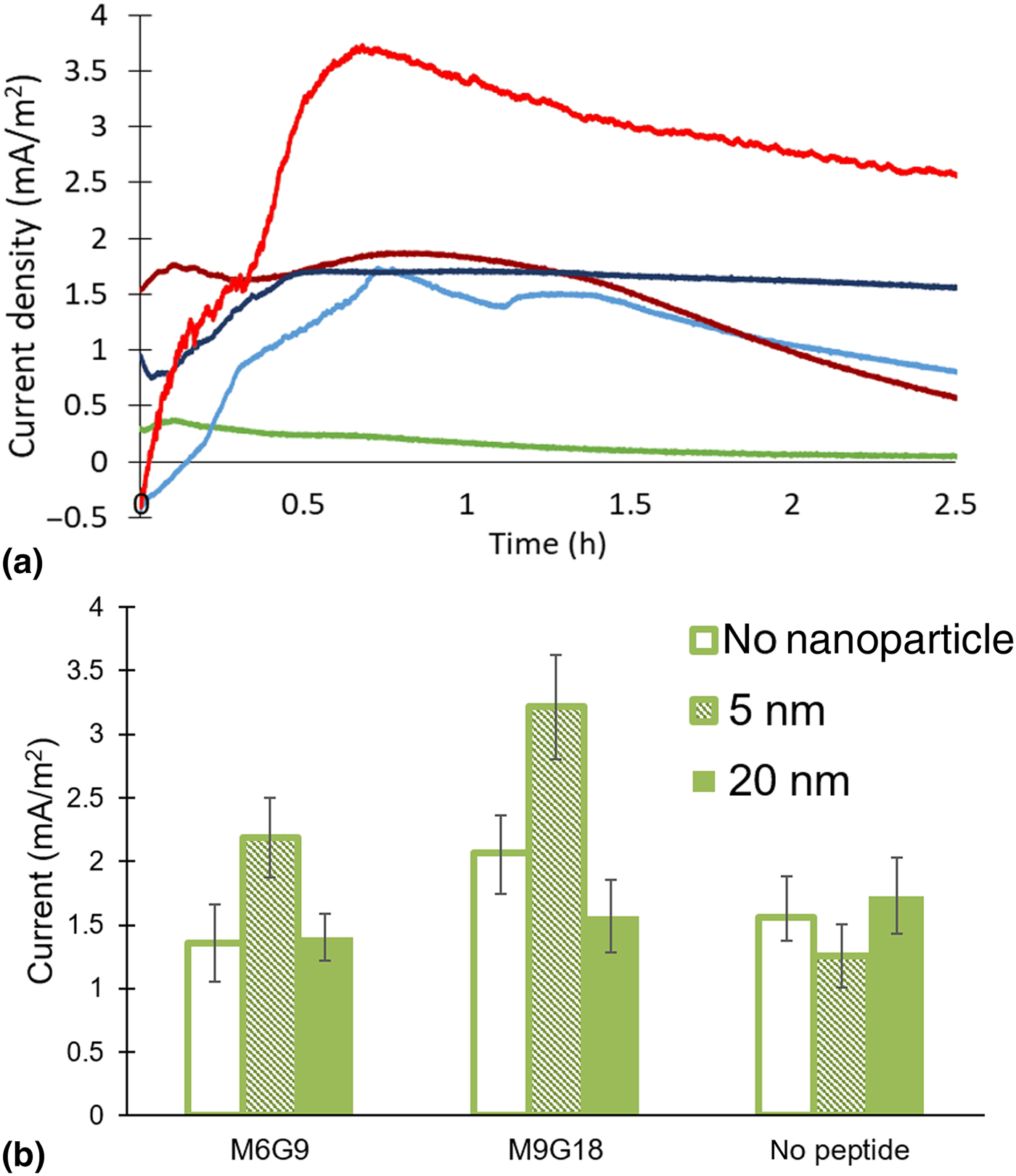
The maximum current was used to quantify the differences between various run conditions of the MFCs undertaken to characterize the effect of peptide length and particle size. These values are plotted for each set of runs in Fig. 1(b). An increase in the maximum current is observed if a peptide is displayed and 5 nm particles are added. However, a larger enhancement in the peak current is shown with the longer M9G18 peptide displayed; this enhancement is significant at the P < 0.025 level. Furthermore, the currents are approximately twice that of the uninduced cells, resulting in a four times power increase due to the combination of the peptide and nanoparticles. To see if the increase in current also decreased the internal resistance of the MFC, the MFCs using M9G18 were run with different external resistances after 5 h of operation. Such a decrease might indicate that the nanoparticles were facilitating the formation of longer range conductive networks, but no such decrease was observed; the internal resistance without nanoparticles was 550 ± 80 kΩ while with nanoparticles it was 860 ± 430 kΩ. Figure 1(b) also shows the importance of nanoparticle size, as 20 nm particles do not show any enhancement in the MFC peak power production. Five nanometer gold particles are barely larger than the gold particle size at which toxicity, due to increased permeability, readily kills E. coli, [Reference Zheng, Setyawati, Leong and Xie22] and increased membrane permeability is known to facilitate the diffusion of redox active species capable of charge transfer out of the cell.[Reference Catania, Ajo-Franklin and Bazan14] Overall, these results show that with an appropriate choice of nanoparticles, the MFC currents can be enhanced with the display of gold-binding peptides.
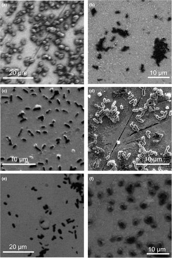
At the end of the runs, SEM images of the electrodes were taken to examine the extent and nature of cell coverage on the electrodes and to determine if there were any differences with or without gold-binding peptides or nanoparticles. Figure 2 shows SEM images for cells not displaying any peptide or displaying either the M6G9 peptide or the M9G18 peptide, both with and without bound 5 nm particles. Because of the large amount of dried salt and media on the electrodes as well as cells, it was not possible to analyze nanoparticle attachment to the cells in the SEM images. Given the short operation of the fuel cells, it was not expected that a biofilm would form, and in all cases, sub-monolayer coverages of cells are observed with some clustering of cells and also many individual cells. With this level of coverage, the increased MFC current observed with 5 nm particles and gold-binding peptides can easily occur by improving the currents generated by cells already in proximity to the electrode, and there is a limited need for long conduction paths for cells far away from the electrodes. The cell densities for all samples were calculated to be around 5 × 10⁶ cells/cm2, with no significant variation due to either the presence or absence of peptides or nanoparticles. This is expected as the cells are not exposed to any shear during the experiments, and the electrodes are rinsed only gently at the end of the experiment. This suggests that the differences in MFC currents are due to differences in how individual cells transport charges to the electrodes, rather than changes in cell numbers.
Representative SEM images of MFCs after completion of the run. The peptides displayed and nanoparticles added are: (a) M6G9, no nanoparticles, (b) M6G9, 5 nm particles, (c) M9G18, no nanoparticles, (d) M9G18, 5 nm particles, (e) no peptide, no nanoparticles, and (f) no peptide, 5 nm particles.

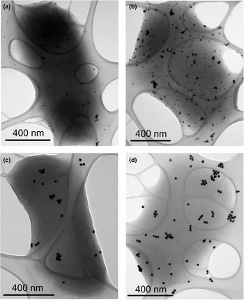
To examine the nanoparticle attachment to the cells, TEM images were acquired under the same conditions used to prepare the cells for use in the MFCs. Figure 3 shows TEM images acquired with both nanoparticle sizes (5 and 20 nm) and for both peptides examined in these experiments (M6G9 and M9G18). No images were taken of the nanoparticle binding to uninduced cells because even under conditions optimized for showing high levels of attachment, the coverages were very low, 5× lower than even the lowest coverage observed here.[Reference Dong, Sarkes, Rice, Hurley, Fu and Stratis-Cullum19] As expected, the nanoparticles are distributed uniformly across the cells, and the smaller 5 nm particles are present in higher numbers than the 20 nm particles. Some clustering of nanoparticles is observed, but no evidence is seen of continuous networks of nanoparticles, even in most densely covered samples. Interestingly, despite the similarity of the peptide sequences, markedly more nanoparticles are bound when M9G18 is expressed when compared with M6G9. The higher nanoparticle coverages with the M9G18 cells may be due to the expected higher affinity of the longer peptide for gold. The longer peptide length may also sterically allow fewer peptides to bind per particle; this allows more particles to be bound for the same fraction of peptides participating in binding.
Representative TEM images of cells displaying either the M6G9 (a,c) or M9G18 peptides (b,d) with either 5 (a,b) or 20 nm (c,d) particles added under identical conditions to those used in the MFCs. Nanoparticles bind under all conditions, but the greatest number of nanoparticles bound occurs with M9G18 and 5 nm particles, where the largest MFC current enhancement was observed.

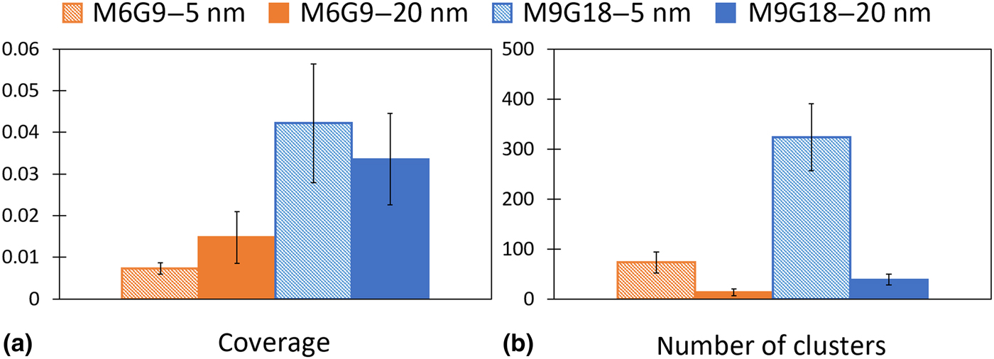
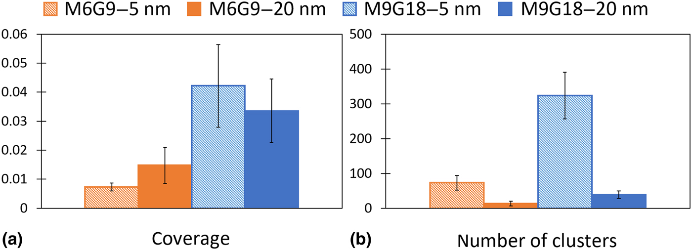
The extent of nanoparticle coverage and the number of nanoparticle clusters are quantified in Fig. 4. The nanoparticle coverage is used to measure the fraction of the cell surface occupied by nanoparticles, while the number of distinct clusters is used as a measure of how the nanoparticles are dispersed on the cell. The details of the analysis are found in Supplementary Material. As shown in Fig. 4(a), the display of the M9G18 peptide results in higher nanoparticle coverages relative to the display of the M6G9 peptide. The nanoparticle-binding behavior of M9G18 was not examined in our previous work, but with the M6G9 the nanoparticle coverage on the cell is significantly lower than when the nanoparticles are added to the cells in phosphate-buffered saline (PBS),[Reference Dong, Sarkes, Rice, Hurley, Fu and Stratis-Cullum19] likely due to nanoparticle aggregation effects in the LB medium. To determine if higher nanoparticle binding would provide an additional benefit to the MFC power generation, runs in PBS were also conducted (Supplementary Fig. S3); a larger effect size is observed, but the overall current density is slightly lower, likely due to the difference between the minimal and rich media. The extent of nanoparticle coverage on each cell does not appear to depend on the nanoparticle size, at least for the two sizes examined here (Fig. 4(a)), with cells displaying M6G9 having higher coverages with 20 nm particles and cells displaying M9G18 having slightly higher coverages with 5 nm particles. This is consistent with our previous results showing that, while weak-affinity gold-binding peptides bind smaller nanoparticles more readily, high-affinity gold-binding peptides also bind both 10 and 20 nm particles.[Reference Dong, Sarkes, Rice, Hurley, Fu and Stratis-Cullum19] Despite the similar coverages, Fig. 4(b) shows that the 5 nm particles are dispersed into more clusters than the larger 20 nm particles. As shown in Supplementary Fig. S2, this is driven by a larger number of nanoparticles on the cell surface with smaller nanoparticles, as even though the clusters are smaller with the smaller nanoparticles the number of nanoparticles per cluster is similar for across all samples. The largest current increase in the MFCs was observed with 5 nm particles and with cells displaying the M9G18 peptide, and these conditions correspond to both the higher nanoparticle coverage on the cells and the higher numbers of nanoparticle clusters. A moderate increase is also observed with the 5 nm particles and cells displaying M6G9; the TEM image analysis shows that these cells have the second highest number of clusters despite a low overall coverage.
TEM image analysis of 5 (dashed boxes) and 20 (solid boxes) nm particles attached to cells displaying either M6G9 (left two colums, orange) or M9G18 (right two columns, blue). (a) Fraction of the cell covered by nanoparticles. (b) Average number of distinct nanoparticle clusters per cell. The error bars show the standard deviations.
Here, we have shown that the current of MFCs could be modulated with an inducible genetically controlled system by which gold-binding peptides displayed on E. coli cell surfaces are used to enhance the current of MFCs by binding gold nanoparticles. The enhancement is up to approximately 2× and corresponds to a 4× increase in power density. No enhancement is observed when no peptide expression has been induced due to a lack of interaction between the E. coli cells and the unfunctionalized gold nanoparticles. The current was also found to be dependent on the nanoparticle size, with 5 nm particles enhancing currents while larger 20 nm particles did not. TEM analysis showed that the extent of nanoparticle binding was correlated with the MFC current enhancement for the 5 nm particle, and this extent could be controlled by varying the peptide design. In addition to potential benefits for common MFC applications such as waste water treatment and remote power generation, these results have implications for whole cell biosensing and cell electronic communications more generally. For example, with future work, it should be possible to take advantage of the inducible nature of peptide surface display within the MFC to demonstrate current enhancement only in the presence of both nanoparticles and a chemical of interest. In addition, the ability to genetically control the peptide sequence should allow both the strength of nanoparticle affinity and nanoparticle material specificity to be controlled, allowing for more fundamental studies of the nanoparticle behavior in bioelectrochemical systems.
Supplementary material
The supplementary material for this article can be found at https://doi.org/10.1557/mrc.2019.81