INTRODUCTION
The long evolutionary history of mollusc parasitism resulted in more than a hundred known nematode species associated to snails and slugs, which act as definitive and intermediate hosts for rhabditids and metastrongyloids, respectively (Grewal et al. Reference Grewal, Grewal, Tan and Adams2003). The main transmission pathway of metastrongyloids to mammals is represented by the ingestion of gastropod intermediate hosts, with some instances of exception of nematodes (e.g. Oslerus osleri) in the family Angiostrongylidae and Filaroididae (Anderson, Reference Anderson2000). However, in the majority of the cases, juvenile first-stage larvae (L1) of metastrongyloids develop into third-stage larvae (L3) in the molluscan tissues (Anderson, Reference Anderson2000). After the infection of proper vertebrate hosts, migrating larvae, adult nematodes and the newborn L1 can produce different degrees of pathogenic outcomes with reported life-threatening conditions (Spratt, Reference Spratt2015). For instance, the zoonotic rat metastrongyloids Angiostrongylus cantonensis and Angiostrongylus costaricensis cause thousands of cases of eosinophilic meningitis and abdominal angiostrongylosis in humans, respectively (Wang et al. Reference Wang, Wu, Wei, Owen and Lun2012; Romero-Alegría et al. Reference Romero-Alegría, Belhassen-García, Velasco-Tirado, Garcia-Mingo, Alvela-Suárez, Pardo-Lledias and Sanchez2014). Additionally, A. cantonensis has been indicated as an agent of neurological disorders in dogs, lemurs and non-human primates and birds (Duffy et al. Reference Duffy, Miller, Kinsella and de Lahunta2004; Lunn et al. Reference Lunn, Lee, Smaller, Mackay, King, Hunt, Martin, Krockenberger, Spielman and Malik2012; Burns et al. Reference Burns, Bicknese, Qvarnstrom, DeLeon-Carnes, Drew, Gardiner and Rideout2014; Kim et al. Reference Kim, Hayes, Yeung and Cowie2014; Spratt et al. Reference Spratt2015). Likewise, A. costaricensis may use dogs as alternative definitive hosts to rats (Alfaro-Alarcón et al. Reference Alfaro-Alarcón, Veneziano, Galiero, Cerrone, Gutierrez, Chinchilla, Annoscia, Colella, Dantas-Torres, Otranto and Santoro2015). Research in feline lungworms has intensified in the last years due to the increased awareness on this group of parasites. Whilst Aelurostrongylus abstrusus has been considered for ages the only metastrongyloid lungworm of cats, an additional species, Troglostrongylus brevior, has been found with increasing prevalence in domestic and wild cat populations (Brianti et al. Reference Brianti, Giannetto, Dantas-Torres and Otranto2014; Falsone et al. Reference Falsone, Brianti, Gaglio, Napoli, Anile, Mallia, Giannelli, Poglayen, Giannetto and Otranto2014). Troglostrongylus brevior was firstly reported from wild cats in Palestine (Gerichter, Reference Gerichter1949) and after six decades, in domestic cats both from Spain (Jefferies et al. Reference Jefferies, Vrhovec, Wallner and Catalan2010) and Italy (Brianti et al. Reference Brianti, Gaglio, Giannetto, Annoscia, Latrofa, Dantas-Torres, Traversa and Otranto2012, Reference Brianti, Giannetto, Dantas-Torres and Otranto2014). In few years, T. brevior has been reported in animals from Italy, Greece, Bulgaria, Spain and Bosnia and Herzegovina (Alić et al. Reference Alić, Traversa, Duscher, Kadrić, Di Cesare and Hodžić2015; Diakou et al. Reference Diakou, Di Cesare, Barros, Morelli, Halos, Beugnet and Traversa2015; Giannelli et al. Reference Giannelli, Capelli, Hinney, Joachim, Losson, Kirkova, Martellet, Papadopoulos, Farkas, Brianti, Tamponi, Varcasia, Alho, Madeira de Carvalho, Cardoso, Maia, Mircean, Mihalca, Mirò, Schnyder, Cantacessi, Colella, Cavalera, Latrofa, Annoscia, Halos, Knaus, Beugnet and Otranto2017) clearly indicating a wider distribution than that initially assumed.
In the same years, another species of metastrongyloid, namely Angiostrongylus chabaudi, has been found affecting the cardiopulmonary system of a domestic cat in insular Italy (Varcasia et al. Reference Varcasia, Tamponi, Brianti, Cabras, Boi, Pipia, Giannelli, Otranto and Scala2014). Firstly reported six decades ago (Biocca, Reference Biocca1957), this nematode was found parasitizing the heart and lungs of 86% of a wildcat population from central Italy. Later on, A. chabaudi has been diagnosed at post-mortem examination of domestic cats from Italy (Varcasia et al. Reference Varcasia, Tamponi, Brianti, Cabras, Boi, Pipia, Giannelli, Otranto and Scala2014; Traversa et al. Reference Traversa, Lepri, Veronesi, Paoletti, Simonato, Diaferia and Di Cesare2015) and wildcats from Germany, Greece, Romania, Italy and Bulgaria (Steeb et al. Reference Steeb, Hirzmann, Eskens, Volmer and Bauer2014; Diakou et al. Reference Diakou, Psalla, Migli, Di Cesare, Youlatos, Marcer and Traversa2016; Gherman et al. Reference Gherman, Ionică, D'Amico, Otranto and Mihalca2016; Giannelli et al. Reference Giannelli, Kirkova, Abramo, Latrofa, Campbell, Zizzo, Cantacessi, Dantas-Torres and Otranto2016; Veronesi et al. Reference Veronesi, Traversa, Lepri, Morganti, Vercillo, Grelli, Cassini, Marangi, Iorio, Ragni and Di Cesare2016). Nonetheless, most of the aspects of the biological life cycle of this parasite (e.g. intermediate host and morphology of larval stages) are unknown, impairing the design of epidemiological investigations and the identification of control strategies. Therefore, the aim of this study was to determine the potential intermediate host in the life cycle of A. chabaudi and to describe the associated developmental larval stages.
MATERIALS AND METHODS
Larval collection
A fresh male road-killed wildcat was found in Bobâlna (Cluj County, Romania; 47.148855′N, 23.640511′E). At necropsy, adult A. chabaudi were found in the pulmonary arteries and identified morphologically according to morphological keys (Gherman et al. Reference Gherman, Ionică, D'Amico, Otranto and Mihalca2016) and molecularly (see below). No other pulmonary or vascular nematodes were found. Further, the cardiopulmonary system was dissected to perform a Baermann examination at the Department of Parasitology and Parasitic Diseases, University of Agricultural Sciences and Veterinary Medicine of Cluj-Napoca (Romania). Tissues were cut in small pieces and placed in a double-layer gauze, secured with a wire and settled into Baermann funnels, filled with 50 mL warm tap water and examined after 24 h. The sediment was poured into a tube and centrifuged at 600 g for 3 min, and the supernatant was discarded. The sediment was placed on a microscope slide for examination under light microscopy (Leica®, DM LB2, Wetzlar, Germany) and larvae isolated and stored in saline solution. The material was sent to the Parasitology Laboratory of the University of Bari (Apulia, Italy), for additional processing. L1 were morphologically identified as A. chabaudi according to their morphology (Gherman et al. Reference Gherman, Ionică, D'Amico, Otranto and Mihalca2016) and further molecularly characterized (see below).
Maintenance and infection of gastropods
Sixty land snails Cornu aspersum were purchased from a farming centre located in Barletta (Puglia, Italy). The snails were housed in a plastic box at controlled room temperature (21 ± 2 °C) and fed every other day with lettuce and water. In order to exclude the presence of helminth infections, 10 randomly selected snails were digested (see below for procedures) and microscopically examined on the arrival and 1 day prior to the experimental infection.
Gastropods (n = 30) were infected with single infective doses of about 100 L1 obtained by centrifugation at 600 g for 3 min of the Baermann sediments and collected under a light microscope (Leica®, DM LB2, Wetzlar, Germany). Cornu aspersum snails were individually placed in the infection chamber consisting of six-well cell culture plate (Corning®; CellBIND®; Sigma-Aldrich®) containing a potato slice (0·3 cm thick) with the infective dose on its surface, and left in the infection chambers for 24 h.
The potential suitability of the gastropod C. aspersum as intermediate host of A. chabaudi was assessed by artificial digestion of five snails at 3, 6, 10, 15, 20 and 30 days post-infection (dpi).
Each snail was digested in a solution of 100 mL HCl (pH 2·2) and 3 mg mL−1 of powder pepsin (⩾250 units mg−1, P700-100G Sigma-Aldrich®, St. Louis, Missouri, USA). The suspension was heated on a magnetic stirrer at 37 ± 2 °C for 90 min, shifted through a 250 µm sieve to remove undigested material, transferred to 50 mL plastic tubes and centrifuged at 600 g for 5 min. At each dpi, the suspension obtained from the gastropod digestion was microscopically examined and larval stages identified according to previous descriptions of metastrongyloid larval stages (Gerichter, Reference Gerichter1949; Ash, Reference Ash1970). The nematodes were preserved in 70% ethanol, subsequently cleared and examined as temporary mounts in glycerol. Drawings were made with a compound microscope Leica DM LB2 (with differential interference contrast) and a drawing tube. Digital images and measurements were taken using Leica LAS® AF 4.1 software. Metrical data are given as the range, with the mean in parentheses.
Molecular identification
Single L1 (n = 10), L2 and L3 suspended in two pools (10 specimens each) were isolated using a 10 µL micropipette, washed and stored in phosphate buffer saline solution for molecular analysis. Genomic DNA was extracted using a commercial kit (DNeasy Blood & Tissue Kit, Qiagen, GmbH, Hilden, Germany), in accordance with the manufacturer's instructions, and a partial fragment of the ribosomal internal transcribed spacer-2 (ITS2) gene was amplified as previously described (Gasser et al. Reference Gasser, Chilton, Hoste and Beveridge1993). The amplicons were purified and sequenced, in both directions using the same primers as for PCR, employing the Taq Dye Deoxy Terminator Cycle Sequencing Kit (v.2, Applied Biosystems) in an automated sequencer (ABI-PRISM 377). Sequences were aligned using the Geneious R9 software package (http://www.geneious.com) and compared (BLASTn) with those available in the GeneBank database (http://blast.ncbi.nlm.nih.gov/Blast.cgi).
RESULTS
Development of A. chabaudi in gastropods
Specimens of C. aspersum digested on the arrival (n = 5) and 1 day prior to the infection (n = 5) were negative for helminths. Larval stages of A. chabaudi were found in 29 out of 30 (96·7%) experimentally infected snails. Number and developmental stages of larvae detected from experimentally infected snails are shown in Table 1.
Number and developmental stages of Angiostrongylus chabaudi larvae collected from five experimentally infected snails at 3 (T1), 6 (T2), 10 (T3), 15 (T4), 20 (T5) and 30 (T6) days post-infection

In brackets mean number of larval stages per snail and standard deviation (s.d.).
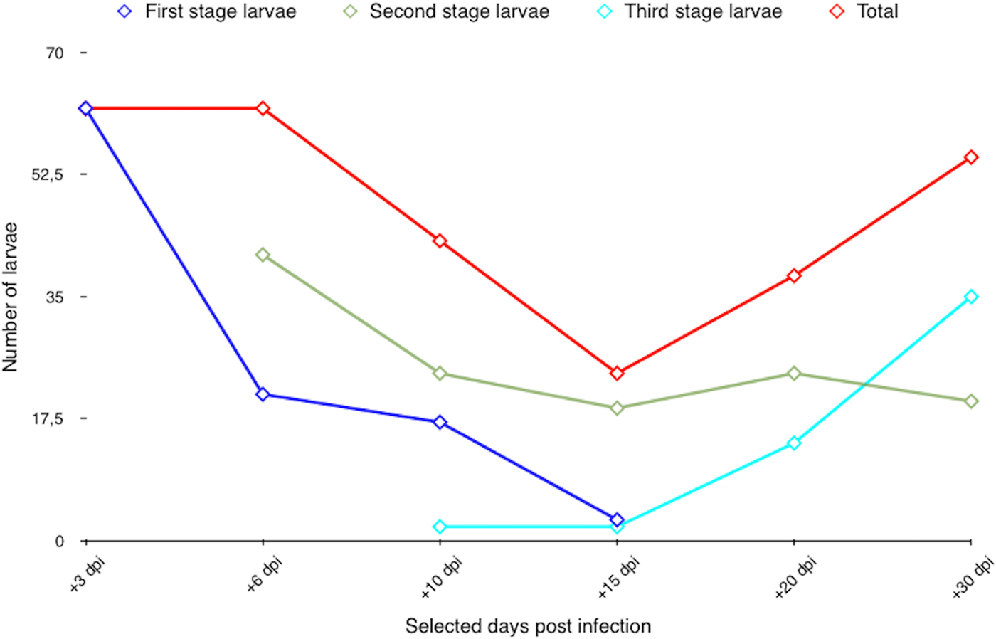
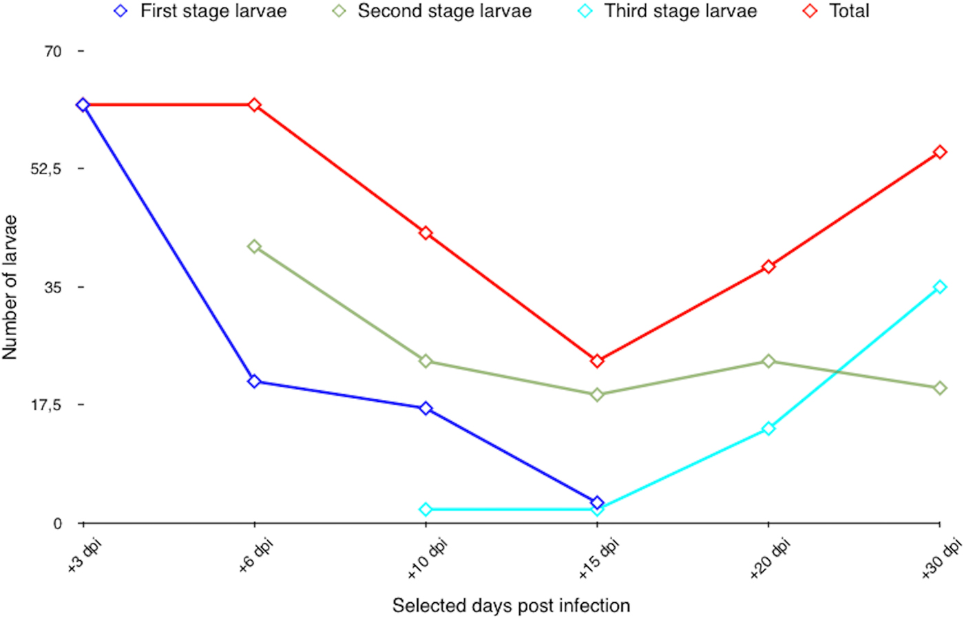
A total of 284 larvae (mean 9·8 ± 3·02 larvae per each specimen) were collected from the gastropods. L2 and L3 were detected at 6 and 10 dpi, respectively (Table 1, Fig. 1). L1 were not detected after 15 dpi, whereas L3 were increasingly detected from 10 dpi until the end of the observational period (i.e. 30 dpi, Fig. 1).
Dynamics of Angiostrongylus chabaudi larval development in Cornu aspersum snails.
Morphological characteristics of larval stages
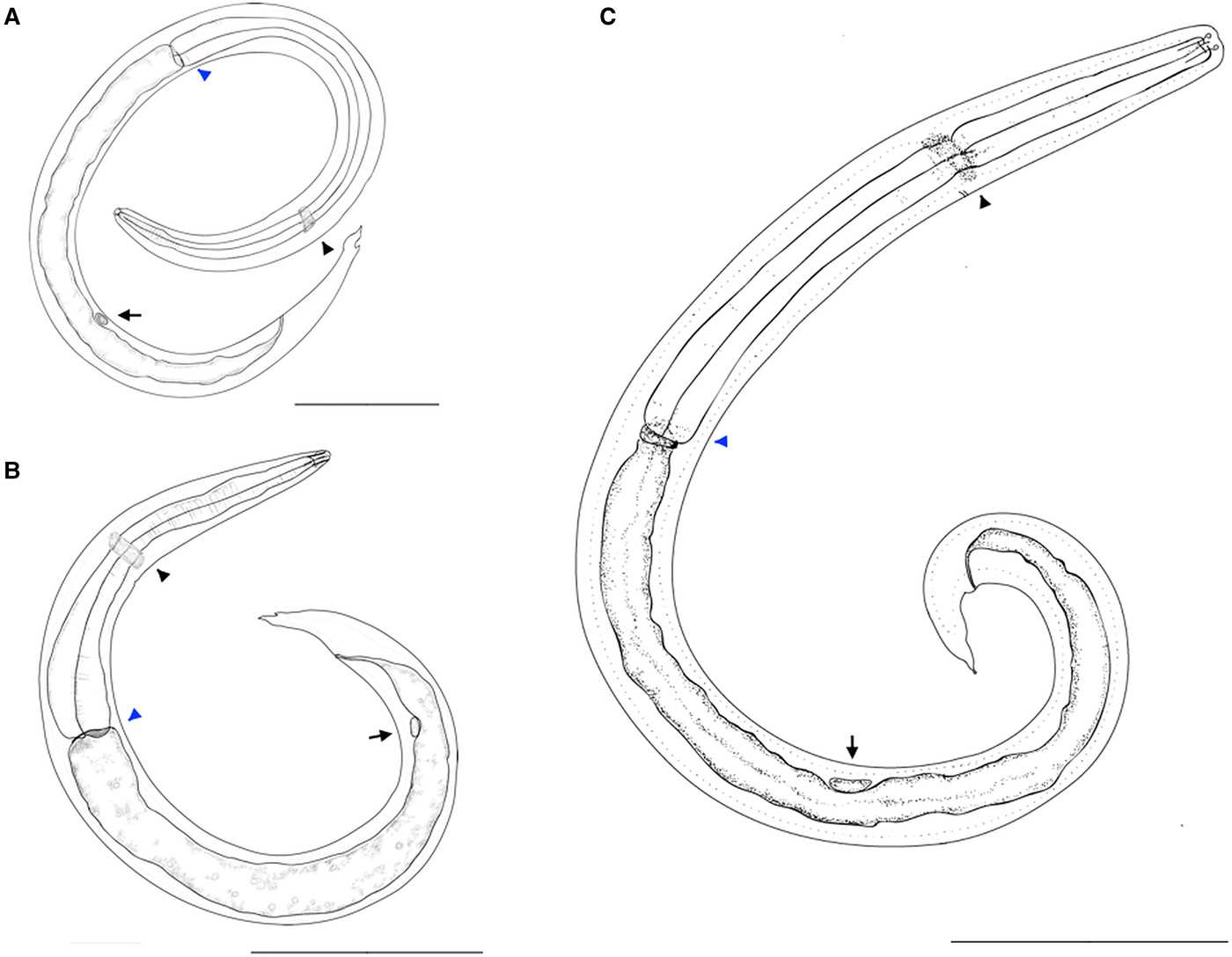
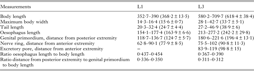
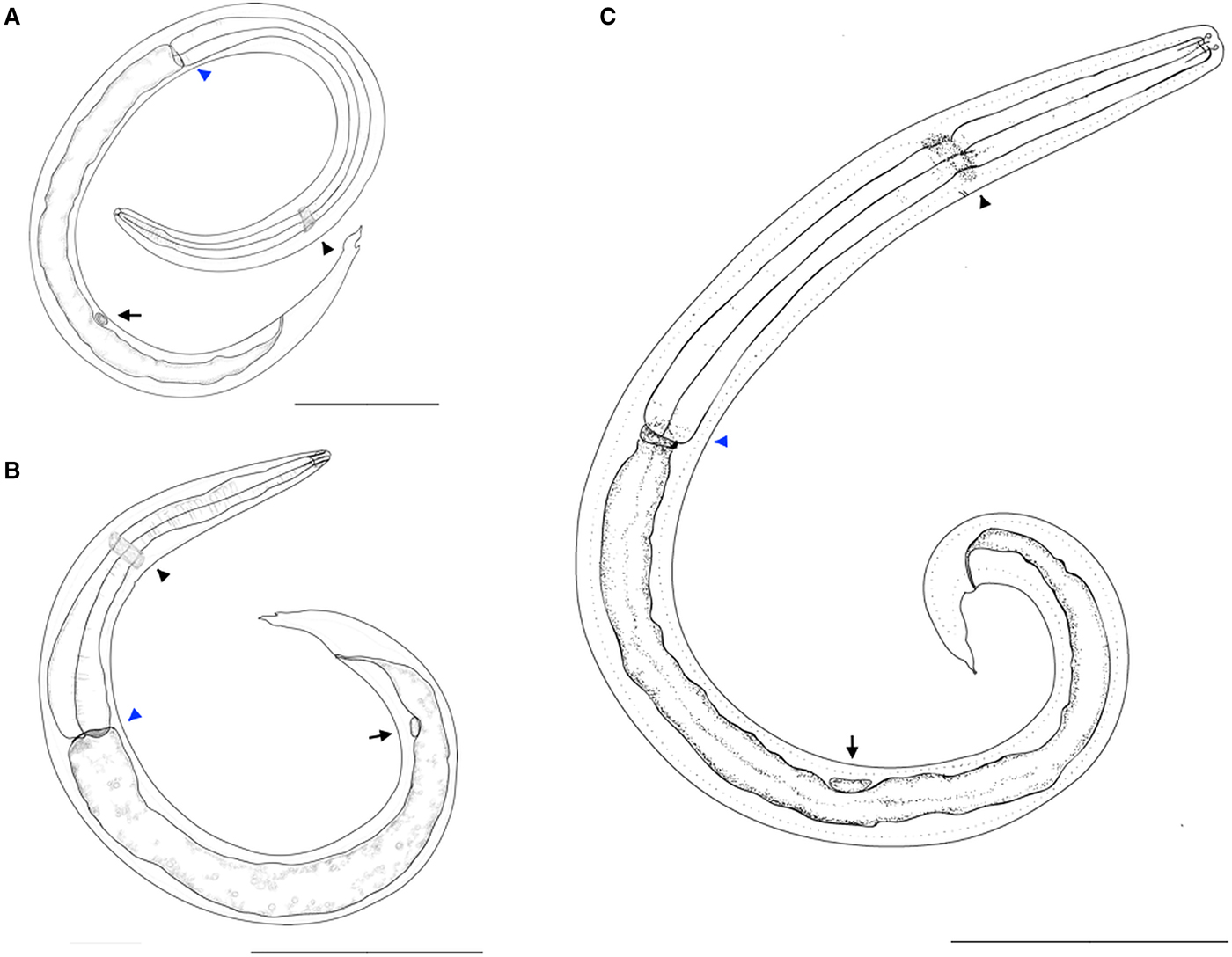
Main measurements of L1 and L3 of A. chabaudi are given in micrometres (μm) and are summarized in Table 2. Morphological characteristics of L1, L2 and L3 are outlined in Fig. 2. The main features of L1 and L3 of A. chabaudi herein described have been compared with those of other feline lungworms (i.e. A. abstrusus and T. brevior) or members of Angiostrongylidae family (i.e. Angiostrongylus vasorum and A. cantonensis; Table 3).
Drawings of Angiostrongylus chabaudi. First-stage larva (A), scale bar 50 µm; second-stage larva (B), scale bar 100 µm; third-stage larva (C), scale bar 100 µm. Note the nerve ring (black arrowhead), oesophago-intestinal junction (blue arrowhead) and genital primordium (arrow).
Measurements (in micrometres) of first- (L1) and third-stage (L3) larvae (n = 10 each) of Angiostrongylus chabaudi
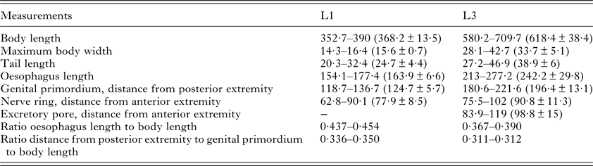
In brackets, mean and s.d. are given.
Main measurements in μm of first- and third-stage larvae of Angiostrongylus chabaudi, Aelurostrongylus abstrusus, Troglostrongylus brevior, Angiostrongylus vasorum and Angiostrongylus cantonensis

First-stage larvae
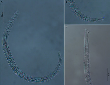
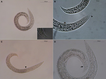
First-stage larvae collected from the wildcat measured 352·7–390 (368·2 ± 13·5) in length and 14·3–16·4 (15·6 ± 0·7) in width (Figs 2A and 3A). The tail was 20·3–32·4 (24·7 ± 4·4) in length, featured by a typical L1 kinked Angiostrongylus morphology with a subterminal dorsal spine separated by a moderately wide and distinct notch (Fig. 3B). The tail had a small ventral indentation, which followed to a slightly short sigmoid ending (Fig. 3B). The shape of the tail was fairly constant in all L1 examined. The anterior extremity was blunted with a terminal buccal opening (3C). The oesophagus was 154·1–177·4 (163·9 ± 6·6) in length and it was divided by a muscular procorpus (Fig. 3C) followed by a metacorpus ending with a gradually widening bulb (Fig. 2A). Nerve ring was situated at 62·8–90·1 (77·9 ± 8·5) from the anterior extremity (Fig. 2A). Excretory pore was noticeable in few specimens and situated slightly posterior to the nerve ring. Genital primordium was small and oval in shape and situated at 118·7–136·7 (124·7 ± 5·7) from the posterior extremity (Fig. 2A). The ratio of the oesophagus length to body length was 0·437–0·454 (0·445). The ratio of the distance from posterior extremity to the genital primordium to body length was 0·336–0·350 (0·343).
First-stage larva of Angiostrongylus chabaudi with body ventrally curved (A). Note the kinked tail with a subterminal dorsal spine (black arrowhead) separated by a notch and a short sigmoid ending (blue arrowhead) (B). Blunted anterior extremity with a terminal buccal opening (C). Note the oesophagus muscular procorpus (arrowhead).

Second-stage larvae
Second-stage larvae collected from experimentally infected snails were coiled C-shaped and featured by numerous granules (Figs 2B and 4A). Larvae measured 434·6–514 (470·3 ± 28·6) in length and 32·5–47·2 (36·9 ± 4·5) in width. Some L2 were encased in the cuticle of the L1 and the space between the cuticle of L2 and the sheath of L1 provided an empty-like appearance at both extremities (Fig. 4B). The oesophagus was 151·9–191·8 (174·6 ± 13·2) in length and the metacorpus filled by numerous granules (Figs 2B and 4C). The anterior extremity was narrowed with a button-like extremity of circa 3·5 in length and 5 in width (Figs 2B and 4D). The anus was well evident, the tail was 34·4–49·2 (40·4 ± 5·9) in length and resembled that of the previous stage with a less wide notch (Fig. 4D). The genital primordium was 152·8–175·3 (164·5 ± 8·5) distant from the posterior extremity (Fig. 2B). The ratio of the oesophagus length to the body length was 0·349–0·373 (0·371). The ratio of the distance from the posterior extremity to the genital primordium to body length was 0·341–0·351 (0·35).
Coiled C-shaped second-stage larva of Angiostrongylus chabaudi (A). Magnification of the tail (insert A); L2 encased in the cuticle of the L1 (arrowhead) (B); oesophago-intestinal junction (arrowhead) (C); well-evident anus (arrow) (D).

Third-stage larvae
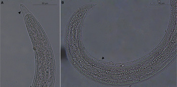
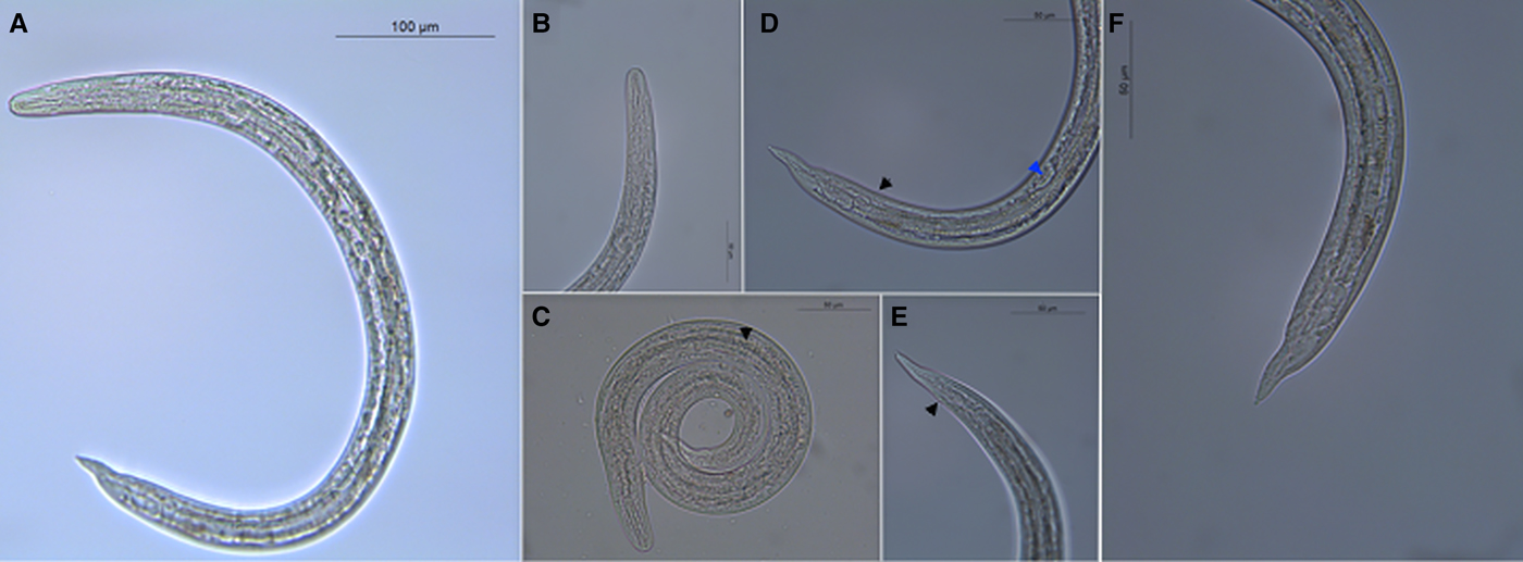
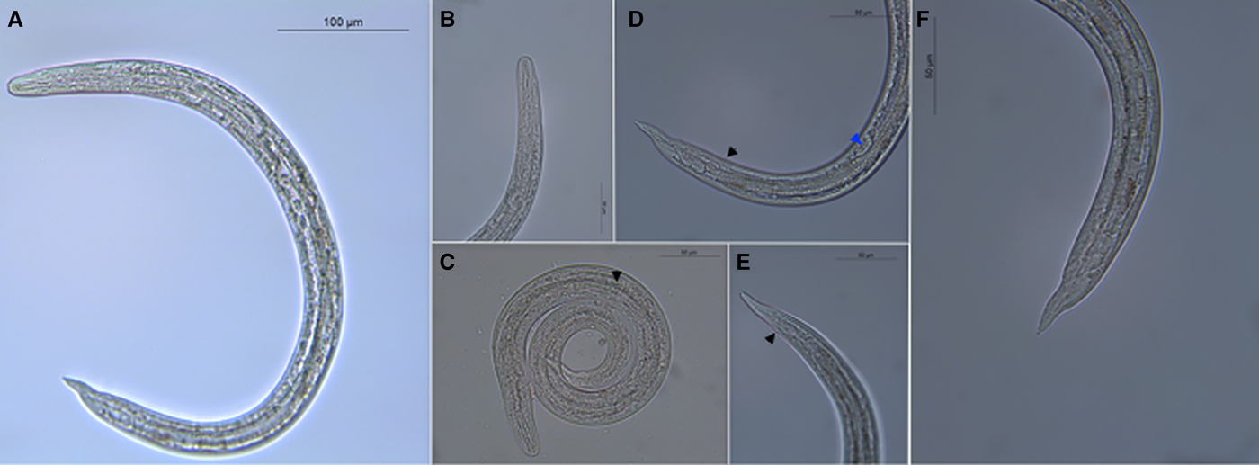
Third-stage larvae (Fig. 2C) were retrieved either enclosed in the cuticle (Fig. 5A and B) of the previous moulting or without the external sheaths (Fig. 6A). The slender body was ventrally curved and measured 580·2–709·7 (618·4 ± 38·4) in length and 28·1–42·7 (33·7 ± 5·1) in width (Fig. 6A). The anterior end was blunt with a distinct buccal cavity featured by two knob-like structures followed by well-developed chitinous rods of circa 6–12 in length (Fig. 6B). The excretory pore was present at 83·9–119 (98·8 ± 15) slightly posterior to the nerve ring, which was at 75·5–102 (90·8 ± 11·3) from the anterior extremity (Fig. 2C). The claviform oesophagus measured 213–277·2 (242·2 ± 29·8) in length, divided in a well-distinct muscular procorpus of circa 28–38 in length followed by a granular metacorpus gradually widening in a terminal bulb (Figs 2C and 6C). The cuticle displayed slight transverse striations more evident in proximity of the middle of the body and the tail (Fig. 6D). The tail was ventrally bent, 27·2–46·9 (38·9 ± 6) in length, with a tiny dorsal subterminal indentation and a ventral notch followed by a small knob-like structure (Fig. 6E and F). The genital primordium was present at 180·6–221·6 (196·4 ± 13·1) from the posterior extremity (Figs 2C and 6D). The ratio of the oesophagus length to the body length was 0·367–0·390. The ratio of the distance from the posterior extremity to the genital primordium to body length was 0·311–0·312.
Anterior (A) and posterior (B) extremities of a third-stage larva of Angiostrongylus chabaudi enclosed in the sheaths (arrowhead) of the previous moults.

Slender body of the third-stage larva of Angiostrongylus chabaudi (A); blunt anterior end with a distinct buccal cavity (B); claviform oesophagus widening in a terminal bulb (arrowhead) (C); slight transverse striations of the cuticle (black arrowhead) and genital primordium (blue arrowhead) (D); detail of the anus (arrowhead) (E); ventrally bent tail featured by a small knob-like structure (F).
Molecular identification
The ITS2 sequences obtained from L1 of the wildcat and L1, L2 and L3 collected after the digestion of experimentally infected snails displayed 100% identity to the nucleotide sequence of A. chabaudi available in GenBank (accession no. KM979214).
DISCUSSION
Data presented indicate that A. chabaudi develops in gastropods with the land snail C. aspersum as the first recorded intermediate host species under laboratory condition. Cornu aspersum is native of Mediterranean regions, and it is one of the most globally widespread snail species (Guiller et al. Reference Guiller, Martin, Hiraux and Madec2012). This gastropod species has already been identified as intermediate host of other metastrongyloids affecting felids (e.g. A. abstrusus and T. brevior; Giannelli et al. Reference Giannelli, Ramos, Annoscia, Di Cesare, Colella, Brianti, Dantas-Torres, Mutafchiev and Otranto2014) and canids (e.g. Crenosoma vulpis; Colella et al. Reference Colella, Mutafchiev, Cavalera, Giannelli, Lia, Dantas-Torres and Otranto2016). Cornu aspersum may be, therefore, a source of multiple infections of feline lungworms for cats in both urban and rural areas, and may represent a model to assess the epidemiology of snail-borne nematodes.
In the experimental infection with A. chabaudi, all but one C. aspersum harboured different developmental larval stages. The failure of the single snail to be infected by A. chabaudi might be due, inter alia, to snail immune responsiveness. Immunological reactions against larval stages of feline and canine lungworms have been demonstrated through the formation of gastropod-derived invertebrate extracellular phagocyte traps (Lange et al. Reference Lange, Penagos-Tabares, Muñoz-Caro, Gärtner, Mejer, Schaper, Hermosilla and Taubert2017).
Availability of this experimental model allowed us to describe the infective stages for the definitive feline hosts; nonetheless, a definitive proof of infectivity of L3 herein described can be achieved by inducing a patent infection in a definitive host. Though different measurements of larval stages may be potentially recorded in other species of intermediate or definitive hosts, the mean length of L1 and L3 of A. chabaudi herein described falls within the ranges of other feline lungworms (i.e. A. abstrusus and T. brevior) or members of Angiostrongylidae family (i.e. A. vasorum and A. cantonensis; Table 3). L3 possess similar morphology to the above mentioned nematodes making the species discrimination ambiguous. For example, the terminal knob-like ending of L3 of A. chabaudi is less pronounced that in A. abstrusus. For diagnostics in definitive hosts, L1 of A. chabaudi share similar morphology of the tail and measurements of main morphological features overlap with those of closely related metastrongyloids. For non-experts, the appreciation of morphological features of larvae from feline (Deak et al. Reference Deak, Gherman, Ionică, Daskalaki, Matei, D'Amico, Domşa, Pantchev, Mihalca and Cozma2017) and gastropod hosts could be difficult. Hence, when performing epidemiological surveys in definitive, paratenic and intermediate hosts, molecular analyses are also required.
Studies focusing on the intermediate hosts will improve current understanding of the snail-borne diseases. The microhabitat is central for the development of the lungworm life cycles and may explain the patchy distribution of snail-borne nematodes among close areas, as demonstrated for A. vasorum (Morgan et al. Reference Morgan, Jefferies, Krajewski, Ward and Shaw2009). The recognition of A. chabaudi as a parasite of wild and domestic cats is a recent finding and should be followed by more in-depth prevalence studies in geographical areas where other lungworms are identified. The occurrence of A. chabaudi in a wildcat population from Romania updates information on the distribution of this parasite with unknown consequences to domestic cats. Indeed, metastrongyloids do not display a very high intermediate or definitive host specificity, and hypothesis of lungworms confined to wildcats (Traversa et al. Reference Traversa, Lepri, Veronesi, Paoletti, Simonato, Diaferia and Di Cesare2015) is unlikely. The lack of a tight association of metastrongyloids to one definitive host species is further embodied by the rat lungworm A. costaricensis, which can occasionally use dogs as definitive hosts (Alfaro-Alarcón et al. Reference Alfaro-Alarcón, Veneziano, Galiero, Cerrone, Gutierrez, Chinchilla, Annoscia, Colella, Dantas-Torres, Otranto and Santoro2015). Additionally, though dogs and wild canids, such as foxes, wolves, coyotes and jackals, are the main definitive hosts for A. vasorum (Morgan and Shaw, Reference Morgan and Shaw2010; Elsheikha et al. Reference Elsheikha, Holmes, Wright, Morgan and Lacher2014), patent infections have been described in animals belonging to the family Mustelidae (e.g. the Eurasian badger and otters; Madsen et al. Reference Madsen, Dietz, Henriksen and Clausen1999; Torres et al. Reference Torres, Miquel and Motjé2001) and Ailuridae (i.e. the red panda; Grøndahl et al. Reference Grøndahl, Monrad, Dietz, Jensen, Johansen and Kapel2005; Patterson-Kane et al. Reference Patterson-Kane, Gibbons, Jefferies, Morgan, Wenzlow and Redrobe2009).
Likely, risk factors, such as living outdoors and/or the higher frequency in hunting behaviour, result in an increased chance of wildlife rather than domestic animals to acquire parasitic infections (Otranto et al. Reference Otranto, Cantacessi, Dantas-Torres, Brianti, Pfeffer, Genchi, Guberti, Capelli and Deplazes2015). The finding of co-infected domestic cats by A. abstrusus, T. brevior and A. chabaudi (Traversa et al. Reference Traversa, Lepri, Veronesi, Paoletti, Simonato, Diaferia and Di Cesare2015) supports the existence of complementary transmission patterns for the complex of feline lungworms sharing the definitive (domestic and wildcats) and intermediate hosts (e.g. the snail C. aspersum).
Wildcats likely act as reservoir hosts of lungworms in given areas (e.g. T. brevior, Falsone et al. Reference Falsone, Brianti, Gaglio, Napoli, Anile, Mallia, Giannelli, Poglayen, Giannetto and Otranto2014), implying the existence of angiostrongylosis by A. chabaudi in domestic cats. Findings of this study fill significant gaps in the understanding of key morphogenetic and ecological features of feline angiostrongylosis by A. chabaudi. Indeed, the first report of snails as intermediate hosts together with the in-depth description of morphological characteristics of L1, L2 and L3 will support the design of epidemiological surveys in gastropods and feline definitive hosts, and the identification of control strategies of A. chabaudi. Future investigations will contribute to understand the actual distribution of this parasite and whether domestic cats can sustain the lifecycle of A. chabaudi in the absence of the wildlife counterpart.
ACKNOWLEDGEMENT
Authors thank Jan Šlapeta (University of Sydney) for critically reviewing the manuscript.
FINANCIAL SUPPORT
This research received no specific grant from any funding agency, commercial or not-for-profit sectors.