INTRODUCTION
Chagas disease is a neglected infectious disease caused by the flagellated protozoan Trypanosoma cruzi (Bern, Reference Bern2015). Due to the lack of prophylactic agents and effective chemotherapy, there is no cure after the parasite spread and install in the organs of the vertebrate host (Novaes et al. Reference Novaes, Sartini, Rodrigues, Gonçalves, Santos, Souza and Caldas2016a , Reference Novaes, Gonçalves, Penitente, Bozi, Neves, Maldonado, Natali and Talvani b ; WHO, 2016). In response to continuous immigration of infected people from Central and South American endemic regions, Chagas disease has also become a serious health problem in non-endemic areas, especially in the USA and European countries (Hotez et al. Reference Hotez, Dumonteil, Woc-Colburn, Serpa, Bezek, Edwards, Hallmark, Musselwhite, Flink and Bottazzi2012). Beyond the 6–7 million people infected with T. cruzi in Central and South America, 70 million people are at risk of contracting the infection worldwide (Bern, Reference Bern2015).
Due to myotropic parasitic behaviour, muscle tissues are intensely parasitized by T. cruzi, generating skeletal myositis, myocarditis, cardiomyocytes atrophy and necrosis in animals and humans (Novaes et al. Reference Novaes, Penitente, Gonçalves, Talvani, Peluzio, Neves, Natali and Maldonado2013; Bern, Reference Bern2015). Cardiac damage is associated with the direct cell parasitism (i.e. cardiomyocytolysis) and unspecific immunomediated mechanisms, in which the exacerbated Th1 inflammatory response against the parasite induces secondary damage to host cells (Marin-Neto et al. Reference Marin-Neto, Cunha-Neto, Maciel and Simões2007; Dutra et al. Reference Gupta, Bhatia, Wen, Wu, Huang and Garg2014). In this mechanism, the overproduction of reactive metabolites (i.e. free radicals) by inflammatory cells and the resultant oxidative tissue damage has been implicated as a pivotal mechanism involved in the pathogenesis of Chagas cardiomyopathy (Gupta et al. Reference Gupta, Wen and Garg2009a , Reference Gusmão, Castanho, Andrade, Farsetti, Mathias, Therezo and Martins b ; Machado et al. Reference Machado, Tanowitz and Ribeiro2013).
It has been indicated that beyond the reactive oxygen (ROS) and nitrogen (RNS) species produced by the inflammatory cells, mitochondrial dysfunction in parasitized cardiomyocytes provides an additional source of reactive mediators (Gupta et al. Reference Gupta, Wen and Garg2009a , Reference Gusmão, Castanho, Andrade, Farsetti, Mathias, Therezo and Martins b ; Wen et al. Reference Wen, Bhatia, Popov and Garg2010). Considering that ROS and RNS exerts a pivotal role in the physiopathology of Chagas cardiomyopathy, there is limited and controversial evidence that antioxidant and anti-inflammatory therapy is beneficial to attenuate reactive tissue damage and the pathological remodelling associated with the myocarditis induced by T. cruzi infection (Gusmão et al. Reference Dhiman, Wan, Popov, Vargas and Garg2012; Marim et al. Reference Marim, Gusmão, Castanho, Deminice, Therezo, Jordão Júnior and Martins2012; Budni et al. Reference Budni, Pedrosa, Dalmarco, Dalmarco, Frode and Wilhelm Filho2013). By using a murine model of T. cruzi infection we compared the relevance of vitamins C, E (abundant and low-cost antioxidants) or ibuprofen (a commercially affordable and widely prescribed non-steroidal anti-inflammatory, NSAID)-based therapy to control the reactive stress and the structural and ultrastructural remodelling associated with acute Chagas cardiomyopathy. Considering that the leucocytes respiratory burst is a primary source of ROS and RNS in inflamed sites (Knight, Reference Knight2000), it is possible that anti-inflammatory therapy is more relevant to restricts reactive cardiac damage compared with the antioxidant therapy, which would be unable to control inflammation and neutralize the pro-oxidant mediators produced in response to infection.
MATERIALS AND METHODS
Animals and infection
Twelve-month-old male Swiss mice (27·39 ± 3·16 g) were provided with rodent chow and water ad libitum and maintained in animal facilities with a controlled temperature at 22 ± 2 °C and 12/12-h light/dark inverted cycles. Seventy animals were equally randomized into five groups: control (CT), uninfected; Infected without treatment (INF vitamin E-/ibuprofen-); and Infected treated with 14·28 × 10−3 mg g−1 vitamin C (INF vitamin C+), 1·14 × 10−2 mg g−1 vitamin E (INF vitamin E+) or 0·4 mg g−1 ibuprofen (INF ibuprofen+). The dose of vitamin C was selected considering the best ascorbic acid concentration in cardiac tissue obtained in a previous study with T. cruzi-infected mice (Marim et al. Reference Marim, Gusmão, Castanho, Deminice, Therezo, Jordão Júnior and Martins2012). In addition, this dose was also based on 50% of tolerable upper intake levels in humans (NIH, 2016). The dose of vitamin E (α-tocopherol acetate) was established according Maçao et al. (Reference Maçao, Wilhelm Filho, Pedrosa, Pereira, Backes, Torres and Fröde2007), which indicated reduced oxidative stress in Chagasic humans exposed to antioxidant therapy. The dose of ibuprofen (0·4 mg g−1) was determined according the human use and the ability to inhibit, in mice, the oxidative stress induced by therapeutic dose of benznidazole (0·1 mg g−1), a pro-oxidant drug used against T. cruzi infection (Santos et al. Reference Santos, Novaes, Bastos, Oliveira, Penitente, Gonçalves, Cardoso, Talvani and Oliveira2015).
The infection was established by intraperitoneal inoculation of blood containing 2000 trypomastigotes forms of T. cruzi Y strain. The treatments were administered after confirm the infection from the microscopic visualization of trypomastigotes in the mice peripheral blood (day 4 after inoculation). After 20 days of inoculation the animals were euthanized under anaesthesia with concentrated isoflurane and the hearts were collected. All analysis included 14 animals per group. This study was approved by the Institutional Animal Ethics Commission (protocol UFV 77/2012).
Vitamin C and E assay
Ascorbic acid (vitamin C) cardiac levels were determined by spectrophotometry (Marim et al. Reference Marim, Gusmão, Castanho, Deminice, Therezo, Jordão Júnior and Martins2012). Briefly, heart fragments were homogenized in 1 mL sodium phosphate buffer (0·1 M, pH = 7·4) and centrifuged at 8000 × g (4 °C, 10 min). 300 µL supernatant was added to 100 µL of solution containing thiourea (5%), copper sulphate (0·6%) and 2,4-dinitrophenylhydrazine (DNPH 2%) in sulphuric acid (25%). After incubation (4 h, 37 °C), 200 µL of sulphuric acid (65%) was added and the optical density (OD) was read in a spectrophotometer at 520 nm (Power Wave X, Bio-Tek Instruments, Inc., Winooski, VT). The results were determined by extrapolating the OD obtained from a standard curve for ascorbic acid.
Vitamin E (α-tocoferol) was quantified by high-performance liquid chromatography (HPLC, Shimadzu, SCL 10AT VP). Lyophilized powder of cardiac tissue (5 mg) was vortexed (45 s) in solution containing 950 µL of bi-distilled water and 50 µL and butylated hydroxytoluene (45 mm L). Homogenates were mixed to 2 mL n-hexane, 1 mL sodium dodecylsulphate (100 mm L) and 2 mL ethanol/isopropanol (95:5 v/v); and vortexed for 2 min. After centrifugation (8000 × g , 5 min), the supernatant (hexane phase) was evaporated by nitrogen flow and resuspended in methanol (800 µL). Methanol (375 µL) and diethyl ether (125 µL) were added and the mixture was injected in HPLC. Determinations were performed in a C18 Phenomenex Gemini chromatographic column (5 µm) by using methanol as eluent at 1 mL min−1 flow rate and fluorometric detection (excitation 286 nm, emission 330 nm) (Kusmic et al. Reference Kusmic, Basta, Lazzerini, Vesentini and Barsacchi2004).
Prostaglandin immunoassay
The prostaglandin F2α (PGF2α ) cardiac levels were quantified by using an enzyme-linked immunosorbent assay (ELISA) kit and the manufacturer's instructions (Cayman Chemical, Ann Arbor, MI, USA). From the same homogenate used in vitamin C quantification, 10 µL supernatant were add to 96-well microplate and incubated with specific antibodies against PGF2α . Prostaglandin levels were determined by spectrophotometry at 412 nm after correction by the protein concentration in the supernatant (Santos et al. Reference Santos, Novaes, Bastos, Oliveira, Penitente, Gonçalves, Cardoso, Talvani and Oliveira2015).
Lipid, protein and DNA oxidation analysis
For analysis of lipid peroxidation, heart samples were homogenized in phosphate buffer, centrifuged at 10 000 × g (10 min) and the homogenate was reacted with thiobarbituric acid solution (trichloroacetic acid 15%, thiobarbituric acid 0·375% and 0·25 N HCl) for 15 min. The formation of thiobarbituric acid-reactive substances (TBARS) was monitored in spectrophotometer at 535 nm as described previously (Buege & Aust, Reference Buege and Aust1978).
Protein carbonyl (PCN) levels were quantified in tissue pellets by using 0·5 mL of DNPH 10 mM. The reaction involved derivatization of the carbonyl group with DNPH, which leads to the formation of a stable 2,4-dinitrophenyl hydrazone product. The OD was measured by spectrophotometry at 370 nm (Levine et al. Reference Levine, Garland, Oliver, Amici, Climent, Lenz, Ahn, Shaltiel and Stadtman1990).
DNA oxidative damage was investigated after nucleic acid extraction and purification (Coombs et al. Reference Coombs, Gough and Primrose1999). Briefly, heart fragments were incubated with proteinase K (10 mg mL−1) at 55 °C. Genomic DNA was extracted by the phenol–chloroform (1:1) method. Tissue levels of 8-hydroxy-2′-deoxyguanosine (8-OHdG) were determined according to the method described by Kakimoto et al. (Reference Kakimoto, Inoguchi, Sonta, Yu, Imamura, Etoh, Hashimoto and Nawata2002). Briefly, DNA was resuspended in 10 mm L−1 Tris–HCl and 0·1 mm L−1 EDTA. Five microlitres of 200 mm L−1 sodium acetate buffer and 5 µg nuclease P1 (Aldrich Chemical Co., Milwaukee, USA) were added to 45 µL DNA samples. The mixtures were incubated at 37 °C for 1 h to digest the DNA to nucleotides. Then, 5 µL of 500 mm L−1 Tris–HCl, 10 mm L−1 MgCl2 and 0·6 units alkaline phosphatase (Aldrich Chemical Co., Milwaukee, USA) were added to the samples. The mixtures were incubated at 37 °C for 1 h to hydrolyse the nucleotides to nucleosides. The nucleoside samples were used for 8-OHdG quantification by ELISA (Cell Biolabs Inc., San Diego, CA, USA).
Antioxidant enzymes assay
Activity of antioxidant enzymes was investigated by using heart samples homogenized in ice-cold phosphate buffer (pH = 7·0) and centrifuged at 3500 × g (5 °C, 15 min). The supernatant was used for the analysis of catalase (CAT), glutathione S-transferase (GST) and superoxide dismutase (SOD). CAT activity was evaluated according to the method described by Aebi (Reference Aebi1984) by measuring the kinetic of hydrogen peroxide (H2O2) decomposition. SOD activity was estimated by a xanthine oxidase method based on the production of H2O2 and the reduction of nitroblue tetrazolium (Sarban et al. Reference Sarban, Kocyigit, Yazar and Isikan2005). GST activity was estimated by spectrophotometer at 340 nm as described by Habig et al. (Reference Habig, Pabst and Jakoby1974) and calculated from the rate of NADPH oxidation. Total protein levels in the supernatant were measured using the Bradford method (Bradford, Reference Bradford1976).
Nitric oxide (NO) analysis
NO was indirectly quantified from the detection of nitrite/nitrate (NO2 −/NO3 −) cardiac levels by the standard Griess reaction (Tsikas, Reference Tsikas2007). Briefly, 50 µL of supernatant from the heart homogenate was incubated with an equal volume of Griess reagent (1% sulphanilamide, 0·1% naphthylene diamine dihydrochloride and 2·5% H3PO4) at room temperature, for 10 min. The absorbance was measured at 550 nm in a microplate scanning spectrophotometer (Power Wave X, Bio-Tek Instruments, Inc., Winooski, VT).
Cytokines immunoassay
Cardiac samples were homogenized in a protease inhibitor cocktail (Sigma-Aldrich, MO, USA) and centrifuged at 3000 × g for 10 min. The supernatant was recovered and the levels of cytokines interferon gamma (IFN-γ), tumour necrosis factor-α (TNF-α), interleukin 10 (IL-10) and the chemokine (C-C motif) ligand 2 (CCL-2) were measured by sandwich ELISA by using commercial kits and the manufacturer's instructions (USCN Life Science Inc., Wuhan, China). The ODs of the samples were detected in a microplate reader at 450 nm, and cytokine concentrations were determined by extrapolating the OD obtained from a standard curve for recombinant cytokines (Santos et al. Reference Santos, Novaes, Bastos, Oliveira, Penitente, Gonçalves, Cardoso, Talvani and Oliveira2015).
Heart histopathology
Heart samples were immersed into histological fixative for 48 h (freshly prepared 10% (w/v) formaldehyde in 0·1 m phosphate buffer, pH 7·2), dehydrated in ethanol and embedded in paraffin. Due to the use of vertical sections for the stereological study, heart samples were placed in paraffin along the vertical axis (Mandarim-de-Lacerda, Reference Mandarim-de-Lacerda2003). Blocks were cut into 4-μm sections and stained by haematoxylin and eosin (H&E). The slides were visualized and the images captured using a light photomicroscope (Olympus BX-60®, Tokyo, Japan). Sixty histological fields were randomly sampled, and a total of 4·37 × 106 µm2 of tissue area were analysed (Novaes et al. Reference Novaes, Penitente, Gonçalves, Talvani, Neves, Maldonado and Natali2011).
Second-order stereological analysis
The stereological parameter of volume density (Vv) was estimated by point counting for muscle fibres [mf], interstitium [int] and blood vessels [bvs] according to the formula Vv[structure] = PP[structure]/PT; where PP is the number of points that hit the structure and PT is the total number of test points. The amount of muscle vascularization was defined as the ratio of Vv[bvs]/Vv[mf]. The number density [QA] of interstitial (inflammatory) cells [inf] was estimated as Q A = Σ [inflammatory cells]/T A . For these analyses, a test system with 72 points was used in a standard test area (T A ) of 73 × 103 µm2 (Novaes et al. Reference Novaes, Penitente, Gonçalves, Talvani, Peluzio, Neves, Natali and Maldonado2013).
Ultrastructural analysis and third-order stereology
Heart fragments were transferred to a fixative solution (2·5% glutaraldehyde, 0·2% picric acid, 3% sucrose and 5 mm CaCl2 in 0·1 m sodium cacodylate buffer [pH 7·2]) for 24 h at 4 °C. The samples were post-fixed in 1% osmium tetroxide, dehydrated in acetone and embedded in Epon 812 resin. Ultrathin sections were stained with 2% uranyl acetate and 0·2% lead citrate in 1 m sodium hydroxide; and observed with a Zeiss EM 109 transmission electron microscope (Basile et al. Reference Basile, Novaes, Marques, Fialho, Neves and Fonseca2012). Third-order stereology was used to estimate the extent of ultrastructural remodelling in cardiomyocytes. The stereological parameter of volume density (Vv) was estimated by counting points hitting intact and degenerated myofibrils. The number density [Q A ] of intact mitochondria and degenerated mitochondria [mitD] were also estimated (Santos et al. Reference Santos, Novaes, Bastos, Oliveira, Penitente, Gonçalves, Cardoso, Talvani and Oliveira2015). The same quantitative principles described to second-order stereology were applied in all quantitative ultrastructural analysis. A test system with 25 points in a standard test area (T A ) with 100 µm2 was used (Mandarim-de-Lacerda, Reference Mandarim-de-Lacerda2003).
Cardiac parasite load
Genomic DNA previously extracted was adjusted to the concentration of 25 ng µL−1. The PCR reaction was established in a 10-μL volume containing 50 ng DNA, 5 µL SYBR green reagent (Applied Biosystems, CA, USA), 0·35 µ m T. cruzi DNA primers (TCZ-F, 5′-GCTCTTGCCCACAMGGGTGC-3′, where M = A or C, and TCZ-R, 5′-C CAAGCAGCGGATAGTTCAGG-3′) or 0·50 µ m murine TNF-α primers (TNF-5241, 5′-TCCCTCTCATCAGTTCTATGGCCCA-3′ and TNF-5411, 5′-CAGCAAGCATCTATGCACTTAGACCCC-3′). Each reaction plate contained a standard curve and two negative controls, which consisted in a mixture of T. cruzi or murine primers without DNA and also tissue DNA from control animals (uninfected). The cycling program was performed as previously reported (Novaes et al. Reference Novaes, Sartini, Rodrigues, Gonçalves, Santos, Souza and Caldas2016a ). T. cruzi DNA values were normalized by the data obtained from murine TNF-α. The efficiencies of amplification were automatically calculated by the StepOne TM software v2·0.
Statistical analysis
Results were expressed as mean and standard deviation (mean ± s.d.). The normality in data distribution was verified using the D'agostino–Pearson test. Parametric data were compared using one-way analysis of variance (one-way ANOVA) followed by the Student–Newman–Keuls post hoc test. Non-parametric data were compared by the Kruskal–Wallis test. Statistical significance was established at P < 0·05.
RESULTS
Infected and untreated mice presented reduced vitamins C and E, and high PGF2α cardiac levels compared with CT animals (Table 1). While specific vitamin supplementation prevented vitamin C and E depletion, ibuprofen administration reduced PGF2α cardiac levels in infected animals (P < 0·05).
Vitamin and prostaglandin concentration in cardiac muscle from uninfected control (CT) and Trypanosoma cruzi-infected (INF) mice without treatment and those treated with vitamins C, E or ibuprofen.

*Indicates statistical difference (P < 0·05).
a vs CT.
b vs INF without treatment.
All analysis included 14 animals per group. Data are presented as mean and standard deviation.
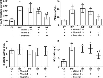
Infected and untreated mice presented high TBARS, PCN and NO levels compared with the group CT (P < 0·05). All these parameters were reduced in the infected animals treated with ibuprofen (P < 0·05). While vitamin C showed no effect, vitamin E was effective only in reduce TBARS levels (P < 0·05). Considering the similar levels of 8-OHdG in all infected and control animals, there is no evidence of DNA oxidative damage (P > 0·05) (Fig. 1).
Markers of reactive cardiac damage in uninfected control (CT, n = 14) and Trypanosoma cruzi-infected (INF) mice without treatment (−, n = 14) and those treated with vitamin C (+, n = 14), vitamin E (+, n = 14) or ibuprofen (+, n = 14). TBARS, thiobarbituric acid-reactive substances; PCN, protein carbonyl; 8-OHdG, 8-hydroxy-2′-deoxyguanosine. Data are presented as mean and standard deviation. *,† Indicate statistical difference (P < 0·05) *vs CT and †vs INF without treatment (– vitamin C, – vitamin E and – ibuprofen).

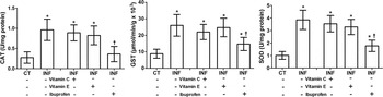
The activity of all antioxidant enzymes investigated (CAT, GST and SOD) was increased in the cardiac muscle from infected untreated animals and those treated with vitamins C or E, compared with CT mice (P < 0·05). Ibuprofen reduced the activity of all antioxidant enzymes compared with the group CT (P < 0·05) (Fig. 2).
Antioxidant enzymes activity in the cardiac muscle from uninfected control (CT, n = 14) and Trypanosoma cruzi-infected (INF) mice without treatment (−, n = 14) and those treated with vitamin C (+, n = 14), vitamin E (+, n = 14) or ibuprofen (+, n = 14). CAT, catalase; GST, glutathione S-transferase; SOD, superoxide dismutase. Data are presented as mean and standard deviation. *,† Indicate statistical difference (P < 0·05) *vs CT and †vs INF without treatment (– vitamin C, – vitamin E and – ibuprofen).

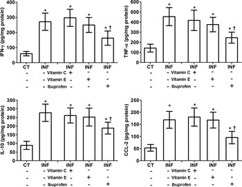
Cardiac levels of both cytokines IFN-γ, TNF-α, IL-10 and the chemokine CCL-2 was increased in all groups infected compared with CT animals (P < 0·05). Only ibuprofen treatment was effective in reduce tissue levels of all these molecules compared with infected and untreated mice (P < 0·05), Fig. 3.
Cytokine levels in the cardiac muscle from uninfected control (CT, n = 14) and Trypanosoma cruzi-infected (INF) mice without treatment (−, n = 14) and those treated with vitamin C (+, n = 14), vitamin E (+, n = 14) or ibuprofen (+, n = 14). Data are presented as mean and standard deviation. *,† Indicate statistical difference (P < 0·05) *vs CT and †vs INF without treatment (– vitamin C, – vitamin E and – ibuprofen).

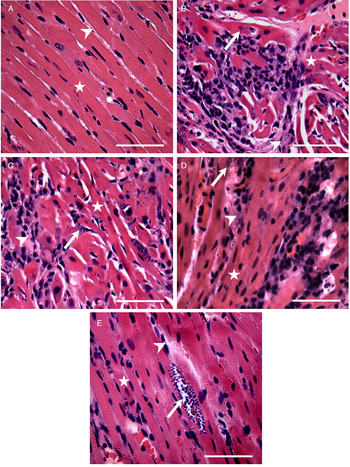
Uninfected control mice (CT) presented a normal cardiac structure, with well-organized cardiomyocytes and evident cross-striation, scarce connective stroma and interstitial cells (Fig. 4A). Infected untreated animals and those treated with vitamin C or E presented diffuse distribution of amastigote nests, intense myocarditis and connective tissue expansion. Loss of cardiomyocytes cross-striation was also observed in these groups (Fig. 4B–D). Infected mice treated with ibuprofen showed marked attenuation myocarditis, and reduced connective tissue expansion, although intracellular T. cruzi amastigote nests have also been observed (Fig. 4E).
Representative photomicrographs of the cardiac muscle from uninfected control (A, n = 14) and Trypanosoma cruzi-infected mice without treatment (B, n = 14) and those treated with vitamin C (C, n = 14), vitamin E (D, n = 14) or ibuprofen (E, n = 14) (haematoxylin and eosin staining, bars = 100 µm). (A) Uninfected animals presented normal tissue structure with well-defined myocytes, reduced area of connective tissue and low interstitial cellularity. Infected animals without treatment (B) and those treated with vitamin C (C) or E (D) presented intense myocarditis, expansion of the stromal connective tissue, and intracellular amastigote nests of T. cruzi. (E) Infected animals treated with ibuprofen showed reduced myocarditis and evident amastigote nests of T. cruzi. Stars = cardiomyocytes, arrows = amastigote nests of T. cruzi; arrowheads = interstitium.

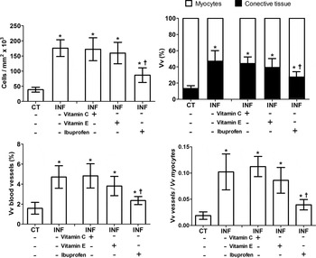
The histopathological findings were confirmed by the quantitative analysis of the cardiac muscle (Fig. 5). Infected untreated animals presented intense inflammatory infiltrate (cells mm−2), higher connective tissue and blood vessels distribution, and myocardial vascularization index (Vv vessels/Vv myocytes) compared with CT animals (P < 0·05). Ibuprofen treatment, but not vitamin C or E administration was able to reduce all these parameters, as well as attenuates parenchymal loss (Vv myocytes) compared to infected and untreated mice (P < 0·05).
Distribution of parenchyma and stroma components in the cardiac muscle from uninfected control (CT, n = 14) and Trypanosoma cruzi-infected (INF) mice without treatment (−, n = 14) and those treated with vitamin C (+, n = 14), vitamin E (+, n = 14) or ibuprofen (+, n = 14). Vv, volume density. Data are presented as mean and standard deviation.*,† Indicate statistical difference (P < 0·05) *vs CT and †vs INF without treatment (– vitamin E and – ibuprofen).

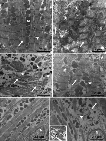
The ultrastructural analysis indicated that in control animals, cardiomyocytes had a typical cytoplasm organization, with compact myofibrils, evident sarcomere banding pattern and well-defined electron-dense mitochondrial profiles (Fig. 6A). Infected and untreated animals and those treated with vitamin C or E showed marked myofibrils and mitochondria degeneration. In these groups, myofibrils sowed reduced myofilaments compaction, while abnormal mitochondria developed irregular crests and reduced electron-density. All ultrastructural changes were attenuated in infected animals treated with ibuprofen.
Representative photomicrographs of the cardiac muscle from uninfected control (A, n = 14) and Trypanosoma cruzi-infected mice without treatment (B, n = 14) and those treated with vitamin C (C, n = 14), vitamin E (D, n = 14) or ibuprofen (E1 and E2, n = 14). (A) Uninfected animals presented a well-organized ultrastructure with well-delimited mitochondrial profiles (M) and compact myofibrils (arrow) with evident pattern of cross-striation (arrowhead). (B–D) Myocytes presenting myofibrils disorganization and fragmentation (arrows) mitochondrial degeneration (asterisks). (E1) Myocytes presenting a well-organized structure with punctual evidences (E2) of myofibrils disorganization (arrow), cytoplasm reabsorption (multilamellar bodies [arrowhead]) and mitochondrial degeneration (highlighted [star]). N = cardiomyocyte nuclei.

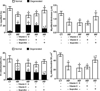
The quantitative ultrastructural analysis indicated reduced distribution and thickness of not disorganized myofibrils, lower distribution and number of mitochondria, as well as increased proportion of degenerated myofibrils and mitochondrial profiles, in all infected animals untreated or treated with vitamins C and E compared with CT animals (P < 0·05). Infected mice treated with ibuprofen but not with vitamin C or E, presented attenuation in all these parameters compared with infected and untreated animals (P < 0·05) (Fig. 7).
Organization of subcellular compartments in cardiac myocytes from uninfected control (CT, n = 14) and Trypanosoma cruzi-infected (INF) mice without treatment (−, n = 14) and those treated with vitamin C (+, n = 14), vitamin E (+, n = 14) or ibuprofen (+, n = 14). Vv, volume density. Data are presented as mean and standard deviation. *,† Indicate statistical difference (P < 0·05) *vs CT and †vs INF without treatment (– vitamin E and – ibuprofen).

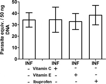
From genomic DNA extraction, quantitative PCR analysis indicated similar parasite load in cardiac muscle in all groups (P > 0·05), Fig. 8.
Cardiac parasite load in uninfected control (CT, n = 14) and Trypanosoma cruzi-infected (INF) mice without treatment (−, n = 14) and those treated with vitamin C (+, n = 14), vitamin E (+, n = 14) or ibuprofen (+, n = 14). Parasitaemia is presented as mean values. Parasite load are expressed as mean and standard deviation and was similar in all groups (P < 0·05).

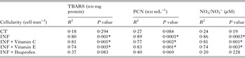
By using regression linear analysis, direct correlations between the intensity of the cardiac inflammatory infiltrate (cells mm−2), TBARS, PCN and nitric oxide (NO2 −/NO3 −) tissue levels were identified (Table 2). In general, infected and untreated animals and those treated with vitamin C or E presented moderated (R 2 > 0·7) or strong (R 2 > 0·8) correlations (P < 0·05). In control animals and those receiving ibuprofen, correlations were not significant (P > 0·05).
Correlations between inflammatory infiltrate, levels of oxidative stress markers and nitric oxide (NO2 –/NO3 –) in the heart from uninfected control (CT) and Trypanosoma cruzi-infected mice (INF) not treated and those treated with vitamin C, vitamin E or ibuprofen.

CT, uninfected control group; TBARS, thiobarbituric acid-reactive substances; PCN, protein carbonyl; NO2 −/NO3 −, nitrite/nitrate.
*Indicated significant correlations in the linear regression analysis (P < 0·05).
All analysis included 14 animals per group.
DISCUSSION
Our experimental model reinforced the relationship between inflammation, oxidative stress and cardiac damage in T. cruzi infection. By using a highly virulent and myotropic T. cruzi strain, infected mice presented reduced vitamin C and E cardiac levels, intense myocarditis and reactive tissue damage, which was correlated with the intensity of the inflammatory infiltrate and diffuse cellular and subcellular pathological remodelling. From our findings, at despite the propositions that antioxidant therapy would be beneficial in Chagas disease (Carvalho et al. Reference Carvalho, Camargos, Almeida, Peluzio Mdo, Alvarez-Leite, Chiari and Reis2006; Budni et al. Reference Budni, Pedrosa, Dalmarco, Dalmarco, Frode and Wilhelm Filho2013), vitamins C and E showed to be irrelevant to counteract tissue reactive stress and myocarditis severity in T. cruzi-infected animals. Conversely, ibuprofen, a NSAID that attenuates prostaglandin biosynthesis from cyclooxygenase-1 and 2 (COX-1 and -2) inhibition (Santos et al. Reference Santos, Novaes, Bastos, Oliveira, Penitente, Gonçalves, Cardoso, Talvani and Oliveira2015), was effective in reduce tissue inflammation, reactive stress and cardiac pathological remodelling, without interfere in tissue parasitism.
Besides the high ROS/RNS molecular reactivity, lipid and proteins seems to be more susceptible to the oxidative/nitrosative stress than DNA, which presented no evidence of reactive damage in our experimental model. Beyond the direct injury triggered by T. cruzi, the pivotal mechanism associated with acute mortality in Chagas disease is the installation of an exacerbated inflammation and immune-mediated morphofunctional cardiac damage (Machado et al. Reference Machado, Tanowitz and Ribeiro2013; Dutra et al. Reference Gupta, Bhatia, Wen, Wu, Huang and Garg2014). Such as leucocytes respiratory burst is a direct source of cytotoxic reactive molecules (i.e. H2O2, hypochlorous acid [HOCl], superoxide anion [O2●-], hydroxyl radicals [OH●-], peroxynitrite [ONOO−] and NO) (Machado et al. Reference Machado, Tanowitz and Ribeiro2013); mitochondrial dysfunction in infected cardiomyocytes also contributes to the biosynthesis of reactive metabolites (Gupta et al. Reference Gupta, Wen and Garg2009a , Reference Gusmão, Castanho, Andrade, Farsetti, Mathias, Therezo and Martins b ; Wen et al. Reference Wen, Bhatia, Popov and Garg2010). However, how much the reactive species produced by the own cardiomyocytes participates in functional and/or morphological heart injury is poorly understood and requires further investigations.
From our results, the close correlation between reactive stress and cardiac inflammation was not surprising, since both processes act in a complex and synergistic way from redundant cross-feedback mechanisms that maintain and amplify the inflammation and the reactive stress (Knight, Reference Knight2000; Machado et al. Reference Machado, Tanowitz and Ribeiro2013). The inflammation-reactive stress coupling was even more evident when the animals were treated with ibuprofen, which drastically reduced prostaglandin biosynthesis, leucocyte infiltration and molecular reactive damage. Although specific vitamin supplementation has prevented vitamin C and E depletion in infected animals, this treatment was ineffective in counteracting reactive stress and cardiac inflammation. Thus, our results do not support the applicability of vitamins C and E in the management of experimental acute Chagas cardiomyopathy. Conversely, control the inflammatory infiltrate appeared to be a more rational strategy to attenuate oxidative/nitrosative cardiac stress than direct antioxidant therapy.
There is limited initiatives investigating the relevance of antioxidant therapy in the treatment of animals (Gusmão et al. Reference Dhiman, Wan, Popov, Vargas and Garg2012; Marim et al. Reference Marim, Gusmão, Castanho, Deminice, Therezo, Jordão Júnior and Martins2012) and humans (Carvalho et al. Reference Carvalho, Camargos, Almeida, Peluzio Mdo, Alvarez-Leite, Chiari and Reis2006; Budni et al. Reference Budni, Pedrosa, Dalmarco, Dalmarco, Frode and Wilhelm Filho2013) infected by T. cruzi. The benefits of antioxidant supplementation in Chagas disease is still controversial, especially for Chagas cardiomyopathy, which was poorly investigated in animal studies and neglected in all human trials. Budni et al. (Reference Budni, Pedrosa, Garlet, Dalmarco, Dalmarco, Lino, Simionato, Amara, Frode and Wilhelm Filho2012) reported that carvedilol, a β-1-adrenergic blocker with antioxidant properties, at 37·5 mg day−1 was effective in attenuating TBARS and PCN plasma levels in humans with chronic Chagas disease; an effect partially mediated by the increased activity of antioxidant enzymes such as SOD, GPX and GST. In addition, Carvalho et al. (Reference Carvalho, Camargos, Almeida, Peluzio Mdo, Alvarez-Leite, Chiari and Reis2006) showed that vitamin E participates in the control of pathological changes triggered by T. cruzi infection, especially myocarditis and sympathetic denervation in rats. Maçao et al. (Reference Maçao, Wilhelm Filho, Pedrosa, Pereira, Backes, Torres and Fröde2007) reported that vitamin C (500 mg day−1) and vitamin E (800 IU day−1) supplementation attenuated the progressive lipid (TBARS) and protein (PCN) oxidation in patients with chronic Chagas cardiomyopathy. Although this finding has been associated with punctual increases in antioxidant enzymes activity (i.e. glutathione peroxidase GPX, GST, and SOD), direct free radical scavenger activity of both vitamins seems to be more realistic to explain the reduced molecular oxidation in these patients. Conversely, Gusmão et al. (Reference Dhiman, Wan, Popov, Vargas and Garg2012) and Marim et al. (Reference Marim, Gusmão, Castanho, Deminice, Therezo, Jordão Júnior and Martins2012) showed that vitamin C (7·14 g/kg) treatment was not effective to attenuate cardiac inflammation and oxidative tissue damage (i.e. TBARS and lipid peroxides) in mice acutely infected by T. cruzi.
At despite the limited evidence, antioxidant therapy seems to be beneficial to increase antioxidant defences in humans with chronic Chagas heart disease (Carvalho et al. Reference Carvalho, Camargos, Almeida, Peluzio Mdo, Alvarez-Leite, Chiari and Reis2006; Budni et al. Reference Budni, Pedrosa, Garlet, Dalmarco, Dalmarco, Lino, Simionato, Amara, Frode and Wilhelm Filho2012, Reference Budni, Pedrosa, Dalmarco, Dalmarco, Frode and Wilhelm Filho2013). However, such as indicated by our findings, similar effects are not manifested in animal models of acute T. cruzi-induced myocarditis treated with vitamin C (Gusmão et al. Reference Dhiman, Wan, Popov, Vargas and Garg2012; Marim et al. Reference Marim, Gusmão, Castanho, Deminice, Therezo, Jordão Júnior and Martins2012) and vitamin E. Apparently, even in the presence of vitamin supplementation, the intense leucocyte infiltrate and reactive stress in acute infection overcome the host ability to restore the redox homoeostasis. This proposition is reinforced considering the high activity of all antioxidant enzymes in both infected groups, indicating that the natural upregulation of endogenous antioxidant mechanisms in response to inflammatory insults (Knight, Reference Knight2000) was not enough to counteract reactive cardiac damage in T. cruzi infection. However, even in the presence of reduced activity of antioxidant enzymes compared with the other infected groups, ibuprofen administration attenuated NO production, lipid and protein oxidation; reinforcing the relevance of anti-inflammatory strategies to control reactive stress in T. cruzi-induced myocarditis.
Considering that the ibuprofen effect was independent of upregulation in antioxidant enzymes activity, the attenuation of cardiac reactive stress and inflammation was potentially related to reduction in prostaglandin biosynthesis (i.e. PDF2α ), a potent proinflammatory mediator. In inflammatory cardiac injuries increased prostaglandin biosynthesis has been associated with upregulation in prostaglandin–endoperoxide synthase (Ptgs-2) expression (Somaio Neto et al. Reference Somaio Neto, Ikejiri, Bertoletto, Chaves, Teruya, Fagundes and Taha2014), an effect potentially induced by pro-inflammatory cytokines such as IL-1β and TNF-α, which lead to NADPH oxidase activation, increased cytoplasmic ROS production followed by COX-2 expression (Korbecki et al. Reference Korbecki, Baranowska-Bosiacka, Gutowska and Chlubek2013). Previous studies indicated that in vitro and in vivo COX activation and prostaglandin production can be triggered by ROS/RNS (Korbecki et al. Reference Korbecki, Baranowska-Bosiacka, Gutowska and Chlubek2013; Rossi et al. Reference Rossi, Windschüttl, Matzkin, Rey-Ares, Terradas, Ponzio, Puigdomenech, Levalle, Calandra, Mayerhofer and Frungieri2016) through nuclear factor κB (NF-κB) activation via NF-κB-inducing kinase, IkB kinase (IKK) or protein kinase B (Korbecki et al. Reference Korbecki, Baranowska-Bosiacka, Gutowska and Chlubek2013). Although prostaglandins (including PDF2α ) levels have been associated with increased reactive stress in cardiac tissue (Wolfram et al. Reference Wolfram, Oguogho, Palumbo and Sinzinger2005; Santos et al. Reference Santos, Novaes, Bastos, Oliveira, Penitente, Gonçalves, Cardoso, Talvani and Oliveira2015), these molecules seem to be a consequence as well as a cause of ROS/RNS production. There is evidence that prostaglandins are direct inducers of cell degeneration, especially by activating cytotoxic mechanisms such as transient decrease in the mitochondrial membrane potential, proteasome inhibition, depletion of antioxidant defences and the own ROS/RNS production and molecular cell damage (Kondo et al. Reference Kondo, Oya-Ito, Kumagai, Osawa and Uchida2001).
The anti-inflammatory effect of ibuprofen was clearly observed due its ability in reduce myocardial leucocytes infiltrate and cytokines tissue levels in infected animals. Molina-Berríos et al. (Reference Molina-Berríos, Campos-Estrada, Henriquez, Faúndez, Torres, Castillo, Escanilla, Kemmerling, Morello, López-Muñoz and Maya2013) indicated that acetylsalicylic acid, an NSAID, was effective in the modulation of inflammation, reducing myocarditis severity and mortality in acute experimental T. cruzi infection. By inhibiting COX-2, NSAIDs attenuated prostaglandin biosynthesis and its pro-inflammatory effects, such as leucocytes chemoattraction, upregulation of inflammatory cytokines (i.e. IL-6 and CCL-2), induction of macrophages, T and dendritic cells differentiation and activation (Kondo et al. Reference Kondo, Oya-Ito, Kumagai, Osawa and Uchida2001). Although the implication of PGF2α on Chagas cardiomyopathy is poorly understood, this molecule has emerged as a potent inducer of cardiac dysfunction, which is produced in high levels in acute and chronic inflammations and can be inhibited by NSAIDs in vitro (Ricciotti and FitzGerald, Reference Ricciotti and FitzGerald2011) and in vivo (Santos et al. Reference Santos, Novaes, Bastos, Oliveira, Penitente, Gonçalves, Cardoso, Talvani and Oliveira2015).
The reduced cardiac cytokines levels in infected animals treated with ibuprofen can also be related to the NSAIDs effect in modulating the inflammation by a COX-independent pathway. It has been proved that NSAIDs can directly inhibit NF-κB activation by blocking ATP cleavage, IKK phosphorylation and activation (Yin et al. Reference Yin, Yamamoto and Gaynor1998). From this molecular interference, there is a cytokines downregulation, which can be also partially linked to our finding of reduced PGF2α cardiac levels in infected animals. This proposition is based on the effect of cytokines such as IL-1, IFNγ and TNF-α in upregulate COX-2 mRNA expression and protein synthesis, as well as induce COX-2 activation and prostaglandin production. While IL-1 and IFNγ induce COX-2 activation by PKA/CREB signalling pathway, TNF-α exerts similar effect acting on extracellular signal-regulated kinases pathway (Tsatsanis et al. Reference Tsatsanis, Androulidaki, Venihaki and Margioris2006). As this issue is extremely complex and poorly understood, further studies are required.
In addition to inflammation, broad cardiac structural and ultrastructural pathological remodelling was observed in T. cruzi-infected animals, which was attenuated by anti-inflammatory but not by antioxidant therapy. Since inflammatory infiltrate and cardiac levels of oxidative/nitrosative markers were closely correlated, our findings were consistent with the pivotal role of reactive stress in cardiac damage. Beyond typical pathological manifestations of Chagas cardiomyopathy (i.e. cardiomyocytolysis, leucocytes infiltration, connective tissue expansion and myofibrils degeneration) (Marin-Neto et al. Reference Marin-Neto, Cunha-Neto, Maciel and Simões2007; Machado et al. Reference Machado, Tanowitz and Ribeiro2013), mitochondrial degeneration seems to be a morphological finding potentially related to cardiac redox imbalance and high ROS production by cardiomyocytes, which are at the same time source and target of these reactive molecules (Wen et al. Reference Wen, Gupta, Guan, Dhiman, Condon, Lui and Garg2006; Dhiman et al. Reference Dutra, Menezes, Magalhães and Gollob2013). Although inflammation and the coupled ROS/RNS production is necessary to control the infection, cardiac parasitic load was not affected by the treatments, indicating that antioxidant and anti-inflammatory therapy did not increase the host's susceptibility to parasitism. Thus, our findings support the evidence that reduction in myocarditis severity occurred as a direct response to ibuprofen anti-inflammatory effect, and not as a secondary response to an improved parasite control and reduced antigenic load. The influence of NSAIDs on the parasite control in Chagas disease is far from being understood, a remarkable point that should be taken into account in further studies, especially considering that the effect of these drugs may be limited to control the consequences but not the cause of Chagas cardiomyopathy.
ACKNOWLEDGEMENTS
We are grateful to the ‘Nucleo de Microscopia e Microanálise’ of the Federal University of Viçosa for assistance in transmission electron microscopy.
FINANCIAL SUPPORT
This study was supported by the Brazilian agencies ‘Fundação de Amparo à Pesquisa do Estado de Minas Gerais – FAPEMIG’ and ‘Conselho Nacional de Desenvolvimento Científico e Tecnológico – CNPq’.