Introduction
In mammalian oocytes, activation occurs when the sperm enters the oocyte at the time of fertilization. The spermatozoon introduces a soluble protein factor, phospholipase C zeta, into the ooplasm (Parrington et al., Reference Parrington, Jones, Tunwell, Devader, Katan and Swann2002; Saunders et al., Reference Saunders, Larman, Parrington, Cox, Royse, Blayney, Swann and Lai2002; Swann et al., Reference Swann, Larman, Saunders and Lai2004; Heytens et al., Reference Heytens, Parrington, Coward, Young, Lambrecht and Yoon2009). This enzyme provokes the generation of inositol 1,4,5-trisphosphate (IP3) by hydrolysis of phosphatidylinositol 4,5-bisphosphate (PIP2). IP3 in turn binds its receptor on the endoplasmic reticulum leading to Ca2+ release. The initiation of the natural Ca2+ oscillations induces further oocyte activating events, such as cortical granule exocytosis, resumption of meiosis and extrusion of the second polar body (Kline & Kline, Reference Kline and Kline1992; Swann & Parrington, Reference Swann and Parrington1999; Swann et al., Reference Swann, Saunders, Rogers and Lai2006).
In vitro, artificial activation of oocytes can be used to obtain blastocysts in an aim to derive embryonic stem cells from their inner cell mass for future stem cell purposes (Brevini et al., Reference Brevini, Pennarossa, Antonini and Ganfolfi2008; Hao et al., Reference Hao, Zhu, Sheng, Yu and Zhou2009). Therefore, different physical and chemical stimuli can be used in an attempt to mimic as closely as possible the natural repetitive Ca2+ increases. However, most artificial activating agents only cause a single and prolonged rise in the cytosolic Ca2+ concentration (Whittingham & Siracusa, Reference Whittingham and Siracusa1978; Kline & Kline, Reference Kline and Kline1992; Alberio et al., Reference Alberio, Zakhartchenko, Motlik and Wolf2001). As it has been shown that preimplantation development can be influenced by the altered calcium patterns caused by the activating agents, their efficiency is still being explored (Ducibella et al., Reference Ducibella, Huneau, Angelichio, Xu, Schultz, Kopf, Fissore, Madoux and Ozil2002; Toth et al., Reference Toth, Huneau, Banrezes and Ozil2006).
For mouse oocytes, parthenogenesis has been reported with a high efficiency (Kline & Kline, Reference Kline and Kline1992; Rybouchkin et al., Reference Rybouchkin, Heindryckx, Van der Elst and Dhont2002). The most used protocol is activation with SrCl2 because this agent is able to induce Ca2+ oscillations to some extent (Bos-Mikich et al., Reference Bos-Mikich, Swann and Whittingham1995; Ma et al., Reference Ma, Liu, Miao, Han, Zhang, Miao, Yanagimachi and Tan2005). Additionally, other activating agents, like ionomycin and electrical pulses, have been proven to be highly effective activators of mouse oocytes (Versieren et al., Reference Versieren, Heindryckx, Lierman, Gerris and De Sutter2010).
Artificial activation in human has been less successful. The use of SrCl2 for parthenogenesis for human oocytes is still under debate. Some studies have shown that treatment with SrCl2 can be an effective method for artificial oocyte activation in case of low fertilization rates after intracytoplasmic sperm injection (ICSI) (Yanagida et al., Reference Yanagida, Morozumi, Katayose, Hayashi and Sato2006; Kyono et al., Reference Kyono, Kumagai, Nishinaka, Nakajo, Uto, Toya, Sugawara and Araki2008). However, others have failed to observe any Ca2+ transients when human oocytes were incubated in SrCl2 containing media (Rogers et al., Reference Rogers, Hobson, Pickering, Lai, Braude and Swann2004). Therefore, alternative activating agents are necessary. There have been a few reports of parthenogenetic blastocyst formation of human in vivo matured oocytes, mostly using ionomycin as activating agent (Cibelli et al., Reference Cibelli, Kiessling, Cunniff, Richards, Lanza and West2001; Lin et al., Reference Lin, Lei, Wininger, Nguyen, Khanna, Hartmann, Yan and Huang2003; Mai et al., Reference Mai, Yu, Li, Wang, Chen, Huang, Zhou and Zhou2007; Paffoni et al., Reference Paffoni, Brevini, Somigliana, Restelli, Gandolfi and Ragni2007; Revazova et al., Reference Revazova, Turovets, Kochetkova, Kindarova, Kuzmichev, Janus and Pryzhkova2007; de Fried et al., Reference de Fried, Ross, Zang, Divita, Cunniff, Denaday, Salamone, Kiessling and Cibelli2008; Heindryckx et al., Reference Heindryckx, De Sutter, Gerris, Simon and Pellicer2009). However, there was still a big variation in the efficiency of the technique as blastocyst formation rate ranged from 0% to more than 50%. Also, these oocytes are normally not available for research, since they are evidently used for infertility treatment of patients.
In human, most research is carried out using in vitro-matured oocytes or aged failed-fertilized oocytes after in vitro fertilization (IVF) or ICSI. Due to the inferior quality of these oocytes, originating from stimulated cycles, they often arrest during preimplantation development (Winston et al., Reference Winston, Johnson, Pickering and Braude1991; De Sutter et al., Reference De Sutter, Dozortsev, Cieslak, Wolf, Verlinsky and Dyban1992; De Sutter et al., Reference De Sutter, Dozortsev, Vrijens, Desmet and Dhont1994; Taylor & Braude, Reference Taylor and Braude1994; Rinaudo et al., Reference Rinaudo, Pepperell, Buradgunta, Massobrio and Keefe1997). Nevertheless, some blastocysts could be obtained (Zhang et al., Reference Zhang, Wang, Blaszcyzk, Grifo, Ozil, Haberman, Adler and Krey1999; McElroy et al., Reference McElroy, Kee, Tran, Menses, Giudice and Pera2008; Yu et al., Reference Yu, Mai, Chen, Wang, Gao, Zhou and Zhou2009). Recently, we have shown that for both in vitro-matured and failed-fertilized oocytes electrical activation is superior to chemical activation with ionomycin (Versieren et al., Reference Versieren, Heindryckx, Lierman, Gerris and De Sutter2010). Still, blastocyst formation was severely compromised so further optimization is still warranted.
In order to improve activation and development after parthenogenesis, we tried to use sperm injection and subsequent removal of the sperm head to mimic more closely the natural Ca2+ increases by release of the oocyte activating factor phospholipase C zeta. As human oocytes donated for research are scarce, we first wanted to develop this technique in a mouse model. Visualization of the sperm could be accomplished by Hoechst staining and UV irradiation. Both Hoechst 33342 and Hoechst 33258 stains are cell-permeable stains that can be used for visualization of the DNA content of living cells (Durand & Olive, Reference Durand and Olive1982; Portugal & Waring, Reference Portugal and Waring1988; Soderlind et al., Reference Soderlind, Gorodetsky, Singh, Bachur, Miller and Lown1999). The main difference between the two types is that Hoechst 33342 has an additional ethyl group in its structure. Therefore, it is more lipophilic than Hoechst 33258 and will more easily cross cell membranes.
Some studies on oocytes and embryos have shown some negative effects of Hoechst staining on development (Critser & First, Reference Critser and First1986; Velilla et al., Reference Velilla, López-Béjar, Rodríguez-González, Vidal and Paramio2002). Also the use of UV light poses the threat of having damaging effects on oocytes and embryos (Yang et al., Reference Yang, Zhang, Kovács, Tobback and Foote1990; Westhusin et al., Reference Westhusin, Levanduski, Scarborough, Looney and Bondioli1992; Smith, Reference Smith1993). However, in non-human primate and human somatic cell nuclear transfer, Hoechst staining and UV irradiation are still frequently used for enucleation purposes since the spindle is not visible under an inverted microscope (Mitalipov et al., Reference Mitalipov, Yeoman, Nusser and Wolf2002; Stojkovic et al., Reference Stojkovic, Stojkovic, Leary, Hall, Armstrong, Herbert, Nesbitt, Lako and Murdoch2005; Heindryckx et al., Reference Heindryckx, De Sutter, Gerris, Dhont and Van der Elst2007). So, in order to fully explore toxicity of this treatment, we first examined the effects of Hoechst 33342 and Hoechst 33258 on artificially activated mouse oocytes. Alternatively, we used MitoTracker staining for removal of the sperm head. MitoTracker stains are mitochondrion-selective stains that are cell permeable and can be used in living cells. As sperm contains a large amount of mitochondria, MitoTracker could be used to visualize the sperm head after ICSI (Bussalleu et al., Reference Bussalleu, Pinart, Yeste, Briz, Sancho, Garcia-Gil, Badia, Bassols, Pruneda, Casa and Bonet2005; Hikichi et al., Reference Hikichi, Kishigami, Thuan, Ohta, Mizutani, Wakayama and Wakayama2005).
Materials and methods
All chemicals and reagents were purchased from Sigma-Aldrich (Bornem, Belgium), unless stated otherwise.
Source of mouse oocytes
Mice were purchased from Charles River Laboratories (Brussels, Belgium) and handled according to the guidelines of the Animal Ethical Committee of the Ghent University Hospital. Mice were kept under controlled temperature and lighting conditions. Food and water were available ad libitum.
Female B6D2F1 hybrid mice aged 7–14 weeks were stimulated to superovulate by intraperitoneal injection of 5 IU equine chorionic gonadotrophin (eCG, Folligon, Intervet, Oss, The Netherlands) followed by 5 IU human chorionic gonadotrophin (hCG, Chorulon, Intervet) 48 h later. Oocytes were collected 14 h post-hCG and freed from cumulus cells by a short incubation in 200 IU/ml hyaluronidase (type VIII). Oocytes were kept in home-made potassium simplex optimized medium (KSOM) supplemented with 0.4% bovine serum albumin (BSA, Calbiochem, Bierges, Belgium) at 37°C under 6% CO2 in air until artificial activation (Lawitts and Biggers, Reference Lawitts and Biggers1991).
Parthenogenetic activation and Hoechst staining
In order to test the effects of Hoechst staining and UV irradiation, in vivo-matured mouse oocytes were activated 16 h post-hCG by incubation in 10 mM SrCl2 in Ca-free KSOM-BSA supplemented with 2 μg/ml cytochalasin D during 4 h. One hour after start of activation, oocytes were stained with Hoechst stain and irradiated with UV light. Non-treated oocytes were used as a negative control to exclude spontaneous activation, activated oocytes that were not stained or irradiated were used as a positive control. In the first experiment, activated oocytes were stained for 10 min with 0.5 μg/ml or 1 μg/ml Hoechst 33258 or Hoechst 33342 in HEPES-buffered KSOM–BSA medium and irradiated with UV light for 10 sec. Oocytes were cultured in KSOM–BSA at 37°C under 6% CO2 and 5% O2 in air and transferred to blastocyst medium (Cook Ireland Ltd., Limerick, Ireland) on day 3.
In a second step, mouse oocytes were activated with sperm as activating agent. For this, sperm heads of male B6D2F1 hybrid mouse sperm were cut off before piezo-driven ICSI. Injected oocytes were incubated in KSOM–BSA supplemented with 2 μg/ml cytochalasin D for 1 h to avoid second polar body extrusion. Oocytes were subsequently stained with 0.5 μg/ml Hoechst 33258 for 10 min and sperm heads were removed under fluorescent light while incubated in HEPES-buffered KSOM-BSA supplemented with 1 μg/ml cytochalasin D. After removal of the sperm heads, oocytes were further incubated in KSOM–BSA with 2 μg/ml cytochalasin D for 3 h. Finally, oocytes were cultured in KSOM–BSA at 37°C under 6% CO2 and 5% O2 in air and transferred to blastocyst medium on day 3.
Because of insufficient staining of the sperm for efficient removal after 10 min of staining, we needed to increase the staining time in the next experiment. To again test for toxicity, oocytes were first activated with SrCl2 as described before, stained with 0.5 μg/ml Hoechst 33258 in HEPES-buffered KSOM–BSA medium for 10, 20 or 30 min and irradiated with UV light for 10 s. Oocytes were cultured as described for the first experiment.
Toxicity of Hoechst staining or UV irradiation
To distinguish between the toxicity of Hoechst staining versus UV irradiation, we artificially activated mouse oocytes with SrCl2 as previously described. One hour after start of activation, oocytes were randomly allocated to different treatment groups: (i) staining with 0.5 μg/ml Hoechst 33258 for 10 min without UV irradiation; (ii) UV irradiation without Hoechst staining; or (iii) staining with 0.5 μg/ml Hoechst 33258 for 10 min followed by UV irradiation. Non-treated oocytes were used as a negative control to exclude spontaneous activation, activated oocytes that were not stained or irradiated were used as a positive control. Activated oocytes were cultured in KSOM–BSA at 37°C under 6% CO2 and 5% O2 in air and transferred to blastocyst medium on day 3.
Blastocyst analysis
To evaluate blastocyst quality, differential staining of inner cell mass (ICM) and trophectoderm cells (TE) of mouse blastocysts was performed (Thouas et al., Reference Thouas, Korfiatis, French, Jones and Trounson2001). Briefly, blastocysts were incubated in 500 μl HEPES-buffered human tubal fluid medium (Lonza, Verviers, Belgium) with 1% Triton X-100 and 100 μg/ml propidium iodide for 10 s, transferred to 1000 μl chilled 100% ethanol with 25 μg/ml bisbenzimide (Hoechst 33258) and stored at 4°C overnight. Blastocysts were mounted onto a glass slide in glycerol and covered with a coverslip. Numbers of ICM (blue), TE (red) and total cell number (TCN) were counted under a fluorescence microscope.
Sperm removal with MitoTracker
Oocytes were activated with sperm as activating agent as described before. However, instead of staining the injected oocytes with Hoechst, sperm heads of male B6D2F1 hybrid mouse sperm were cut off and stained with 10 μM MitoTracker Green FM (Invitrogen, Merelbeke, Belgium) for 10 min before piezo-driven ICSI. Sperm heads were removed under fluorescent light 1 h after ICSI.
Statistical analysis
Statistical analysis was performed with InStat from GraphPad Software. Activation, cleavage and preimplantation developmental data were analysed by contingency table analysis followed by chi-squared test or Fisher's exact test for independence. The level of significance was set at P < 0.05. Parameters of blastocyst quality (ICM, TE, TCN and ICM/TE ratios) were compared using one-way analysis of variance (ANOVA) followed by Tukey post-test when the level of significance reached P < 0.05.
Results
Parthenogenetic activation and Hoechst staining
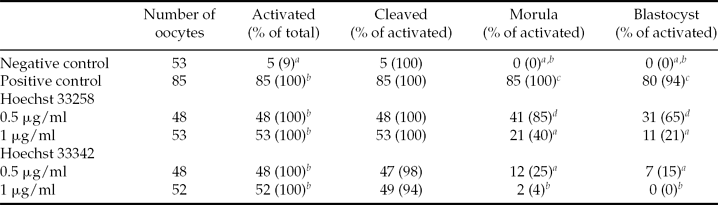
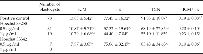
Table 1 presents the preimplantation development of mouse oocytes artificially activated with SrCl2 after Hoechst staining for 10 min and UV irradiation. Although activation and cleavage rate were not affected by the staining procedure, morula and blastocyst development were significantly decreased in all groups of stained oocytes compared to the positive control group. The effects were dependent on the concentration and the type of Hoechst that was used. Staining with Hoechst 33258 resulted in 65% blastocyst formation with 0.5 μg/ml and 21% with 1 μg/ml. Hoechst 33342 staining lead to only 15% blastocysts with 0.5 μg/ml while no blastocysts could be obtained when 1 μg/ml was used. Differential staining was performed on all obtained blastocysts (Table 2). Hoechst 33258 staining did not affect the number of cells in the ICM, but TE was significantly decreased in comparison with the positive control group. In contrast, Hoechst 33342 staining resulted in a significant reduction in ICM compared to the positive control, but TE was not influenced. Still, the ICM/TE ratios of all groups of stained oocytes were comparable to the positive control group.
Preimplantation development of activated mouse oocytes after Hoechst staining for 10 min and UV irradiation

a ,b,c,d Different superscripts within a column indicate significant difference (P < 0.05).
Differential staining of mouse blastocysts after Hoechst staining for 10 min and UV irradiation
Values are expressed as mean ± standard deviation (SD).
a ,b,c Different superscripts within a column indicate significant difference (P < 0.05).
ICM: inner cell mass, TCN: total cell number, TE: trophectoderm.
So, although staining with Hoechst 33258 decreased blastocyst development compared to the positive control group, blastocyst rate was still relatively high when 0.5 μg/ml was used (65%). Therefore this concentration and type of Hoechst was used to remove the sperm head after ICSI. However, since the staining of the sperm was too rapidly fading away for efficient removal after 10 min of staining, we needed to increase the staining time in the next experiment.
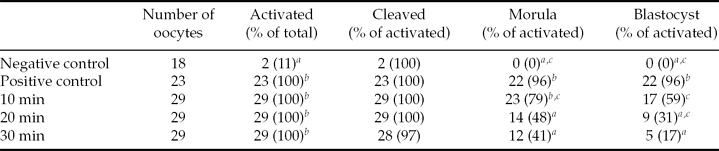
In Table 3 the preimplantation development of artificially activated oocytes after staining with 0.5 μg/ml Hoechst 33258 for 10, 20 or 30 min and UV irradiation is presented. Although activation and cleavage rate were again not affected, morula and blastocyst percentage decreased drastically with longer staining times. A 10 min staining resulted in only 59% blastocysts compared with 96% in the positive control group. An extended time interval of 20 or 30 min of staining reduced the blastocyst rate further to 31% or 17% respectively. Differential staining of the obtained blastocyst revealed no differences in ICM, TE, TCN or ICM/TE ratios (data not shown). Since Hoechst staining had a significant negative effect on blastocyst formation, we searched for an alternative staining method to remove the sperm head.
Preimplantation development of activated mouse oocytes after staining with 0.5 μg/ml Hoechst 33258 and UV irradiation
a ,b,c Different superscripts within a column indicate significant difference (P < 0.05).
Toxicity of Hoechst staining or UV irradiation
In order to further examine toxicity of staining and irradiation, activated oocytes were exposed to either Hoechst staining or UV irradiation or both (Table 4). Our results show that there was no difference in blastocyst formation rate between the oocytes that were only stained (88%) or only exposed to UV light (92%) compared with the positive control group (96). However, when oocytes were both stained and irradiated, blastocyst development significantly decreased to 54%, similar to the previous experiment.
Toxicity of Hoechst staining or UV irradiation
a ,b,c Different superscripts within a column indicate significant difference (P < 0.05).
Sperm removal with MitoTracker
In a next experiment, sperm heads were stained with 10 μM MitoTracker Green FM for 10 min before ICSI. However, MitoTracker staining was almost not visible anymore in all oocytes 1 h after ICSI. In case the sperm head, visualized by MitoTracker, could be removed, 95% of oocytes died after 1 day. No blastocysts could be obtained. As most oocytes died the next day, no other concentrations or staining times were investigated.
Discussion
Since parthenogenetic activation of human oocytes is still not very efficient, we aimed to improve activation rate and development by using sperm injection and subsequent removal of the sperm head. This way we tried to mimic more closely the natural Ca2+ increases by releasing the oocyte activating factor phospholipase C zeta. Because of the scarce availability of human oocytes for research, we first tested the technique on a mouse model. To remove the sperm after ICSI we needed to visualize the sperm head. We first examined the toxicity of Hoechst 33342 and Hoechst 33258 staining and UV irradiation. Our results show that both types of Hoechst combined with UV irradiation have toxic effects on the development of parthenogenetically activated mouse oocytes. Although activation and cleavage rate were not affected, blastocyst formation was significantly reduced. The effects are more severe with Hoechst 33342 than with Hoechst 33258. A higher concentration of Hoechst or longer staining times also significantly reduced blastocyst formation.
In this study, we attempted to find a method for artificial oocyte activation that closely resembles the physiological activation pattern induced by the sperm during fertilization. One possible way to achieve this goal is to use microinjection of cRNA encoded for the oocyte activating factor phospholipase C zeta. This technique has already been used effectively for parthenogenetic activation and development in mouse oocytes and could be used to further optimize parthenogenetic activation of human oocytes (Saunders et al., Reference Saunders, Larman, Parrington, Cox, Royse, Blayney, Swann and Lai2002; Yu et al., Reference Yu, Saunders, Lai and Swann2008). However, phospholipase C zeta cRNA is very unstable during injection and it is difficult to control the amount of cRNA that will get into the oocyte and that will be expressed. Since the creation of stable recombinant phospholipase C zeta protein has been unsuccessful until now, the use of sperm for activation might be a simpler technique and might more resemble the natural pattern of phospholipase C zeta expression.
The results of our study question the use of Hoechst staining and UV irradiation for parthenogenesis or somatic cell nuclear transfer purposes. During nuclear transfer in human and non-human primates, Hoechst staining is frequently used for enucleation of recipient oocytes as the spindle is not visible under normal inverted microscopy (Mitalipov et al., Reference Mitalipov, Yeoman, Nusser and Wolf2002; Stojkovic et al., Reference Stojkovic, Stojkovic, Leary, Hall, Armstrong, Herbert, Nesbitt, Lako and Murdoch2005; Heindryckx et al., Reference Heindryckx, De Sutter, Gerris, Dhont and Van der Elst2007). However, we have shown that even at low concentrations Hoechst staining and UV irradiation have detrimental effects on parthenogenetically activated mouse oocytes. Therefore, the use of this technique should be completely avoided when working with oocytes and embryos. One alternative for enucleation during nuclear transfer is to use non-invasive polarized microscopy to remove the spindle (Keefe et al., Reference Keefe, Liu, Wang and Silva2003; Montag and van der Ven, Reference Montag and van der Ven2008). Using this technique, nuclear transfer has already been successful in several species (Byrne et al., Reference Byrne, Pedersen, Clepper, Nelson, Sanger, Gokhale, Wolf and Mitalipov2007; Nandedkar et al., Reference Nandedkar, Chohan, Patwardhan, Gaikwad and Bhartiva2009).
Previously, some studies have shown that the use of Hoechst staining and UV irradiation during experiments with oocytes and embryos could have detrimental effects on further development. When prepubertal goat oocytes were stained with 0.5 μg/ml Hoechst 33342 and irradiated with UV light at different time intervals during in vitro maturation, the percentage of mature metaphase II oocytes and the fertilization rate of stained oocytes decreased significantly (Velilla et al., Reference Velilla, López-Béjar, Rodríguez-González, Vidal and Paramio2002). So, Hoechst staining reduces oocyte viability when it is used during the early stages of in vitro maturation. Another study showed that when porcine and mouse embryos were stained with Hoechst 33342 there was a significant reduction in in vitro development (Critser & First, Reference Critser and First1986). For mouse embryos, formation of morula and blastocyst stage decreased to 56% compared with 77% in the control group. Our results have shown that the type of Hoechst used in that study is the most toxic and may be better replaced by Hoechst 33258. The fact that in our experiments we observed a more drastic decrease of the blastocyst formation rate (from 65% to 0%) could be because they only used the Hoechst staining and UV irradiation from the 2-cell embryo stage on, so after embryo genome activation (Goddard & Pratt, Reference Goddard and Pratt1983). However, for parthenogenesis and somatic cell nuclear transfer purposes, staining and UV irradiation is already necessary at the zygote stage, which can result in a higher sensitivity to the treatment, as seen in our study.
The toxic effects of the treatment could be due to both the Hoechst staining and the UV irradiation. Most likely, they are the result of an interaction between the two as mouse oocytes exposed to Hoechst staining without UV-exposure develop normally in vitro (Critser and First, Reference Critser and First1986). Our results also demonstrated that when Hoechst staining or UV irradiation were used separately, embryos continued to have a normal preimplantation development. Only a combined treatment with Hoechst staining and UV irradiation decreased significantly the blastocyst rate. One possible explanation is that the interaction between the stain and the UV light leads to free radical formation which might contribute to the detrimental effects (Hamdoun & Epel, Reference Hamdoun and Epel2007). Also other studies have reported that Hoechst staining could enhance the cytotoxic effects of UV and γ-irradiation, for example by formation of reactive oxygen species or blocking DNA repair mechanisms (Singh et al., Reference Singh, Dwarakanath and Mathew2004; Athar et al., Reference Athar, Chaudhury, Hussain and Varshney2010).
It has also been demonstrated that the effects of UV light are largely dependent on the time interval of the exposure. For example, exposure of bovine oocytes to UV irradiation for 10 s has no effect on embryo viability and production of live calves after nuclear transfer (Westhusin et al., Reference Westhusin, Levanduski, Scarborough, Looney and Bondioli1992). On the other hand, exposure for more than 30 s induced loss of membrane integrity, decrease of methionine incorporation into proteins and altered protein synthesis (Smith, Reference Smith1993). Also in rabbit oocytes longer exposure times lead to decreased viability (Yang et al., Reference Yang, Zhang, Kovács, Tobback and Foote1990). For that reason we limited the exposure of UV light in our study to max 10 s to minimize damaging effects and we could show that no decrease in blastocyst formation was seen when oocytes were only exposed to UV light without staining.
Studies in mice have shown that there is a different sensitivity of the pronuclei and the cytoplasm to Hoechst staining and UV irradiation (Tsunoda et al., Reference Tsunoda, Shioda, Onodera, Nakamura and Uchida1988). The exposure of zygotes stained with Hoechst 33342 to UV irradiation for 20–30 s completely inhibited in vitro blastocyst development. However, when stained oocytes were enucleated and injected with untreated pronuclei, a significant decrease in blastocyst formation could only be observed when the cytoplasm was exposed for 40 s. This demonstrates that the cytoplasm of mouse oocytes is more resistant to the irradiation treatment than the pronuclei. Consequently, this might also explain why nuclear transfer could have been successful in some species using Hoechst staining and UV irradiation as enucleation technique (Forsberg et al., Reference Forsberg, Strelchenko, Augenstein, Betthauser, Childs and Eilertsen2002; Liu et al., Reference Liu, Jiang, Yan, Jiang, Ouyang, Sun and Chen2005; French et al., Reference French, Adams, Anderson, Kitchen, Hughes and Wood2008). During this procedure the stained chromosomes are removed from the oocyte and only the recipient cytoplast, which is less sensitive, has been exposed for a short time (Li et al., Reference Li, White and Bunch2004). However, in our study, both chromosomes and cytoplasm were stained and irradiated, so both could be affected by the treatment. Still, even for nuclear transfer purposes, it might be best to avoid Hoechst staining and UV irradiation and use alternative enucleation techniques, like polarized microscopy, to avoid damaging effects of the treatment on the recipient cytoplast.
We have shown that staining of activated mouse oocytes with Hoechst 33258 resulted in 65% and 21% blastocyst formation when respectively 0.5 μg/ml and 1 μg/ml was used. Hoechst 33342 staining lead to only 15% blastocysts with 0.5 μg/ml while no blastocysts could be obtained with 1 μg/ml. Thus, our results confirm that Hoechst 33258 is less cytotoxic than Hoechst 33342 (Soderlind et al., Reference Soderlind, Gorodetsky, Singh, Bachur, Miller and Lown1999; Zhang & Kiechle, Reference Zhang and Kiechle2003). Both Hoechst 33342 and Hoechst 33258 are cell-permeable DNA stains that bind in the minor groove of double-stranded AT-rich regions (Portugal and Waring, Reference Portugal and Waring1988). Because they bind to DNA, they can disrupt DNA replication during cell division and as a result they can be mutagenic and carcinogenic. Their difference in toxicity can be attributed to their structural difference, their affinity to bind to DNA and their ability to inhibit different proteins and enzymes (Soderlind et al., Reference Soderlind, Gorodetsky, Singh, Bachur, Miller and Lown1999).
Despite the fact that using Hoechst staining and UV irradiation decreased blastocyst development, still a 65% blastocyst rate could be obtained with 0.5 μg/ml Hoechst 33258. However, staining with this concentration of Hoechst was too weak for removal of the sperm head after ICSI. Therefore, in a next experiment staining times were increased to 20 or 30 min to obtain a clearer signal, though blastocyst development was further reduced to 31 and 17% respectively. As Hoechst staining did not seem to be a good technique to remove the sperm head, we searched for alternative staining methods.
As a next option, we chose the MitoTracker Green FM staining. As both the oocyte and the sperm contain a large amount of mitochondria, we chose to stain the sperm head before ICSI so we would be able to distinguish clearly the sperm head from the oocyte (Joshi et al., Reference Joshi, Medina, Colasante and Osuna2000). This way, we were also able to avoid staining of the oocyte so less damaging effects could be expected. It has been shown that the interaction leading to release of the oocyte activating factor from the spermatozoon takes place within 30 min after injection (Dozortsev et al., Reference Dozortsev, Qian, Ermilov, Rybouchkin, De Sutter and Dhont1997). To make sure that the oocyte activating factor was released completely and that the oocyte had enough time to recover from ICSI, we removed the sperm heads 1 h after injection. However, the MitoTracker staining faded quickly so that 1 h after injection the staining was not visible anymore in all oocytes. Moreover, when the sperm heads could be removed, 95% of oocytes died after 1 day. The reason for this low survival is still unclear. One possibility is that the oocytes were damaged too much from the double manipulation and they could not restore membrane integrity. However, this is unlikely because the oocytes were still alive when they were put into culture 3 h later. Additionally, when nuclear transfer is performed also two manipulations can be sequentially performed with a high success rate (Heindryckx et al., Reference Heindryckx, Rybouchkin, Van Der Elst and Dhont2002; Rybouchkin et al., Reference Rybouchkin, Heindryckx, Van der Elst and Dhont2002). Another possibility is that the damaging effects resulted from the irradiation of the oocytes during removal of the sperm head. Since most oocytes died the next day, no other concentrations or staining times were investigated. Also, as no blastocysts could be obtained from the oocytes that had undergone successful removal of the sperm, maybe also other important factors were affected. Therefore, alternative artificial activating techniques should be explored.
In conclusion, our results have shown that both types of Hoechst combined with UV irradiation have toxic effects on the development of parthenogenetically activated mouse oocytes. The effects are dependent on the type of Hoechst and the concentration of the stain. Consequently, the use of Hoechst staining and UV irradiation should be avoided when working with oocytes and embryos. As MitoTracker staining was also not successful for removal of the sperm, other non-invasive and non-toxic methods for sperm identification could be investigated.
Acknowledgements
K.V. is supported by the Special Research Foundation (BOF No.: 01D32707) of the Ghent University, Belgium. P.D.S. is holder of a fundamental clinical research mandate by the Flemish Foundation of Scientific Research (FWO-Vlaanderen), Belgium.