Both hypertrophic cardiomyopathy and Wolff–Parkinson–White syndrome are common causes of sudden cardiac death in the young. Here, we present a young soldier who survived sudden cardiac death, and was found to have concomitant hypertrophic cardiomyopathy and Wolff–Parkinson–White syndrome.
Case report
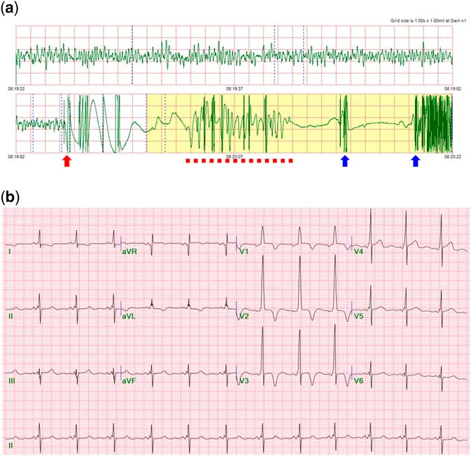
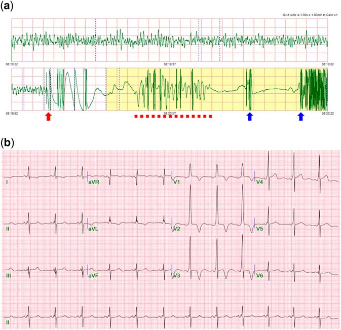
A 19-year-old male soldier with no prior medical history suddenly collapsed during morning reporting after overnight sentry duty. Immediate bystander cardiopulmonary resuscitation followed by a single automated external defibrillator shock (Fig 1a) successfully resuscitated him. Upon arrival at our institution, he was alert, albeit with some retrograde amnesia, and had stable vital signs. Physical examination was unremarkable except for a grade II ejection systolic murmur. His baseline electrocardiogram was notable for augmented QRS voltages with inverted T in V1-3 and short PR interval with slurred upstroke of QRS, indicative of ventricular pre-excitation (Fig 1b). Echocardiographic findings were consistent with hypertrophic cardiomyopathy with dynamic obstruction (Fig 2, upper panel). However, the patient had no family history of inheritable heart disease or sudden cardiac death, and his parents’ electrocardiograms and echocardiographies were also normal.
( a ) Automated external defibrillator record at the time of cardiac arrest. Ventricular fibrillation (upper panel) was terminated by a single shock of 188.3 J (red arrow). A short run of self-terminating polymorphic ventricular tachycardia (red dashed line) and subsequent sinus rhythm (blue arrow) were recorded. ( b ) The 12-lead standard electrocardiography revealed ventricular pre-excitation.
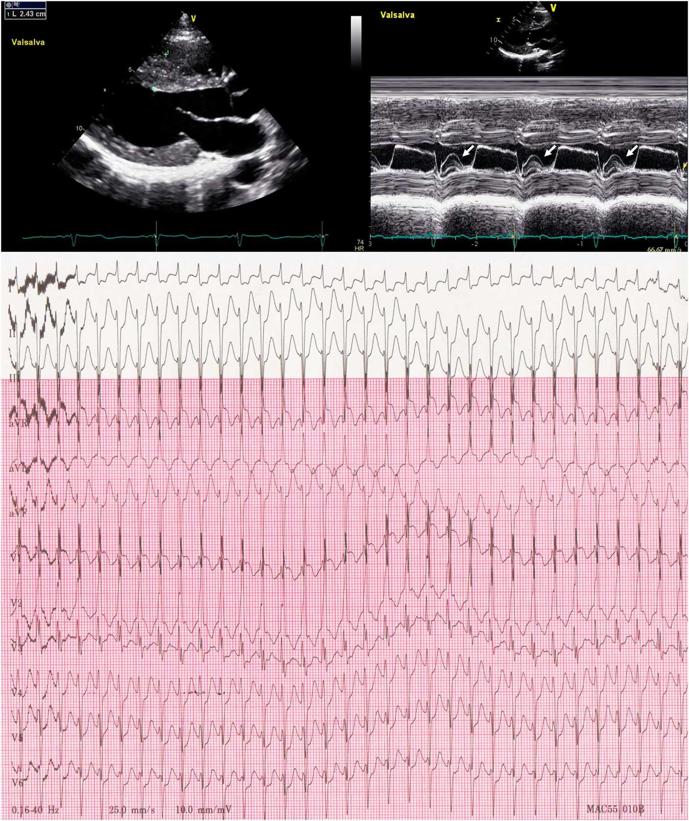
(Upper panel) Transthoracic two-dimensional echocardiography showed marked intraventricular septal thickening (24 mm, left) and systolic anterior motion of mitral valve (arrows, right) during a Valsalva manoeuvre, causing a mild pressure gradient (16 mmHg) across the left ventricular outflow tract. (Lower panel) Narrow QRS tachycardia at the cycle length of 300 ms was induced during isoproterenol infusion. Note the significant ST segment depressions suggesting ischaemia.
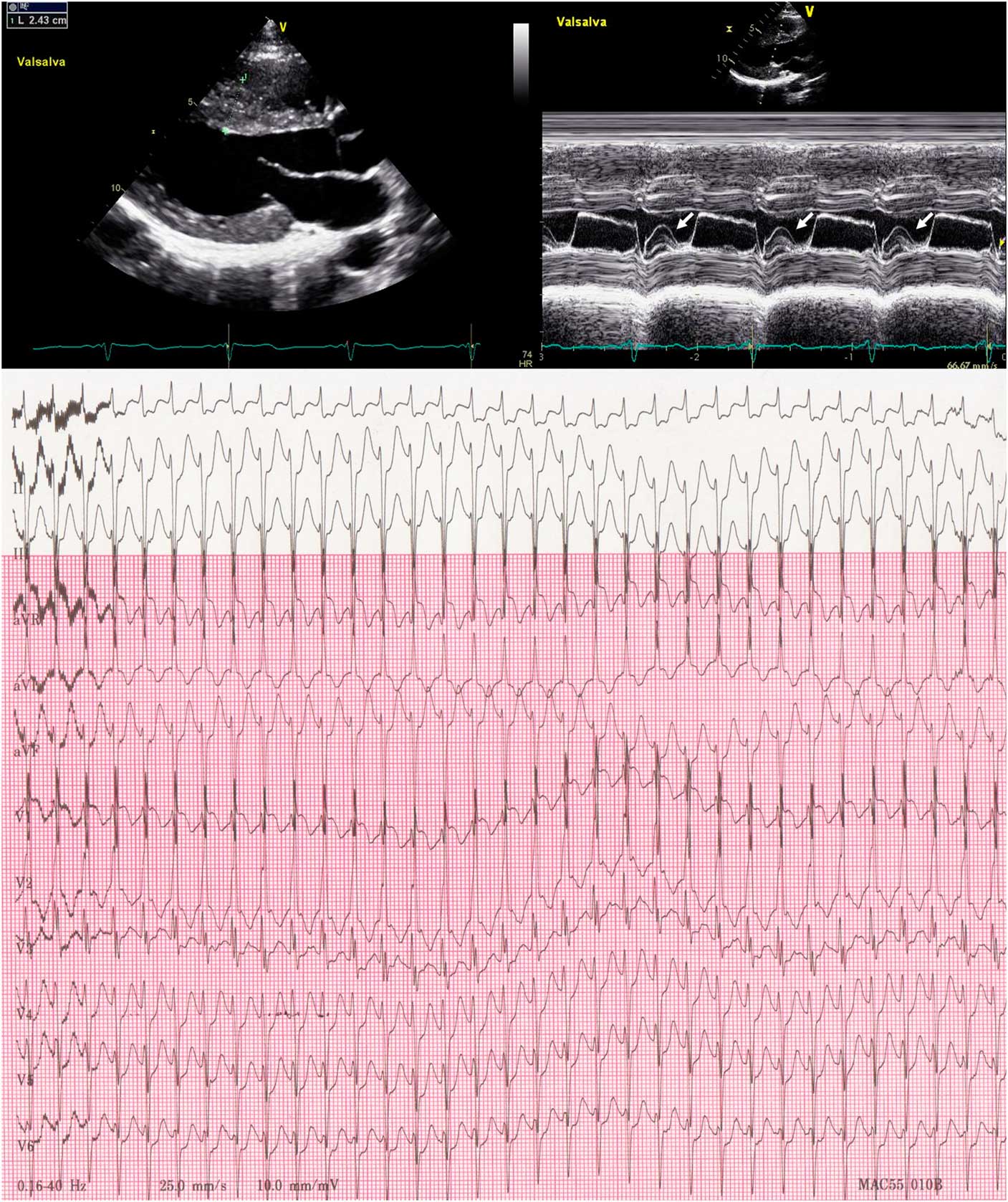
Since high school years, the patient has experienced episodic palpitations accompanied by chest pain, which were self-limiting but poorly tolerated on occasion, particularly in an upright position. During isoproterenol infusion test, narrow QRS tachycardia (Fig 2, lower panel) resulting in a significant drop of systolic blood pressure of >20 mmHg in supine position occurred and reproduced his episodic symptoms. Also, abnormal vascular response to exercise with a systolic blood pressure drop of 44 mmHg was noted during a treadmill test. On the basis of patient’s history and obtained findings, we postulated that the sudden death might be precipitated by supraventricular tachycardia causing haemodynamic compromise, subsequently degenerating into ventricular fibrillation. An electrophysiology study revealed a left posterolateral accessory pathway Kent bundle and reproducibly inducible orthodromic atrioventricular re-entrant tachycardia, utilising the atrioventricular node for anterograde and Kent bundle for retrograde conduction (Supplemental Fig 1). Anterograde and retrograde effective refractory period of Kent bundle were 320 and 300 ms, respectively. After successful Kent bundle ablation, triple ventricular extra-stimulation (400/220/200/200 ms) produced a single episode of very fast polymorphic ventricular tachycardia, but the rhythm was not reliably inducible despite repeated attempts. Mutations in the myosin-binding protein C (MYBPC3, NM_000256.3:c.26-1G>T, splicing aberration, pathogenic) and cardiac troponin T (TNNT2, NM_000364.3:c.281G>C, p.Arg94Thr, variant of uncertain significance) genes were detected in a diagnostic exome sequencing test. His hospital course was unremarkable, and abnormal vascular response to exercise was no longer present after β-blockade (propranolol 120 mg a day). The patient has remained uneventfully so far for more than 3 years.
Discussion
Concomitant hypertrophic cardiomyopathy and Wolff–Parkinson–White syndrome is a very rare condition, with most reports in the literature on familial form related to a PRKAG2 mutation.Reference Porto, Brun and Severini 1 Our patient presented with aborted sudden death due to ventricular fibrillation, and was found to have sporadic hypertrophic cardiomyopathy and Wolff–Parkinson–White syndrome with clearly documented Kent bundle-mediated supraventricular tachycardia.
We found that this is the first description of either TNNT2 variant or double mutation in a patient with concomitant hypertrophic cardiomyopathy and Wolff–Parkinson–White syndrome. The p.Arg94Thr variant in TNNT2 was an unregistered de novo mutation predicted to be deleterious by in silico analysis. So far, cardiac troponin I, MYBPC3, and β-myosin heavy chain (MYH7) mutations have been identified in this rare condition, but their causal association with Wolff–Parkinson–White syndrome itself remained undetermined because pre-excitation wave was not present in all affected family members of cardiac troponin I and MYBPC3,Reference Kimura, Harada and Park 2 and MYH7 mutation case was a sporadic disease without familial involvement.Reference Bobkowski, Sobieszczanska and Turska-Kmiec 3 Considering the well-established pathogenic role of MYBPC3 and TNNT2 genes in the development of hypertrophic cardiomyopathyReference Elliott and Anastasakis 4 and the sporadic nature of this case, we could not conclude whether the observed double mutation was responsible for both diseases, or it was an incidental Wolff–Parkinson–White syndrome co-existing with hypertrophic cardiomyopathy.
Several mechanisms can be proposed for the development of the ventricular fibrillation in this patient: 1) supraventricular tachycardia-related haemodynamic compromise, 2) pre-excited atrial fibrillation, and 3) primary ventricular tachyarrhythmia. An association between supraventricular tachycardia and sudden cardiac death in hypertrophic cardiomyopathy patients has been well acknowledged for years. Furthermore, there is direct electrophysiological evidence documenting its fatal consequence; rapid repetitive conduction to ventricles during supraventricular tachycardia, for example re-entrant tachycardia, atrial fibrillation, and even sinus tachycardia, resulting in spontaneous rhythm degeneration into ventricular fibrillation.Reference Munclinger, Thornton, Schutte and Sliwa 5 – Reference Elliott, Sharma, Varnava, Poloniecki, Rowland and McKenna 7 Aggravating factors responsible for this phenomenon include inherent vulnerability to ischaemia due to myocardial mass, left ventricular outflow tract obstruction, and abnormal vascular response, which were all present in our case and might play a synergistic role.Reference Stafford, Trohman, Bilsker, Zaman, Castellanos and Myerburg 6 , Reference McKenna and Behr 8 Fast pre-excited atrial fibrillation is also a well-known cause of sudden cardiac death in the young; however, our electrophysiology study revealed a Kent bundle refractory period of more than 300 ms and no inducible atrial fibrillation; thus the ventricular fibrillation seemed less likely to be related to atrial fibrillation.Reference Priori, Blomstrom-Lundqvist and Mazzanti 9 Lastly, fast polymorphic ventricular tachycardia was induced once during the test. However, programmed extra-stimulation-induced ventricular arrhythmia has limited prognostic significance in hypertrophic cardiomyopathy,Reference Priori, Blomstrom-Lundqvist and Mazzanti 9 and the ventricular tachycardia was not reproducible despite multiple repeated stimulation; therefore it could be regarded as idiosyncratic reaction to aggressive stimulation protocol. Nevertheless, the possibility of primary ventricular tachyarrhythmia could not be ruled out.
Although implantable cardioverter/defibrillator is considered the first-line treatment option for sudden cardiac death survivors with hypertrophic cardiomyopathy, current guideline, on the contrary, recommends electrophysiology study for those with ventricular pre-excitation to identify and treat an ablatable substrate.Reference Priori, Blomstrom-Lundqvist and Mazzanti 9 Proper discretion is particularly required when implanting the implantable cardioverter/defibrillator in the young, because they are more likely to suffer from inappropriate shock, psychological distress, and poorer quality of life than elderly, which can paradoxically potentiate sudden death risk.Reference Priori, Blomstrom-Lundqvist and Mazzanti 9 – Reference Lampert, Salberg and Burg 12 Indeed, our patient refused to undergo implantable cardioverter/defibrillator therapy due to concern about its impact on his future employment. Implantable cardioverter/defibrillator therapy should be the first choice for secondary prevention, however, upon his refusal, we decided to eliminate the anatomical arrhythmogenic substrate, which we thought could potentially reduce the risk of new events. Nevertheless, the importance of lifelong monitoring and periodic risk re-evaluation can never be emphasised enough for this young man. Further studies using global registry are required to determine the optimal strategy for the sudden cardiac death survivors who have multiple predisposing conditions and correctable causes.
Supplementary Material
To view supplementary material for this article, please visit https://doi.org/10.1017/S1047951118002123
Acknowledgements
The authors thank Jinyoung Park, the chief technical staff of the electrophysiology laboratory, for their support during the last 7 years.
Financial Support
This research received no specific grant from any funding agency, commercial or not-for-profit sectors.
Conflicts of Interest
None.