Introduction
The use of three-dimensional (3D) printing has been rapidly expanding over the last several decades, and its utilisation has been demonstrated across many different fields. Also known as additive manufacturing, 3D printing started in the early 1980s and was commercialised by Charles W Hull in 1988.Reference Gross, Erkal, Lockwood, Chen and Spence1 The process involves the creation of a digitised 3D model using computer-aided design, which is then read by a printer. An object is created by the deposition of material layer by layer.
In otolaryngology, 3D printed models have been used in the pre-operative planning for complex reconstructive defects. In vivo and in vitro studies have been published that utilise 3D printing in various aspects of airway surgery, including a bioresorbable extraluminal tracheobronchial splint for severe tracheobronchomalacia.Reference Kaye, Goldstein, Zeltsman, Grande and Smith2,Reference Zopf, Flanagan, Wheeler, Hollister and Green3 The 3D printed models have also been used for multidisciplinary planning for tracheal reconstruction in conjunction with congenital cardiac defects.Reference Balakrishnan, Cofer, Matsumoto, Dearani and Boesch4 Balakrishnan et al. described the use of 3D printed models in developing management strategies in cases of congenital long-segment tracheal stenosis associated with complete tracheal rings.Reference Balakrishnan, Cofer, Matsumoto, Dearani and Boesch4
The demonstration of acquired (as opposed to congenital) laryngotracheal stenosis in 3D printed models might present a different scenario. The present case series aimed to describe the use of 3D printed airway models to depict high-grade acquired laryngotracheal stenosis in the paediatric airway, and to discuss the potential applications of such 3D printed models in paediatric airway surgery.
Materials and methods
This paper describes a retrospective case series from a tertiary care paediatric airway centre of two patients with high-grade laryngotracheal stenosis, in whom 3D printed models of the laryngotracheal complex were developed. Institutional Review Board approval was obtained prior to the review of these cases.
After bronchoscopic confirmation of laryngotracheal stenosis, and computed tomography (CT) scanning with 1 mm cuts, a standard protocol was used to obtain the dimensions for the 3D printed airway. Raw Digital Imaging and Communications in Medicine (‘DICOM’) images were securely uploaded for image processing and model creation. Mimics Medical software, version 19.0 (Materialise, Plymouth, Missouri, USA), was used to perform segmentation of the airway using a threshold-based algorithm. Segmentation of the airway was obtained with the proximal extent being the superior border of the epiglottis and the distal extent being 1.5 cm below the carina including the proximal bronchi. The 3D images were subsequently created and exported to 3-matic Medical software, version 11.0 (Mimics Innovation Suite; Materialise).
A hollow operation was then performed, which took the outer surface of the solid airway model and made it the inner surface of the hollowed model, with a constant thickness that was set to 1.5 mm. The inner wall of the model simulated the interior of the windpipe, with an outward arbitrary thickness of 1.5 mm.
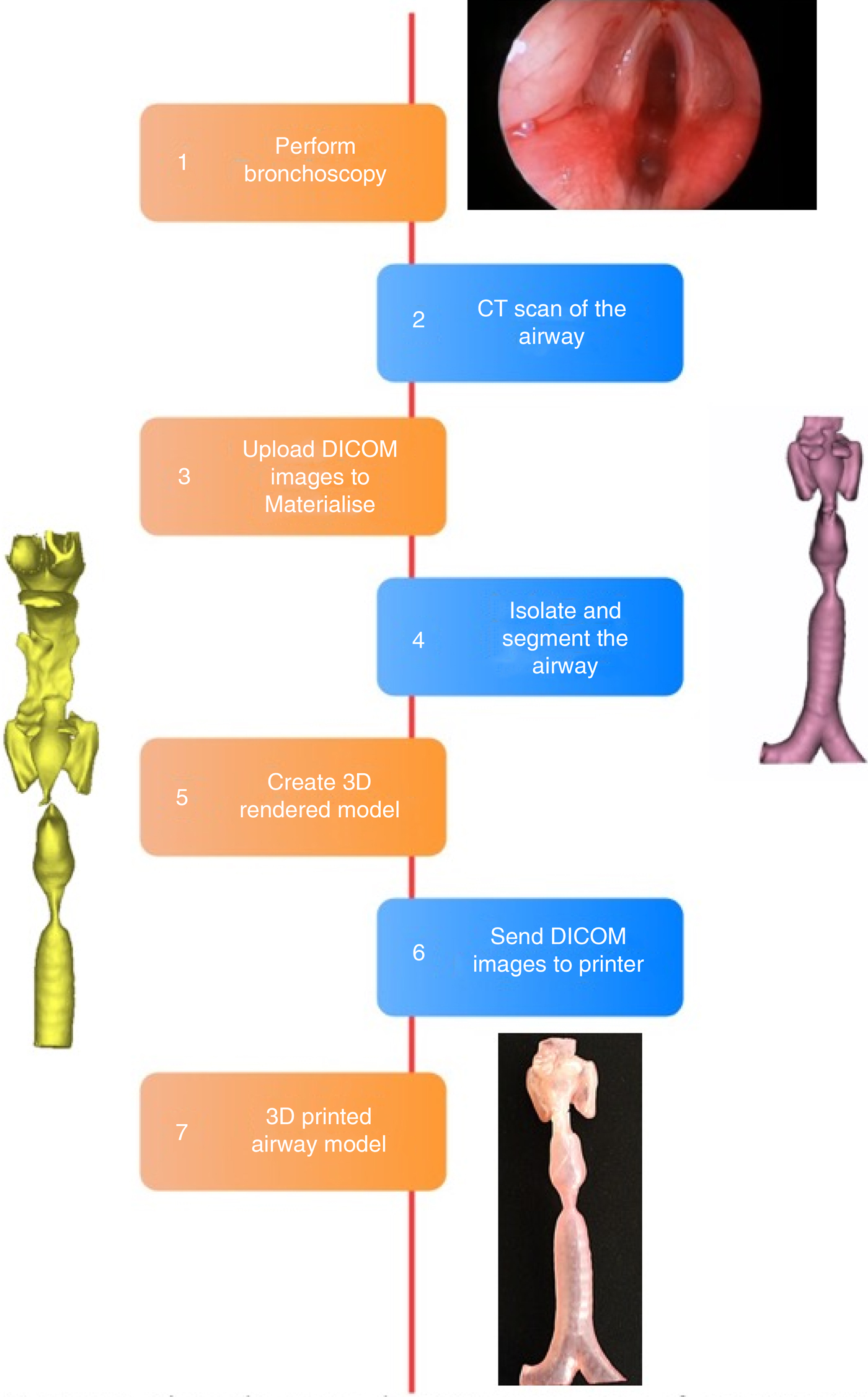
A standard tessellation language (‘STL’) file of the final model was exported and printed using a Polyjet Connex 3D printer (Stratasys, Eden Prairie, Minnesota, USA). PolyJet technology works by laying a 16 µm thick sheet of liquid photopolymer on a build tray that is cured via ultraviolet light. The liquid polymer used was a proprietary material known as Materialise Heartprint (the actual polymer is not disclosed by the company). The process resulted in the formation of soft and flexible 3D models that depicted the area of stenosis. The process is sequentially depicted in Figure 1.
Flow diagram depicting process of creating three-dimensional printed model of airway. CT = computed tomography; DICOM = Digital Imaging and Communications in Medicine; 3D = three-dimensional

Case reports
Case one
A six-year-old child was referred from a facility at another centre for rehabilitation following a road traffic accident. The child had been intubated for one month and was subsequently tracheostomised with a 3.5 Bivona® tracheostomy tube at the other centre's facility for extubation failure.
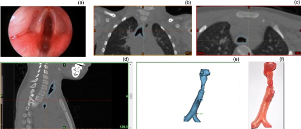
A direct laryngoscopy and bronchoscopy revealed Cotton–Myer grade 4 subglottic stenosis (100 per cent), with posterior glottic involvement (Figure 2a). A CT scan with 1 mm cuts was obtained to evaluate the stenosis; this revealed grade 4 subglottic stenosis with posterior glottic involvement (Figure 2b–d). The Digital Imaging and Communications in Medicine images were used to create a 3D printed model, using the process described above (Figure 2e and f).
Tracheal stenosis in a six-year-old. (a) Bronchoscopy showing grade 4 subglottic stenosis. (b) Coronal, (c) axial and (d) sagittal computed tomography scans. (e) Three-dimensional (3D) rendered model of airway. (f) Finished 3D printed model.

The child was a candidate for extended partial cricotracheal resection with placement of a stent. Prior to the surgery, the 3D printed model was used to educate the family regarding the nature and extent of the stenosis, and the details of the surgical procedure. The model helped in explaining the complex reconstruction to the family, and clarified the surgical steps for the resident and the surgical team. The child subsequently underwent extended partial cricotracheal resection with thyrotracheal anastomosis.
Case two
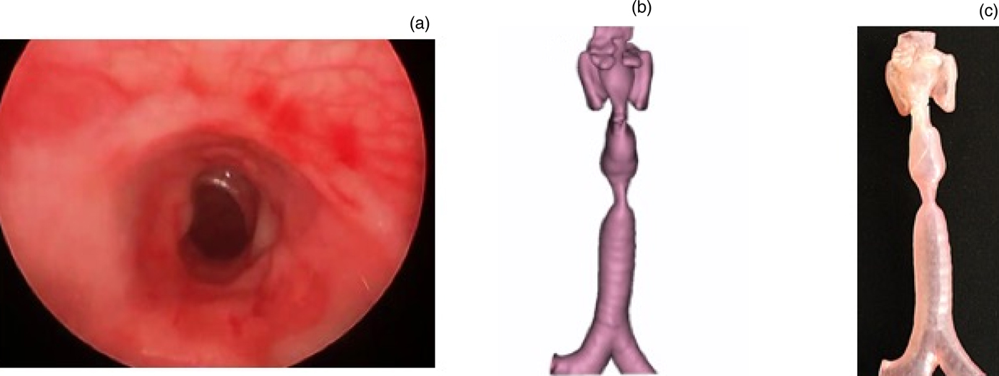
A 16-year-old patient was referred for persistent biphasic stridor following intubation. The patient underwent a bronchoscopy which revealed grade 3 tracheal stenosis (80 per cent) (Figure 3a). The stenosis recurred even after two sessions of laser incision of the tracheal stenosis and balloon dilatation, prompting a decision for tracheal resection and end-to-end anastomosis.
Tracheal stenosis in a 16-year-old. (a) Bronchoscopy showing grade 3 tracheal stenosis. (b) Three-dimensional (3D) rendered model from Digital Imaging and Communications in Medicine (‘DICOM’) images. (c) Finished 3D printed model.

Prior to the surgery, a 3D model of the trachea was printed that depicted the tracheal stenosis (Figure 3b and c). The model was used to educate the family regarding the surgical procedure. The model was also used by the resident for surgical simulation. The 3D model trachea was used to ‘resect’ the depicted stenosis, and to achieve end-to-end anastomosis using running, double-armed, size 5-0 PDS® polydioxanone sutures. The patient subsequently underwent tracheal resection and end-to-end anastomosis.
Discussion
Applications of 3D printing have been described in various specialties, including cardiothoracic, orthopaedic and craniofacial surgery.Reference Tack, Victor, Gemmel and Annemans5 Complex anatomy can be clinically demonstrated using 3D printing, and the models can be used for pre-operative surgical planning and surgical simulation.Reference Itagaki6,Reference Hermsen, Burke, Seslar, Owens, Ripley and Mokadam7 In otolaryngology, 3D printed models have been used in the pre-operative planning for complex mandibular reconstruction.Reference Crafts, Ellsperman, Wannemuehler, Bellicchi, Shipchandler and Mantravadi8 Otolaryngology residents have used 3D printed temporal bones to practise their surgical skills outside of the operating theatre. The efficacy of 3D printing in airways has been demonstrated using resorbable airway splints in patients with severe tracheobronchomalacia.Reference Crafts, Ellsperman, Wannemuehler, Bellicchi, Shipchandler and Mantravadi8
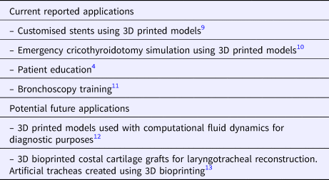
The various reported uses of 3D printing that specifically relate to the paediatric airway are described in Table 1.Reference Balakrishnan, Cofer, Matsumoto, Dearani and Boesch4,Reference Han, Yang, Huang, Wang and Li9–Reference Taniguchi, Matsumoto, Tsuchiya, Machino, Takeoka and Elgalad13 Although use of 3D printed models has been described in the planning of complex tracheal resections in cases of complete tracheal rings, according to our knowledge, the representation of paediatric airway stenosis with 3D printed models has not been described.
Uses of 3D printing in airway surgery

3D = three-dimensional
The present case series includes two cases where airway stenosis could be depicted in 3D printed models. The models were used primarily for patient education, and for discussing the surgery with the family. The models were also brought to the operating theatre to educate the operating theatre staff. In one of the cases, the model was used by the resident for pre-operative surgical simulation. Surgical simulation is an exceedingly important part of residents’ and fellows’ training.
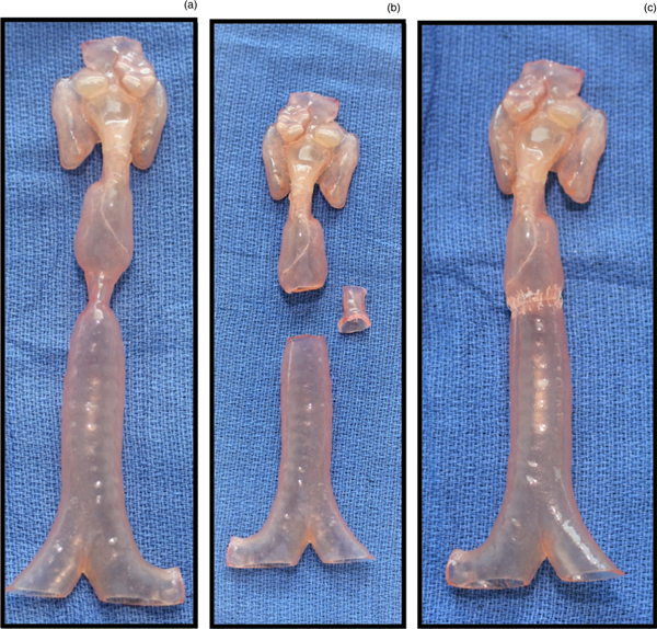
Airway surgical simulation has traditionally been practised on animal models. The models in our case series were printed using a proprietary material that resulted in the production of soft, pliable and flexible models. These models can be used to ‘resect’ the stenosis and practise end-to-end anastomosis (Figure 4a–c). This eliminates the need for animal models and allows the surgeon to practise the surgery on models representing the actual pathology. This paper presents, to our knowledge, the first reported use of 3D printed models for surgical simulation of tracheal resection and end-to-end anastomosis.
(a–c) Surgical simulation prior to actual tracheal resection and end-to-end anastomosis. The pliable three-dimensional printed model makes it easier to practise surgery prior to actual resection.

The average cost for these 3D printed models was US$300. In this case series, the 3D printing was outsourced, which accounts for the price of the models. However, most hospitals have their own 3D printer, which is shared amongst specialties. Having a 3D printer within the hospital decreases the cost required to create the models. These models are a beneficial use of resources, increasingly so as 3D printing becomes more efficient and less expensive. If the patient and caregivers understand the pathology and the surgery, this could lead to better post-operative outcomes, with fewer adverse events following discharge from the hospital.
Future applications of 3D printed models could include coupling with computational fluid dynamics for a potential diagnostic and planning tool. Currently, bronchoscopy is still considered the ‘gold standard’ when it comes to diagnosing laryngotracheal stenosis. However, computational fluid dynamics has been shown to be effective in demonstrating the degree of airflow obstruction,Reference Zdanski, Davis, Hong, Miao, Quammen and Mitran14 and is useful in quantifying the improvement of airflow post-operatively compared to pre-operatively.Reference Chen, Horng and Shih12 The hollowed 3D printed model could determine the improvement in airflow following open airway reconstruction. This information will allow the surgeon to plan accordingly, to achieve optimal results in the actual patient.
• The use of three-dimensional (3D) printing has rapidly expanded
• Three-dimensional tracheal printing was undertaken in two paediatric laryngotracheal stenosis cases
• The 3D printed models can be used in pre-operative planning, patient education and surgical training
• Parents and children can hold the 3D printed model and visualise the complex surgery procedure
• The models can also be used as an education tool for surgical teaching, and in practising surgical resection given the models’ pliability
• To our knowledge, this is the first report of 3D models for acquired laryngotracheal stenosis and the first to discuss 3D printing models in airway surgical simulation
For more complex laryngotracheal reconstructions, there is an expansion of research looking at alternatives to the commonly used autologous costal cartilage grafts. With the expected increased duration of surgery and the possibility of more complications or morbidity associated with costal cartilage grafts, there is an interest in 3D-printed bioresorbable materials that can accomplish the same goals with a reduction in complication rate. The merger of the 3D printing and tissue-engineering fields creates a new and exciting area of research called 3D bioprinting. In the realm of paediatric airway reconstruction, this would entail the creation of a patient-specific bioprinted tracheal construct to replace long-segment tracheal stenosis.
Conclusion
Three-dimensional printed models serve as an educational tool and a visual aid for parents when discussing surgery in complex laryngotracheal stenosis cases. Parents and children can hold the 3D printed model and can visualise what is going to be done during the complex surgery. The models can also be used as an education tool for residents and fellows, in surgical teaching. The pliability of the models also means that they can be used to practise surgical resection. Overall, these models add another dimension in the education of patients and residents.
Competing interests
None declared