Introduction
Experimental findings and epidemiological and clinical observations consistently support the hypothesis of the impact of the intrauterine environment on the structural and functional changes in the fetal organs, increasing the likelihood of certain diseases in adulthood.Reference Skogen and Øverland 1 – Reference Olsen and David 3 Therapeutic decisions after birth would be easier if the adverse effects of the intrauterine environmental factors on the organ function and metabolic processes in the developing fetus could be properly documented by detection of specific proteins. Novel noninvasive biomarkers to be used by neonatologists not only might confirm exposure of the fetus to a variety of hazards but also predict the potential impact of the resulting abnormalities on the health in adulthood.Reference Skogen and Øverland 1 , Reference Ng and Lam 4
The clinical material used in this study in search for novel biomarkers for diagnostic uses in neonatology is meconium, the fetal bowel contents which can take up a wide array of substances and may serve as a source of information expressed as laboratory parameters that describe the intrauterine environment.Reference Ostrea 5 – Reference Lisowska-Myjak and Żytyńska-Daniluk 9
The aim of the study was proteomic analysis of proteins contained in meconium of healthy full-term neonates and classification of the proteins according to their biological properties and potential to produce clinical symptoms. Taking into consideration that the protein composition of meconium may change in the course of intrauterine development, the protein composition was assessed in all meconium portions passed after birth, distinguishing the proteins present exclusively in the first meconium (FM) portions and those found in later meconium (LM).
Material and methods
Material
The study material consisted of all consecutive meconium portions collected serially from the nappy with a disposable spatula and transferred into 50 ml plastic containers. The date, time and weight of each collection were recorded. Meconium collection was considered complete when on inspection a dark-greenish-black color of this material with a characteristic gel-like consistency had changed to the yellowish-brown characteristic of feces. Passing of meconium stopped between 14 and 52 h postnatally. Four to five meconium portions were collected from each neonate. The collected material was frozen at −20°C for approximately 3–5 days and subsequently stored at −80°C for up to 3 months.
Newborns
A total of 10 healthy neonates, born in the Clinical Department of Obstetrics, Female Diseases and Gynecological Oncology, Central Clinical Hospital of the Ministry of the Interior, Warsaw, were included in the study.
Neonate profile: gender: four females and six males; gestational age [weeks]: mean±SD: 38.7±1.5weeks (range 36–41; birth weight [g]: mean±SD: 3244±648.1 (range 2040–4280); Apgar score 1/3/5/10, 10/10/10/10 (n=8); 9/9/10/10 (n=1); 8/9/10/10 (n=1).
Meconium proteome preparation
Before protein assay, meconium samples were thawed over 12 h at 4°C. Analytical grade distilled water was added to the container with meconium to a final homogenate volume of 45 ml. The material was thoroughly shaken using a 358 S Shaker at 350 cpm in a horizontal position for 30 min. The homogenate was subsequently poured into 2 ml deep-freeze test tubes and stored at −80°C.
For protein extraction methanol/chloroform method was used.Reference Wessel and Flügge 10 The pellet was dissolved in 100 mM NH4HCO3. The protein concentrations in meconium before and after methanol/chloroform extraction were measured with the Bradford method. Samples were stored in siliconized microtubes (Sigma-Aldrich) at −80°C for further use.
For reduction of proteins in the individual or pooled samples (see Sample pooling section), 100 mM dithiothreitol was added to the final concentration of 10 mM and samples were incubated at 56°C for 40 min. To block reduced cysteines, 0.5 M iodoacetamide at a final concentration of 50 mM was added and the sample was incubated at room temperature for 30 min in the dark. Proteins were digested with trypsin (Promega) added in 1:20 vol./vol. ratio, with incubation at 37°C overnight.
Sample pooling and isoelectrofocussing fractionation of peptides
In total, 50 samples of meconium from 10 healthy newborns were analyzed, 10 samples of the FM and 40 samples of the remaining meconium passed later (LM). Samples from the same analytical groups (FM or LM) were pooled to compare protein identifications from FM and LM. The samples were pooled to the final volume corresponding to 340 µg of protein.
After the standard procedure of reduction, blocking and trypsin digestion (see Meconium proteome preparation), the resulting peptides were separated on the isoelectrofocussing (IEF) strips as follows: 340 µl buffer was added to the sample [8 M urea, 0.2% IPG (Immobilized PH Gradient) buffer pH 3–11 NL (GE Healthcare), 0.002% bromophenol blue in 50 mM Tris-HCl, pH 8.0]. The solution was applied to an 18 cm IPG strip with 3–11 NL pH gradients (GE Healthcare) for IEF: 340 µl of sample (corresponding to 340 µg protein) per strip. The IPG strip was rehydrated overnight in an IPG box (GE Healthcare). Subsequently, the strips were isoelectrofocussed using the Ettan IPGphor 3 electrophoresis system (GE Healthcare). Two steps of electrophoresis were used. The first step consisted of a 5 h pre-run at 500 V. During this step, the conductivity decreases, and salts and other highly conductive compounds move towards the electrode (anode). Second, a long gradient focussing program was used: 1 h at 500 V, 9 h at 1000 V and 30 h at 8000 V (the final current was 3 µA).
After focusing, the strip was removed from the tray and the overlay oil was blotted with paper tissue. The strip was wrapped in a parafilm and stored at –80°C. The strip was placed on a tray cooled with dry ice and cut into 10 mm sections. The sections were transferred into individual 1.5 ml siliconized tubes. In all, the 18-cm long gel strips were sliced into 18 sections. Peptides were extracted from gel sections by two cycles of 60 µl 0.1% TFA, 2% acetonitrile addition and vortexing for 1 h at room temperature. Aliquots with extracted peptides were stored at −80°C for LC-MS/MS analysis.
Mass spectrometry: LC-MS/MS settings
The peptide mixture (20 µl) was applied to the nanoACQUITY UPLC Trapping Column (Waters) using water containing 0.1% formic acid as the mobile phase and then transferred to the nanoACQUITY UPLC BEH C18 Column (Waters, 75 µm inner diameter; 250-mm long) using an acetonitrile gradient (3–33% acetonitrile over 150 min) in the presence of 0.1% formic acid with a flow rate of 250 nl/min. The column outlet was directly coupled to the ion source of the LTQ-OrbitrapVelos mass spectrometer (Thermo Scientific) working in the regime of data-dependent MS to MS/MS switch. Higher-energy collisional dissociation was used. Other Orbitrap parameters were as follows: one MS scan was followed by maximum 5 MS/MS scans, capillary voltage was 1.5 kV, data were acquired in positive polarity mode.
To increase the number of peptide identifications, three LC-MS/MS runs were performed per individual sample, each covering one of the three ranges of m/z values: 300–600, 500–900 and 800–2000. This approach was found to improve substantially the coverage of protein identification.
Mass spectrometry: qualitative MS/MS data processing and database search
The acquired MS/MS raw data files were processed to peak lists with the Mascot Distiller software (version 2.3.2.0, Matrix Science). The resulting ion lists were searched using the Mascot search engine (version 2.4.1, Matrix Science) against a database composed of all human protein entries from the Swissprot database (20306 sequences) and their reversed versions. The search parameters were set as follows: enzyme specificity, semitrypsin; fixed modification, carbamidomethylation; variable modifications, oxidation; protein mass, unrestricted. The peptide and fragment ions mass tolerances of the database search were established separately for each subfraction LC-MS/MS runs after mass measurement error recalibration, as previously described.Reference Mikula, Gaj and Dzwonek 11 For rescoring database search results Mascot Percolator was used.Reference Brosch and Choudhary 12 , Reference Käll, Canterbury, Weston, Noble and MacCoss 13
Results processing was carried out using MScan, a proprietary software tool implemented in the Java programming language (available at http://proteom.ibb.waw.pl/). The statistical significance of peptide identifications was assessed using target and decoy database search approach and a procedure that provides q-value estimates for each peptide spectrum match (PSMs) in the data set.Reference Mikula, Gaj and Dzwonek 11 , Reference Käll, Storey, MacCoss and Noble 14 Only PSMs with q-values ⩽0.01 were regarded as confidently identified.
Ethical approval
A written informed consent was obtained from the parents of all the newborns before they were included in the study. The Local Committee for Human Experiments, Central Clinical Hospital of the Ministry of the Interior approved the study and the procedures were in accordance with the ethical standards.
Results
Qualitative analysis of meconium
Qualitative analysis of both mix samples: FM and LM yielded a list of 4231 peptides assigned to 946 proteins, of which 430 were identified by at least two peptides (Supplementary Material Data).
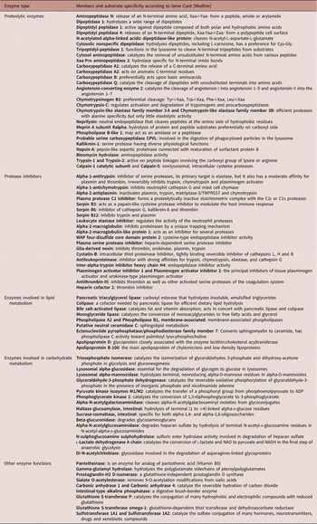
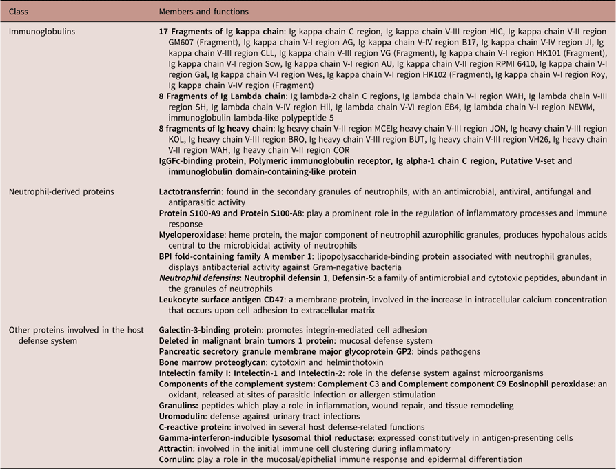
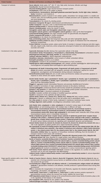
In order to simplify the complex picture of proteins found in the meconium, Tables 1, 2 and 3 present their classification based on a literature search for each protein in the PubMed database. The meconium proteins included enzymes with complex specificity for substrates (Table 1), proteins which play a role in the nonspecific immunity (Table 2), panels of proteins with a common biological function, such as transport and structural proteins or redox and apoptosis proteins and proteins with multiple intracellular functions (Table 3)Footnote a .
Classification of enzymes found in meconium

Meconium proteins which have a role in innate immunity

Panels of meconium proteins with a common biological function

Comparison of protein identifications in FM and LM pooled samples
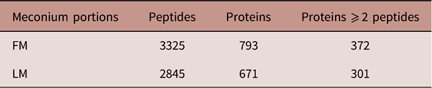
Taking into consideration the sequence at which meconium portions were passed after birth, meconium portions collected from individual neonates were divided into the FM and the LM. Table 4 compares the numbers of proteins identified in FM and in LM.
Number of proteins identified in first meconium (FM) and in later meconium (LM) portions

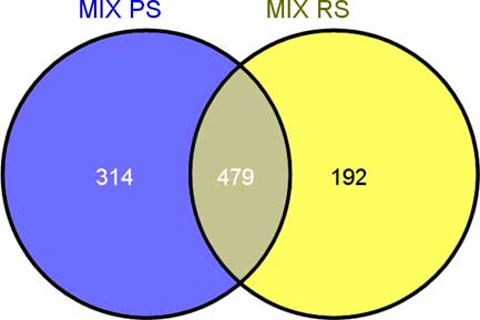
Figure 1 is a graph presenting the differences in the number of proteins found in the FM only (n=314), LM (n=192) only and identified in both the FM and LM portions (n=479).
Venn diagram showing the number of proteins identified in both first meconium portions and later meconium portions.

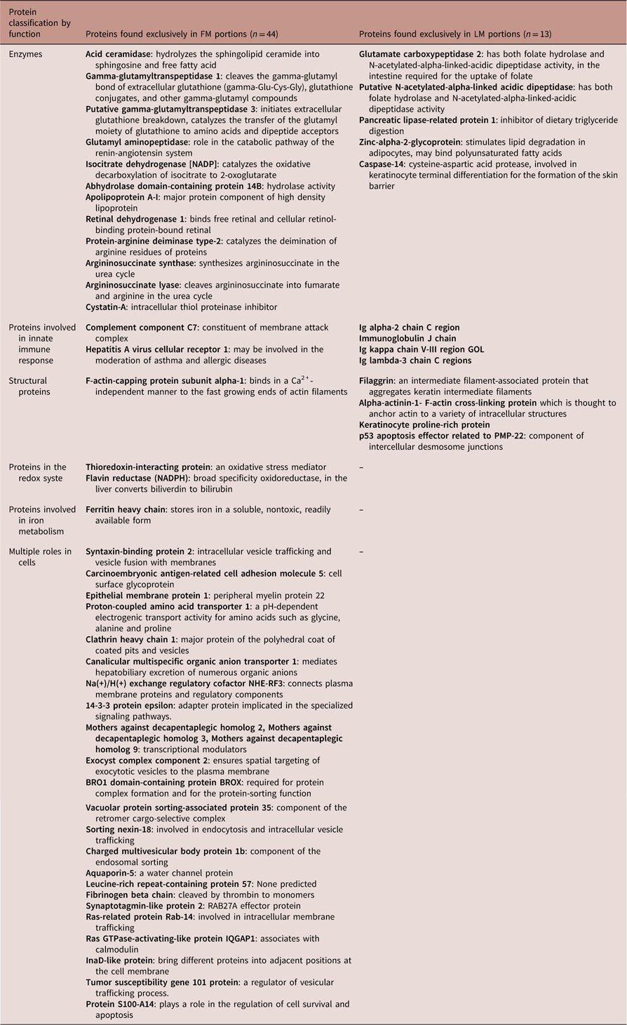
Table 5 lists the proteins represented by two or more peptides identified exclusively in either the FM (n=44) or LM portions (n=13). The proteins are classified by their biological role as enzymes, proteins with a role in the innate immune response, structural proteins, with a role in iron metabolism and other multiple intracellular functions.
Classification by function of proteins found exclusively in first meconium (FM) portions and exclusively in later meconium (LM) portions, identified by at least two peptides

Discussion
Proteomic analysis of the meconium portions passed by healthy full-term neonates demonstrated the presence of 946 proteins, including 430 proteins represented by two or more peptides. A question arises whether these proteins can be mediators of organ- and/or disease-specific metabolic processes in the fetus.
The fetal origins of adult disease hypothesis suggests that a variety of intrauterine environmental hazards to which a fetus is exposed during the vulnerable phases of its development may produce irreversible structural and functional changes which increase the susceptibility to certain diseases in later life.Reference Skogen and Øverland 1 – Reference Olsen and David 3 , Reference Stewart and Malhotra 15
The presence of numerous proteins in meconium is an obvious proof of their direct participation in many processes in the immediate environment of a developing fetus. It is to be hoped that meconium proteins may be of use as novel specific biomarkers employed in noninvasive laboratory diagnostic tests in neonatology.Reference Drozdowski, Clandinin and Thomson 16
Based on the literature search for the function of each protein in the PubMed database, the proteins identified in the meconium samples were classified by their biological function(s).
A large group of enzymatic proteins identified in the meconium was found to include proteolytic, lipolytic and amylolytic enzymes. The human exocrine pancreas is functionally immature at birth with low trypsin activity in the meconium.Reference Drozdowski, Clandinin and Thomson 16 , Reference Neu 17 Interestingly, this study found a large number of proteolytic enzymes with varied protein substrate affinity. Similarly, a large group of protease inhibitors identified in the meconium confirms their role in maintaining the protease-antiprotease balance in the intrauterine environment.
The development of the innate immune system in the intestine may be critical not only during gestation but also play a role in later life. The finding of antibacterial neutrophil-derived proteins, numerous immunoglobulin fragments, proteins involved in the host defense, intestine-specific structural proteins and a large family of mucins suggests their multifaceted potential as a noninvasive diagnostic tool to assess the intrauterine environment and the role of the intestine. The literature data indicate the association between other inflammatory mediators and the development of meconium aspiration syndrome.Reference Laforgia, Baldassarre and Pontrelli 8 , Reference Lisowska-Myjak and Żytyńska-Daniluk 9 , Reference Lee, Romero and Lee 18 Considering that meconium-stained amniotic fluid produces fetal inflammatory response in some neonates only, a question arises which of the components of meconium would play a critical role in the pathogenesis of meconium aspiration syndrome.Reference Kääpä and Soukka 19 , Reference De Luca, Minucci and Tripodi 20
Transport proteins present in meconium and responsible for binding and transport of some vitamins, hormones and microelements constitute important evidence of adequate supply of these substances to the developing fetus. Iron-regulating proteins form a large group with a role in maintaining systemic iron homeostasis.Reference Lou, Mai and Lozoff 21 Elucidation of the role of specific meconium proteins which may be involved in the regulation of iron levels and iron utilization by the fetus may lead to their use as biomarkers for fetal iron status with its consequences for health and disease after birth.
As normally meconium is not excreted by the fetus during gestation, the composition of serial meconium layers deposited in the fetal intestine throughout gestation changes. The differences between these two groups of proteins probably reflect their involvement in specific metabolic processes at different stages of the fetal development. Interestingly, urea-cycle enzymes were found in the FM portions only.
To our knowledge these are the first published findings which indicate the usefulness of meconium of the identification and measurements of meconium proteins for the comprehensive laboratory assessment of the fetal development.
Investigations of meconium in search for novel biomarkers to characterize the intrauterine environment must be strictly controlled. Meconium must not be contaminated by feces formed after birth. Investigators must be aware that the levels of specific proteins measured in consecutive meconium portions may differ. The reference levels of specific proteins which are candidate biomarkers should be established by comparing their levels in meconium from healthy full term neonates to those in infants born prematurely and after complicated pregnancies. Also, the effect of perinatal factors such as the gestational age, mode of delivery or Apgar score on the meconium protein levels should be investigated. Limitations of this study include the small size of the study population. Also, in this study neither MS-friendly protease inhibitors nor antibiotics were added to the meconium samples at the time of collection.
Summing up, many aspects of fetal developmental pathology remain poorly understood. Specific biomarkers to be found in meconium may help to clarify the etiology and pathogenesis of many disorders or defects of fetal organs as well as have a prognostic role in the case of disorders which start in utero but continue after birth and may possibly predispose to disease in adulthood. Meconium naturally passed by neonates can be easily and noninvasively obtained for use in laboratory investigations. The application of proteomics to identify meconium proteins opens the way to novel biomarkers for specific clinical conditions/diseases in neonates, with additional applications as potential predictors of much later health outcomes to be observed in adults.
Acknowledgments
None.
Financial Support
This study was supported by a grant ‘Research Project funded by the National Center for Science, No DEC-2011/01/B/NZ7/00648.’
Conflicts of Interest
None.
Ethical Standards
A written informed consent was obtained from the parents of all the newborns before they were included in the study. The Local Committee for Human Experiments in Central Clinical Hospital of the Ministry of Interior approved the study and procedures were in accordance with the ethical standards.
Supplementary materials
To view supplementary material for this article, please visit https://doi.org/10.1017/S2040174418000028