Introduction
Tularaemia is a rare zoonosis caused by the Gram-negative coccobacillus Francisella tularensis. Human disease is primarily associated with two subspecies: F tularensis tularensis (type A), which is highly virulent and found only in North America; and the less virulent F tularensis holarctica (type B), which is thought to be endemic throughout the northern hemisphere.Reference Ellis, Oyston, Green and Titball1, Reference Schmid, Kornblatt, Connors, Patton, Carney and Hobbs2 In Germany, there are an average of three new cases of tularaemia per year.3 Depending on the route of infection, different forms of tularaemia can be distinguished. After animal bites (from rodents or cats) or transmission by ticks or mosquitoes, the ulceroglandular form occurs, involving skin lesions, fever and lymphadenopathy.Reference Capellan and Fong4 Glandular tularaemia is characterised by lymphadenopathy without an identifiable skin lesion. Airborne infection can cause pneumonic tularaemia,Reference Gill and Cunha5 while inoculation of the conjunctiva leads to the oculoglandular form.Reference Peter and Banyai6 Oropharyngeal tularaemia only accounts for 1–4 per cent of all cases and can be acquired by exposure to contaminated water or consumption of poorly cooked meat (e.g. hares and rabbits).Reference Luotonen, Tapiainen, Kallioinen and Luotonen7 Typical manifestations include painful pharyngitis, fever and cervical lymphadenopathy. Acute otitis media is an extremely rare presentation of this disease. To the best of our knowledge, only a single case of tularaemia in the middle ear has been reported to date.Reference Luotonen, Tapiainen, Kallioinen and Luotonen7
Here, we describe the case of a 27-year-old woman who presented with acute otitis media and prolonged suppurative cervical lymphadenopathy due to infection with F tularensis holarctica.
Materials and methods
Pus and aspirate taken from several suppurative lesions were cultured on cysteine heart agar, according to standard protocols for this fastidious pathogen.Reference Splettstoesser, Matz-Rensing, Seibold, Tomaso, Al Dahouk and Grunow8 Specimens from assumed sterile sites were additionally incubated in a blood culture system (Bactec BD, Heidelberg, Germany) for 21 days. Deoxyribonucleic acid (DNA) was extracted from aspirate using the DNeasy blood and tissue kit (Qiagen, Hilden, Germany). Polymerase chain reaction amplification and product detection were performed in a LightCycler instrument (Roche, Mannheim, Germany). For 16S rDNA polymerase chain reaction amplification, the oligonucleotide primers fD1 (5′ AGAGTTTGATCCTGGCTCAG 3′; where A = adenine, G = guanine, T = thymine and C = cytosine) and 800R (5′ GAGTACCAGGGTATCTAATCC 3′) were used, which target a region of the 16S ribosomal ribonucleic acid gene of eubacteria. In order to identify the resulting 800 bp amplicon, the product was purified and subjected to automated sequencing using primer 800R (Agowa GmbH, Berlin, Germany). The nucleotide sequence of the amplicon was matched to existing GenBank sequences by nucleotide–nucleotide Basic Local Alignment Search Tool analysis, which allowed identification of francisella species specific DNA up to species level. Real-time polymerase chain reaction protocols targeting the 16S rRNA gene and fopA gene were performed as recently described.Reference Splettstoesser, Matz-Rensing, Seibold, Tomaso, Al Dahouk and Grunow8, Reference Fujita, Tatsumi, Tanabayashi and Yamada9 Conventional polymerase chain reaction protocolsReference Bystrom, Bocher, Magnusson, Prag and Johansson10, Reference Broekhuijsen, Larsson, Johansson, Bystrom, Eriksson and Larsson11 were used to confirm the presence of the holarctica subspecies. Serum samples from the patient were examined by enzyme-linked immunosorbent assay and immunoblot, as recently described.Reference Schmitt, Splettstoesser, Porsch-Ozcurumez, Finke and Grunow12
Case report
A 27-year-old, previously healthy woman was admitted to the ENT clinic at the Munich university hospital complaining of a feeling of pressure in the left ear, with accompanying hearing loss and periauricular swellings.
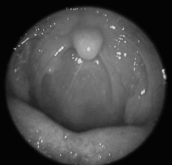
On physical examination, tender, fluctuating masses in the left preauricular and laterocervical regions were found (Figure 1), along with a fluid accumulation, possibly suppurative, behind the tympanic membrane. Oral examination revealed a bulging, retropharyngeal mass behind the right posterior palatine arc (Figure 2).
Preauricular and laterocervical swellings found on initial presentation.

Oral endoscopic view of right retropharyngeal abscess. Note the bulging of the right posterior pharyngeal wall, displacing the right posterior palatine arc craniolaterally.

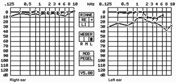
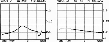
Audiometric tests showed a conductive hearing loss of 15–30 dB (Figure 3). Tympanometry revealed reduced compliance of the sound conduction apparatus (Figure 4). Laboratory tests revealed a white blood cell count of 9.2 × 10⁹/l and a C-reactive protein (CRP) concentration of 36 mg/l (reference range: <5 mg/l). Ultrasound examination of the neck revealed a phlegmoneous inflammation of the soft tissue around the left sternocleidomastoid muscle. Tuberculosis screening was negative.
Pure tone audiogram showing conductive hearing loss of the left ear.

Tympanogram showing a type B pattern in the left ear, suggesting tympanic effusion.

Thirty-five days prior to presentation, the patient had begun to have symptoms of a sore throat (which had lasted three days, days zero to three), ear pain (which lasted four weeks, days zero to 28) and a feeling of pressure in the ear. One week after onset of symptoms, fever occurred (days seven to 14), and another week later (day 14) laterocervical and periauricular swellings developed which were only mildly painful.
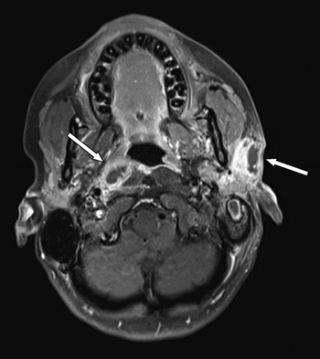
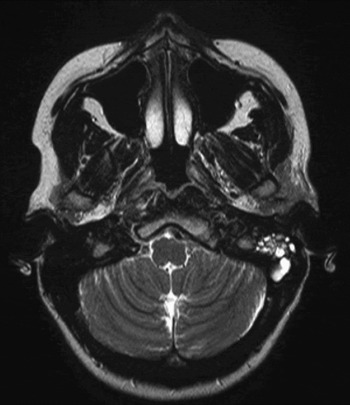
Before the patient had been referred to our hospital, her local ENT specialist had performed computed tomography and magnetic resonance imaging (MRI) scans. These scans had revealed multiple suppurative lesions, including mastoiditis, a preauricular and an infra-auricular abscess, phlegmoneous inflammation of the laterocervical soft tissue (all on the left side), and a retropharyngeal abscess on the right side (Figures 5 and 6).
Axial, T1-weighted, contrast-enhanced magnetic resonance imaging scan, showing suppurative lesions in the left preauricular and right retropharyngeal regions (arrows).

Axial, T2-weighted magnetic resonance imaging scan showing left mastoid effusion.

The patient had been commenced on oral amoxicillin (days seven to 16) and oral cefuroxim (days 17 to 34). On day 25, the patient had noticed multiple, tender, bruise-like, erythematous eruptions on her left shin, probably representing erythema nodosum, which lasted for 10 days.
In our hospital, the patient received intravenous ceftriaxone (days 35 to 44) as empirical therapy. As there was no regression in the lymphadenopathy, as evidenced by repeated ultrasound examination, the enlarged cervical lymph nodes were aspirated on day 41 and the aspirates sent for microbiological analysis. Since conventional microbiological cultures were negative, the aspirate specimens were subjected to eubacterial polymerase chain reaction analysis, which yielded a positive result. By using 16S DNA sequencing, the polymerase chain reaction amplicon was identified up to genus level as francisella species specific DNA. For definitive species identification, the DNA extract and aspirate were subjected to real-time polymerase chain reaction, resulting in the diagnosis of Francisella tularensis subspecies holarctica.
Serological detection of high immunoglobulin (Ig) M and IgG titres against F tularensis Lipopolysaccharide (1:12 800 and 1:6400, respectively) confirmed a diagnosis of acute tularaemia in our patient. Therefore, the patient was commenced on intravenous ciprofloxacin combined with oral doxycycline (days 44 to 50), followed by oral ciprofloxacin (days 51 to 69). Repeated punctures of the suppurative cervical lymph nodes were performed. On day 45, the CRP level was 8 mg/l.
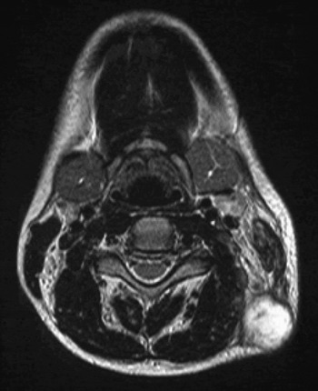
Despite intensive anti-bacterial therapy and repeated needle aspiration of the suppurative lesions, no clear improvement was seen on repeated MRI scans, ultrasound examinations, audiometric tests and tympanometry. On the contrary, the laterocervical cellulitis evolved into an abscess (Figure 7). Consequently, intravenous gentamicin therapy was commenced (days 55 to 69). Even with such therapy, the patient's symptoms were not relieved to a satisfactory degree. Thus, definitive, open surgical drainage of all the suppurative lymph nodes was undertaken, followed by daily irrigation. Paracentesis was conducted in order to relieve the feeling of pressure in the middle ear, revealing no significant fluid collection in the tympanic cavity.
Axial, T2-weighted magnetic resonance imaging scan taken four weeks after presentation, showing left laterocervical abscess formation.

On day 69, the patient was discharged with a CRP level of 3 mg/l (reference range: <5 mg/l).
Over the following six months, the patient was seen several times for follow up. Repeated audiometry revealed a persistent, mild, conductive hearing loss of about 15 dB, corresponding to an unchanged, mild, subjective auditory impairment, although otomicroscopy showed no pathological findings. At the time of writing, the patient did not wish to undergo further tympanoscopic exploration.
Diagnosis of tularaemia
In any infection of the head and neck area with associated lymphadenopathy, the clinician should bear in mind not only common agents but also rare pathogens such as F tularensis. The incubation period of tularaemia is two to six days on average, with a range of one to 20 days. An excessive, unilateral lymphadenopathy is typical. The clinician should be especially alerted by the presence of multiple, suppurative lymph nodes in a patient of good general condition, along with slow clinical progression, similar to cervical tuberculosis. In contrast to invasive bacterial diseases, tularaemia is generally not associated with dramatic changes in blood chemistry (e.g. changes in CRP and white blood cell count).
The mainstay of tularaemia diagnosis is detection of antibodies against F tularensis by microagglutination assay or enzyme-linked immunosorbent assay. The antibody response appears at the end of the second week after onset of disease. Upon clinical recovery, titres decline slowly, but antibodies may persist for several years. For diagnostic purposes, preferably two serum samples are taken, one in the acute phase and another three weeks later. A four-fold increase in titre will confirm the diagnosis. However, patients may seek attention after their antibody level has peaked. A single sample showing a titre of >160 is regarded as confirmatory. In addition, a subsequent sample taken several months later showing a fourfold reduction of antibody titre can also be taken as confirmatory.
Since F tularensis is fastidious and relatively slow-growing, cultures are often unsuccessful. When sample tissue or aspirate is available, polymerase chain reaction should also be performed, since it is the more powerful diagnostic tool, with a sensitivity of approximately 80 per cent and a rapid processing time.Reference Johansson, Berglund, Eriksson, Goransson, Wollin and Forsman13
Discussion
Tularaemia is a very rare disease in central Europe. The oropharyngeal form accounts for only 1–4 per cent of all tularaemia cases and is usually characterised by pharyngitis and lymphadenopathy. Here, we present a unique case with acute otitis media as the primary manifestation and long-lasting conductive hearing loss as a consequence. A medical medline database search yielded only one article on tularaemia affecting the middle ear; a 2002 report of a 10-year-old Finnish boy with tularaemia of the middle ear.Reference Luotonen, Tapiainen, Kallioinen and Luotonen7
Due to its rarity and variable presentation, the diagnosis of tularaemia is difficult and therefore often delayed. Many differential diagnoses need to be considered for symptoms such as fever, sore throat and lymphadenopathy. These include mononucleosis, acute bacterial tonsillitis, bartonellosis, tuberculosis and others. To complicate matters further, our patient spent the first 15 days after clinical manifestation of the disease abroad, and was referred to a hospital only on day 35 of the clinical course. Thus, it took six weeks for our patient to be correctly diagnosed and commenced on specific antibiotic treatment. Tularaemia may well be generally underdiagnosed, because the ample use of antibiotics in patients with the above mentioned symptoms may conceal the typical disease course in many cases.
During our patient's long period of hospitalisation, she was in excellent general condition, which may suggest that infection with F tularensis is often not detrimental. Nevertheless, timely commencement of adequate therapy is important to prevent serious complications. Tularaemia is a potentially fatal disease with an estimated mortality rate of between less than 1 and 14 per cent, depending on the subspecies involved.Reference Staples, Kubota, Chalcraft, Mead and Petersen14 In the pre-antibiotic era, as many as 33 per cent of infected patients died.Reference Wills, Gedosh, Smith and Nichols15 Without appropriate treatment, our patient's inflammation of cervical soft tissue could have developed into life-threatening mediastinitis. However, despite intensive, specific antibiotic therapy, there was no satisfactory resolution of our patient's cervical swelling, such that surgical treatment of the multilocular abscesses was necessary.
In order to clarify the possible route of transmission in our patient, we questioned her thoroughly about her activities before the onset of disease. The first symptoms occurred on 31 July. Between 1 July and 15 August, the patient had stayed in Lyon, France. There, she had been exposed to mosquito bites, and had also had contact with both a domestic rabbit and a cat known to hunt rats. Neither the rabbit, the cat nor any other individuals in contact with these two animals had shown any symptoms suggesting infection. The patient was not aware of any tick bites or erythema. Poorly cooked wild animal meat was excluded as a source of infection. However, one week prior to the first manifestation of tularaemia, the patient had been actively involved in canyoning in the east of France. Consequently, a possible route of infection may have been accidental ingestion of contaminated water during this outdoor activity. Three other persons who also took part in the water sports activities were in good health, but serological investigations excluding subclinical infection were not performed. Considering all the facts, the patient's oropharyngeal contact with water seems to have been the most likely route of infection, but a definitive statement cannot be made.
• Tularaemia is a rare zoonosis caused by the Gram-negative coccobacillus Francisella tularensis
• Following an animal bite (from rodents or cats) or transmission by ticks or mosquitoes, the ulceroglandular form of the disease occurs, with skin lesions, fever and lymphadenopathy
• This paper describes an extremely rare case of otitis media due to F tularensis, complicated by multiple suppurative cervical lesions and a lasting conductive hearing loss
• Otitis media unresponsive to conventional therapy and accompanied by unusually pronounced lymphadenopathy should prompt the clinician to consider tularaemia as a differential diagnosis, in order to initiate timely, specific therapy
According to most authors, the aminoglycosides streptomycin and gentamicin are the drugs of choice for tularaemia treatment.Reference Everett16, Reference Enderlin, Morales, Jacobs and Cross17, Reference Tärnvik and Chu18 One of the most important side effects of these antibiotics, however, is ototoxicity. Since our patient was a music teacher, we were reluctant to risk damaging her inner ear. Therefore, we started treating her with ciprofloxacin, which has recently been reported as suitable for tularaemia.Reference Johansson, Berglund, Gothefors, Sjöstedt and Tärnvik19, Reference Limaye and Hooper20 However, neither ciprofloxacin nor gentamicin, which was added later, could sufficiently relieve the patient's symptoms, probably because antibiotic treatment was initiated too late.Reference Tarnvik and Berglund21 Only after open surgical incision and drainage did the lesions start to resolve. They continued to secrete small amounts of serous fluid for several months before healing spontaneously. Surgical incision and drainage is not always necessary in oropharyngeal tularaemia. It seems to be required when specific treatment is delayed and when manifest suppuration has developed. During an outbreak of oropharyngeal tularaemia in Turkey, 66 per cent of patients required lymph node excision or drainage. In this patient group, the average delay before initiation of an appropriate antibiotic (streptomycin) was eight weeks.Reference Celebi, Baruönü, Ayoğlu, Cinar, Karadenizli and Uğur22
Conclusion
Tularaemia should be taken into consideration in patients presenting with prolonged otitis media associated with lymphadenopathy which is unresponsive to antibiotic treatment, especially when common pathogens cannot be found.
Acknowledgement
The first author was supported by a scholarship of the Programm zur Förderung von Forschung und Lehre of the University of Munich.