Myocarditis is an inflammatory disease of the myocardium caused mainly by a viral infection. Coxsackie viruses, adenoviruses, human herpes virus-6 type B, and parvovirus B19 are frequent causes of viral myocarditis. Despite being an uncommon cause, influenza A and B viruses are a well-documented aetiology. In reported series of patients with myocarditis, influenza was detected in 2% of the patients. Reference Bowles, Ni and Kearney1 On the other hand, myocarditis may complicate about 0–10% of influenza virus infections Reference Ukimura, Satomi and Ooi2 (0.4–5% in paediatric cases). Reference Moore, Vaudry and Scheifele3,Reference Bratincsák, El-Said and Bradley4
The clinical presentation varies widely, from ischaemic-like chest pain to fulminant myocarditis with acute haemodynamic compromise, requiring mechanical circulatory support, with high mortality in the acute phase. Reference Ukimura, Satomi and Ooi2,Reference Hékimian, Jovanovic and Bréchot5
Endomyocardial biopsy (EMB) Reference Dominguez, Kühl and Pieske6 and histologic examination are the gold standard technique for an accurate diagnosis. However, its use is limited in paediatric patients as it is an invasive and risky procedure with its possible complications such as ventricular perforation, tricuspid valve damage, and arrythmias. For this reason, cardiac MRI could be a useful diagnostic tool in stable patients.
We report a series of paediatric patients in our centre with myocarditis due to influenza infection, to emphasise the importance of considering this uncommon aetiology.
Case report
From March, 2007 to March, 2020, 49 paediatric patients (0–16 years) were diagnosed with acute myocarditis at our institution. The diagnosis was established based on histological examination (EMB or necropsy) in 16 patients (16/49, 32.7%) and on cardiac magnetic resonance imaging (MRI) in 33 (33/49, 67.3%). The isolated virus was influenza in four patients (4/49, 8.1%). Patient data are described in Table 1.
The four girls presented with cardiogenic shock 2–4 days after starting with fever and unspecific symptoms of upper respiratory tract infection. No respiratory compromise was reported initially, but all patients developed respiratory failure eventually. All four patients needed intravenous inotropes and three of them required mechanical circulatory support with veno-arterial extracorporeal membrane oxygenation (ECMO). ECMO support was initiated during the first 24 hours of admission in all three patients through right cervical cannulation. After initial stabilisation on ECMO, an atrial septostomy was carried out in the catheterisation laboratory to decompress left cavities. Reference Dolader, Betrian Blasco and Balcells7 During the same procedure, an EMB was performed, obtaining six samples from the right side of the interventricular septum. Reverse transcription polymerase chain reaction (RT-PCR) test for influenza virus were carried out in heart (n = 3), blood (n = 4) and respiratory (n = 4) samples. Only respiratory samples tested positive. Interestingly, despite the severe presentation, histological findings revealed mild inflammatory infiltration (Table 1) and necrosis with oedema just in one patient (patient 1). In patient 4, the diagnosis of myocarditis was achieved by MRI.
Patient data.

a Months.
b Days of symptoms are the days between first day of symptoms and the cardiac symptoms.
c LVEF: left ventricle ejection fraction on admission.
d LVEDV: left ventricle end-diastolic volume.
e PE: pericardial effusion.
f DT: diagnostic test to support myocarditis diagnosis, EMB endomyocardial biopsy, MRI: magnetic resonance imaging.
g CD3/mm Reference Ukimura, Satomi and Ooi2 : positive CD3 lymphocytes per square millimeter at biopsy.
h RT-PCR: reverse transcription polymerase chain reaction test for influenza virus ARN detection at myocardial and respiratory tract samples, Neg: negative.
i RT-PCR: reverse transcription polymerase chain reaction test for influenza virus ARN detection at myocardial and respiratory tract samples, Inf B: influenza B virus.
j Troponin I assay (normal values: <0.04) measures before February, 2020.
k High-sensitivity cardiac troponin T assay (normal values: <45) measured after February, 2020.
l CK: creatine-kinase (normal values: <145).
m P-BNP: pro-brain natriuretic peptide (normal values: <300).
n LVEF: left ventricle ejection fraction at hospital discharge.
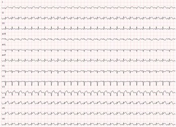
From an echocardiographic point of view, there was a normal or reduced left ventricle end-diastolic volume, septal or posterior wall hypertrophy, and systolic dysfunction. All patients, excluding patient 4, had significant right ventricle dysfunction. Moderate to severe pericardial effusion was present in three patients. From an electrocardiographic perspective, sinus tachycardia and low voltages were present in all patients and non-specific repolarisation abnormalities with ST elevation were present in patient 4 (Fig 1). Although arrythmias in myocarditis may be frequent and severe, our patients presented none. All patients had significant troponin, creatine kinase, and natriuretic peptide elevation. Patients 1 and 3 had the most elevated creatinine kinase values due to severe rhabdomyolysis and required transient renal replacement therapy. All patients recovered myocardial function during the first 6–10 days but two patients experienced severe complications. Patient 2, developed a disseminate invasive aspergillosis, with cerebral, pulmonary, and cardiac involvement (vegetation located at the mitral valve). This girl followed a protracted course, needing prolonged antifungal therapy, prolonged ECMO support (73 days) due to respiratory failure and neurosurgical evacuation of a cerebral haemorrhage. Patient 3 presented a pulmonary and cerebral (intraventricular) haemorrhage while on ECMO. Both patients were eventually discharged with mild and moderate to severe neurological sequelae, respectively.
ECG from patient 4.

As it relates to specific medical treatment and according to our protocol, all patients received steroids (2mg/kg/day), intravenous immunoglobulin, and oseltamivir.
Discussion
Influenza accounts for 3–5 million cases of severe illness in general population and myocarditis is a possible complication that can be present in 0.4–5% of paediatric patients. Reference Moore, Vaudry and Scheifele3,Reference Bratincsák, El-Said and Bradley4 On the other hand, in reported series of myocarditis, Influenza was responsible of the disease in 2% of the patients. Reference Bowles, Ni and Kearney1
Myocarditis has a broad spectrum of clinical manifestations including fulminant presentation with severe cardiac dysfunction, that can require mechanical circulatory support. Reference Bratincsák, El-Said and Bradley4 As described previously, Reference Estabragh and Mamas8 sustained sinus tachycardia, electrocardiographic changes, or increased troponin levels, were documented in all our patients and should alert about potential cardiac involvement and should prompt a formal cardiac evaluation. Transfer of the patient to a tertiary centre with ECMO capability should be considered early in the course of the disease. Fatal arrhythmias are also described, but were not present in our patients. Reference Ukimura, Satomi and Ooi2
EMB and histological examination are the gold standard technique for the diagnosis of myocarditis. However, it is an invasive procedure with its own risks and its use is limited. Cardiac MRI has become the reference non-invasive diagnostic tool allowing identification of the hallmarks of myocardial inflammation. Sensitivity defined for the diagnosis of myocarditis is 87.5% and specificity 96.2%. Nevertheless, the sensitivity of the examination may be influenced by the pattern and extent of myocardial involvement, which actually reflects clinical presentation of the disease. As previously reported, MRI sensitivity is high for infarct-like presentation (80%), low for cardiomyopathy and heart failure presentation (57%), and very low for arrhythmic presentation (40%). Reference Francome, Chimenti and Galea9 On the other hand, cardiac MRI can only be performed in stable patients and is not useful for establishing aetiologic diagnosis. Three of our patients required ECMO and underwent EMB, whereas the fourth patient was stable and an MRI was performed to confirm the diagnosis.
Strikingly in all of the patients presented, we found mild myocardial histological changes compared with other types of fulminant myocarditis. As previously described, there are viruses, as enteroviruses, that cause direct myocardial damage, as viral replication can cause direct cardiomyocyte injury. However, respiratory viruses, such as Influenza, are examples of viruses that can trigger an immune-mediated lymphocytic myocarditis in the absence of viral genome in the myocardium. Reference Ammirati, Frigerio and Adler10 For that reason, it is unlikely to isolate viral genetic material at the myocardium Reference Ukimura, Satomi and Ooi2–Reference Ammirati, Frigerio and Adler10 and in most cases, myocardial dysfunction can be related with specific cytokines and endothelial dysfunction. Reference Ukimura, Satomi and Ooi2
Regarding treatment, there are no specific therapies that have been proven to improve outcome in Influenza myocarditis and the mainstay of treatment appears to be supportive. However, immunosuppressive therapy in virus-triggered myocarditis, in which inflammatory response appears to be the cause of myocardial damage, may play an important role in the treatment of these patients. Reference Ammirati, Frigerio and Adler10 Our usual strategy is to prescribe steroids on patients with fulminant myocarditis presentation. Antiviral treatment is reserved to those patients with a viral infection confirmed by a positive RT-PCR result, in these patients with oseltamivir. Cardiac function recovery is common, Reference Estabragh and Mamas8 but complications are frequent and can include rhabdomyolysis, bleeding complications, and opportunistic infections.
Acknowledgements
This paper and the research behind it would not have been possible without the exceptional support of all our colleagues at the Paediatric Cardiology Department and Paediatric Intensive Care Unit.
Financial support
This research received no specific grant from any funding agency, commercial, or not-for-profit sectors.
Conflicts of interest
The authors declare that they have no conflict of interest
Ethical standards
This research study was conducted retrospectively from data obtained for clinical purposes. Ethical approval was not required.