INTRODUCTION
Toxoplasma gondii has a global distribution and infects a wide range of mammals and birds (Dubey and Beattie, Reference Dubey and Beattie1988). Even though there is a well-documented sexual phase in felids in the life-cycle of T. gondii (Dubey et al. Reference Dubey, Miller and Frenkel1970; Hill et al. Reference Hill, Chirukandoth and Dubey2005) it is not obligatory and 3 clonal lineages are predominant among humans and domestic animals in Europe and North America (Howe and Sibley, Reference Howe and Sibley1995). Interestingly, much greater genetic variation has been found among T. gondii strains from South America, especially in forest regions where wild cats remain (Carme et al. Reference Carme, Bissuel, Ajzenberg, Bouyne, Aznar, Demar, Bichat, Louvel, Bourbigot, Peneau, Neron and Darde2002, Reference Carme and Demar-Pierre2006), and it was recently proposed that the species originated there and only spread to other continents through human activities in the past 500 years (Lehmann et al. Reference Lehmann, Marcet, Graham, Dahl and Dubey2006). In mouse models, the parasite genetic lineage is a crucial determinate of pathogenesis with Type I strains being highly virulent and the others largely apathogenic (Sibley and Boothroyd, Reference Sibley and Boothroyd1992). Similarly, acute infection in immunocompetent adults is more frequently associated with Type I or atypical parasite genotypes (Grigg et al. Reference Grigg, Ganatra, Boothroyd and Margolis2001; Carme et al. Reference Carme, Bissuel, Ajzenberg, Bouyne, Aznar, Demar, Bichat, Louvel, Bourbigot, Peneau, Neron and Darde2002; Khan et al. Reference Khan, Jordan, Muccioli, Vallochi, Rizzo, Belfort, Vitor, Silveira and Sibley2006). In immunocompromised patients overt toxoplasmosis is primarily associated with the decline of CD4-positive T-cells, but the parasite genotype may also be of importance. Type I (Khan et al. Reference Khan, Su, German, Storch, Clifford and Sibley2005) and Type II (Howe et al. Reference Howe, Honore, Derouin and Sibley1997; Honoré et al. Reference Honore, Couvelard, Garin, Bedel, Henin, Darde and Derouin2000) strains have both been cited as the main cause of disease reactivation in HIV-patients; however, this may reflect variations in the general distribution of strains in the region.
Around two thirds of all HIV-patients live in sub-Saharan Africa (WHO, 2006), but little is known about the distribution of T. gondii genotypes in this part of the world. A small number of strains with presumed African origin have been found to possess unusual or mixed genotypes (Ajzenberg et al. Reference Ajzenberg, Banuls, Su, Dumetre, Demar, Carme and Darde2004; Khan et al. Reference Khan, Su, German, Storch, Clifford and Sibley2005). In Uganda, we have previously found evidence that all three lineages can cause reactivation in HIV-patients but that the majority belonged to genotype II (Lindström et al. Reference Lindström, Kaddu-Mulindwa, Kironde and Lindh2006). However, typing of parasites from human blood samples can only reveal disease-causing strains; in order to assess the diversity of strains in Uganda it is important to sample an unbiased population.
Free-range chickens feed from the ground and are thus good indicators of the oocyst contamination (Dubey et al. Reference Dubey, Venturini, Venturini, Piscopo, Graham, Dahl, Sreekumar, Vianna and Lehmann2003a); furthermore, they may play a role in direct transmission to humans. There are no epidemiological studies examining risk factors for T. gondii infection in Uganda, but a serological study from Nairobi, Kenya (Bowry et al. Reference Bowry, Camargo and Kinyanjui1986) suggests that oocysts from environmental sources like soil and water are of importance for transmission in this part of Africa. Chickens are also popular food animals and even though meat is generally eaten well cooked in Uganda, handling practices may allow for cross-contamination of pathogens from raw to cooked chicken meat (Wanyenya et al. Reference Wanyenya, Muyanja and Nasinyama2004). Isolation and genotyping of T. gondii in chickens thus provide important background information on the strains that may infect humans, either through oocyst-contaminated food/water or through bradyzoites in meat. To date one study has been completed on chickens from sub-Saharan Africa. Here genotyping of 16 isolates at the SAG 2 locus revealed a very high proportion of Type III strains (Dubey et al. Reference Dubey, Karhemere, Dahl, Sreekumar, Diabaté, Dabiré, Vianna, Kwok and Lehmann2005a). In the current study we assessed the distribution and diversity of parasite strains in 85 free-range chickens from Kampala, Uganda. To our knowledge this is the first study to isolate and genetically characterize a set of African T. gondii strains on multiple genetic loci.
MATERIALS AND METHODS
Chickens
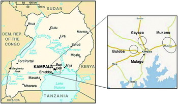
Free-range chickens (n=85) were obtained from households in 4 different areas in and around the capital Kampala (latitude: 0°19′North, longitude: 32°35′East). Of these, 25 chickens originated from Gayaza north of the city, 21 came from Buloba in the west, 19 from rural environments in Mukono district, east of Kampala and finally, 20 chickens were obtained from Mulago, close to the city centre (Fig. 1). The chickens ranged in age from approximately 6 months to 2 years and were classified into 4 age groups based on the length of the tarsometatarsus and the size of the comb. Group 1: 6–9 mo (n=27), 2: 10–13 mo (n=13), 3: 14–17 mo (n=32), 4: ⩾18 mo (n=13). Symptomatic toxoplasmosis is rare in chickens (Dubey et al. Reference Dubey, Webb, Sundar, Velmurugan, Bandini, Kwok and Su2007a) and all the birds in this study appeared healthy. The chickens were purchased and brought to the Veterinary Faculty of Makerere University where they were bled and killed. The blood was allowed to clot, centrifuged and 0·5–1·5 ml of serum was stored at 4°C until use. Whole head, heart and serum from each chicken were transported on cold packs to the Animal Parasitic Diseases Laboratory, U. S. Department of Agriculture, Beltsville, MD, for T. gondii evaluation. Five to seven days elapsed between the death of the animals and the arrival of tissues in Beltsville.
Map of sampling areas.

Serological examination
Sera of all chickens were tested for anti-T. gondii antibodies with the modified agglutination test (MAT) as described previously (Dubey and Desmonts, Reference Dubey and Desmonts1987). Control sera came from a seropositive pig with a titre of 1:200. Briefly, different dilutions of sera (ranging from 1:5 to 1: 640) were incubated overnight with T. gondii antigen (formalin-fixed parasites), borate buffer, Evans blue dye and β-mercaptoethanol in round-bottomed microtitre plates. A positive reaction leads to the formation of antibody-antigen precipitates, while negative reactions result in an accumulation of antigen at the bottom of the well.
Bioassay and isolation of T. gondii
Tissues of all chickens were bioassayed for T. gondii infection. Birds with serological titres of 1:20 or higher were bioassayed individually while tissues from chickens with MAT-titres of 1:5 or 1:10 were pooled in groups of 3–5. The brains and hearts were homogenized with 0·85% NaCl in sterile blenders, digested in pre-warmed acidic pepsin at 37°C for 45 min, filtered, washed and finally resuspended in 0·85% NaCl with 1000 U penicillin and 100 μl of streptomycin per ml. This digest was subcutaneously inoculated in 4 out-bred female Swiss Webster mice. Six weeks post-inoculation the mice were bled for serological testing and thereafter killed. The brains were examined microscopically for T. gondii, used for DNA-extraction and also cryopreservation of live parasites. Brains and hearts from 45 seronegative chickens (MAT-titres of <1:5) were pooled in 3 groups of 15 and fed to 3 T. gondii-free cats. Faeces of cats were examined for shedding of T. gondii oocysts 3–14 days after ingestion of chicken tissues, as previously described by Dubey (Reference Dubey1995).
Genetic characterization for T. gondii
T. gondii DNA was extracted with the DNeasy-kit (QIAgen) from the pepsin-digested homogenates of chicken tissues and also from the brains of all infected mice (as determined by MAT–titre ⩾1:25). Strain typing was performed by PCR amplification of 5 genetic markers (SAG1, SAG2 (5′ and 3′ amplified separately), SAG3, BTUB and GRA6) and thereafter analysed by restriction fragment length polymorphism (RFLP) with the appropriate enzymes as described previously (Dubey et al. Reference Dubey, Patitucci, Su, Sundar, Kwok and Shen2006a). Samples were sequenced to resolve strain type whenever there were unclear enzyme digestion results and sequencing was also performed on selected samples to screen for possible new polymorphisms.
RESULTS
Antibodies to T. gondii were detected in 40 of 85 chickens (47%) at the following titres; 1:5 (n=9), 1:10 (n=11), 1:20 (n=5), 1:40 (n=12), 1:80 (n=1), 1:160 (n=1) and 1:320 (n=1). The chickens from the Buloba area showed a markedly lower seroprevalence (9·5%) and were on average younger than chickens from the other areas, 76% compared to 17% belonged to age group 1. Among the 64 chickens from Gayaza, Mukono and Mulago the total infection rate was 59% and 20 of 64 had titres of ⩾1:20. An increase in seropositivity over the four age categories was observed and the infection rate in age group 4 was 69%.
All chickens with MAT ⩾1:20 were bioassayed individually in mice and 9 of 20 samples were found to contain infectious parasites, as indicated by seropositive mice. None of the strains were mouse virulent but appeared to be unusually slow growing, producing only a small number of tissue cysts in brain. In several cases parasites were not visible by careful microscopic examination, but T. gondii DNA was still detected through PCR of infected mouse brains. Pooled tissues from chickens with titres of 1:5 or 1:10 did not result in any seropositive mice and none of the cats fed tissues from seronegative chickens shed oocysts.
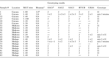
A multiplex external PCR followed by separate amplifications with internal primers for 5 different genes was applied to all chicken tissue homogenates. Only 1 of the pooled samples was PCR-positive, it originated from 3 Mulago chickens with MAT 1:10 and was found to be a Type I based on SAG1, SAG2 and GRA6 digestion patterns. All the chickens with MAT ⩾1:20 were found to harbour parasites through positive PCR-amplifications of at least 2 markers, including samples from 11 chickens which did not infect mice. Direct genotyping of these by PCR-RFLP revealed that 6 chickens were infected with Type I parasites, 8 with Type II and 1 with a Type III strain. In addition, tissues from 5 of the chickens gave mixed genotype results with different alleles at different markers, either I/II (samples 62, 68 and 78) or II/III (2 and 70). Genotyping of parasites from infected mouse brains for some of these samples did, however, indicate infections with multiple T. gondii strains in the same host. For example, tissue from sample 2 appears to contain alleles from all 3 genotypes. Three out of 4 mice injected with this sample became infected, parasites from 2 of these mice had alleles typical of Type II and Type III strains while the third mouse gave a Type I/II mix. In cases where mixed genotypes were detected via RFLP, sequencing was performed to verify the results. Analysis of these sequences frequently revealed double peaks at known polymorphic sites indicating the presence of multiple alleles (Fig. 2), which suggests multiple infections rather than recombination. The genotyping results are shown in Table 1.
Sequence showing multiple infection.

Serology titre, bioassay and genotyping result from all chickens positive by PCR

1 Number of mice infected (as determined by MAT-titre ⩾1:25) out of number of mice inoculated.
2 The SAG1 locus can only differentiate between two types. 1=type I; 2=type II or III.
3 One of the mice died before the end of the experiment.
The short gene fragments utilized for genotyping in this study appear to be fairly well conserved. On the basis of PCR-RFLP only 1 sample, from chicken 70, was found to possess an atypical allele. This showed an unusual digestion pattern for SAG1; being sensitive to both Sau96I that cuts Type I strains and HaeII, which cuts Type II and III. Sequencing confirmed that this digestion pattern was due to a polymorphism in a single strain rather than double digestion caused by multiple strains being amplified. Overall, 47 sequences were retrieved (GenBank Accession EF585673-EF585715) from different strains, mostly from the SAG1, SAG3 and GRA6 loci, but only 5 additional SNPs were found; 3 in the SAG3 sequence from the Type III strain from chicken 70 and 1 each for samples 25 (SAG1) and 83 (SAG2 3′), both of which are Type I strains.
DISCUSSION
Direct genotyping of T. gondii from 20 asymptomatic Ugandan chickens showed a predominance of Types I and II. Only 1 pure Type III strain was detected, but Type III alleles were also found in 2 samples with mixed infections. This pattern differs from the previous studies on chickens from Africa, Europe, USA, Middle East and Asia (Sreekumar et al. Reference Sreekumar, Graham, Dahl, Lehmann, Raman, Bhalerao, Vianna and Dubey2003; Dubey et al. Reference Dubey, Graham, Dahl, Hilali, El-Ghaysh, Sreekumar, Kwok, Shen and Lehmann2003b, Reference Dubey, Graham, Dahl, Sreekumar, Lehmann, Davis and Morishitac, Reference Dubey, Salant, Sreekumar, Dahl, Vianna, Shen, Kwok, Spira, Hamburger and Lehmann2004a, Reference Dubey, Karhemere, Dahl, Sreekumar, Diabaté, Dabiré, Vianna, Kwok and Lehmann2005a, Reference Dubey, Edelhofer, Marcet, Vianna, Kwok and Lehmannb, Reference Dubey, Rajapakse, Ekanayake, Sreekumar and Lehmannc; Reference Dubey, Vianna, Sousa, Canada, Meireles, Correia da Costa, Marcet, Lehmann, Dardé and Thulliez2006b), where Types II and III were common and Type I very rarely found. It is also very different from the distribution in chickens from Central and South America (Dubey et al. Reference Dubey, Venturini, Venturini, Piscopo, Graham, Dahl, Sreekumar, Vianna and Lehmann2003a, Reference Dubey, Graham, Silva, Lehmann and Bahia-Oliveirad, Reference Dubey, Navarro, Graham, Dahl, Freire, Prudencio, Sreekumar, Vianna and Lehmanne, Reference Dubey, Levy, Sreekumar, Kwok, Shen, Dahl, Thulliez and Lehmann2004b, Reference Dubey, Morales and Lehmannc, Reference Dubey, Bhaiyat, de Allie, Macpherson, Sharma, Sreekumar, Vianna, Shen, Kwok and Lehmann2005d, Reference Dubey, Gomez-Marin, Bedoya, Lora, Vianna, Hill, Kwok, Shen, Marcet and Lehmanne, Reference Dubey, Lopez, Alveraz, Mendoza and Lehmannf, Reference Dubey, Marcet and Lehmanng, Reference Dubey, Lenhart, Castillo, Alvarez, Marcet, Sreekumar and Lehmannh, Reference Dubey, Patitucci, Su, Sundar, Kwok and Shen2006a, Reference Dubey, Sundar, Pineda, Kyvsgaard, Luna, Rimbaud, Oliveira, Kwok, Qi and Suc, Reference Dubey, Su, Oliveira, Morales, Bolaños, Sundar, Kwok and Shend), where Types I, III and recombinant or atypical strains dominate and Type II is uncommon. The genotype distribution found in this study bears more resemblance to what has been seen in human patients from Europe (Fuentes et al. Reference Fuentes, Rubio, Ramirez and Alvar2001; Ajzenberg et al. Reference Ajzenberg, Banuls, Tibayrenc and Darde2002; Aspinall et al. Reference Aspinall, Guy, Roberts, Joynson, Hyde and Sims2003). The lineage distribution in Ugandan chickens is also similar to our previous data generated from HIV-patients from the same part of Uganda (Lindström et al. Reference Lindström, Kaddu-Mulindwa, Kironde and Lindh2006), with the main difference being a somewhat higher proportion of Type II in the human patients. We did not find any evidence of recombinant T. gondii strains in the chickens and only a limited number of SNPs; most notably the atypical digestion pattern seen at SAG1 for chicken 70, which has previously been found in T. gondii from different host species in Asia and South America (“u-1” in Dubey et al. Reference Dubey, Cortés-Vecino, Vargas-Duarte, Sundar, Velmurugan, Bandini, Polo, Zambrano, Mora, Kwok, Smith and Su2007b, Reference Dubey, Rajapakse, Wijesundera, Sundar, Velmurugan, Kwok and Suc, Reference Dubey, Sundar, Gennari, Minervino, Farias, Ruas, dos Santos, Cavalcante, Kwok and Sud, Reference Dubey, Zhu, Sundar, Zhang, Kwok and Sue). However, we cannot exclude the presence of recombinants and more detailed genotyping of isolated clones is needed to find out if and how the T. gondii strains in Uganda differ from strains found elsewhere.
An unexpected finding in this study was the preferential selection of genotype II in the mouse bioassay. Mice from 9 out of 20 groups showed serological evidence of infection 42 days post-inoculation. Six of these were pure Type II strains while 3 were samples from chickens infected with Type II and at least 1 other strain. Among the 11 strains that failed to grow in mice 6 belonged to genotype I, 2 were Type II, 2 were mixed I/II infections and 1 was Type III. The explanation for this divergence is unknown; however, both the parasite burden and the ratio of tachyzoites to encysted bradyzoites may vary between strains and thus their survival during transport and susceptibility to acidic pepsin digest may differ (Dubey, Reference Dubey1998; Fux et al. Reference Fux, Nawas, Khan, Gill, Su and Sibley2007). It is also possible that strains may vary in their capacity to infect mice or in transmissibility through subcutaneous inoculation.
An interesting finding was that 5 out of 20 chickens appeared to be infected by 2 or 3 genetically distinct strains of T. gondii. Mouse experiments have shown that previous infections only confer partial protection towards reinfection with strains of other lineages (Araujo et al. Reference Araujo, Slifer and Kim1997; Dao et al. Reference Dao, Fortier, Soete, Plenat and Dubremetz2001), but multiple infections in naturally infected hosts have only been reported occasionally (Aspinall et al. Reference Aspinall, Guy, Roberts, Joynson, Hyde and Sims2003; Dubey et al. Reference Dubey, Vianna, Sousa, Canada, Meireles, Correia da Costa, Marcet, Lehmann, Dardé and Thulliez2006b). However, considering the high prevalence of infection in chickens and the presence of multiple parasite genotypes within the relatively small geographical areas of this study, it is not surprising that some individuals have been exposed to more than one parasite genotype during their lifetime. Consistent with this, we found that all chickens with multiple infections belonged to age group 3 or 4, and both the individuals with triple infections were ⩾18 months old.
Re-infection with new strains may also be of importance for latently infected humans, perhaps even more so in the case of immunocompromised individuals. In this study, the use of multiple markers and combination of direct genotyping and bioassay revealed infection with strains of different genotypes in 5 chickens. With closer typing, for example microsatellite analysis, it might also be possible to detect multiple infections with different strains from the same lineage. Studies investigating the presence of multiple infections in HIV-patients would be important in order to consider superinfection relative to reactivation for toxoplasmosis in AIDS.
We would like to thank William Muyombya, Maria Nakamya, Katherine Hopkins and Luciana Bandini for excellent technical assistance. Irene Lindström is funded by a Marie Curie Fellowship for Early Stage Training.