Cardiac rhabdomyomas in tuberous sclerosis complex usually occur as single or multiple tumours detected antenatally or in infancy, with subsequent stabilisation or spontaneous regression.Reference Jóźwiak, Kotulska and Kasprzyk-Obara 1 – Reference Smythe, Dyck, Smallhorn and Freedom 6 Development of a new cardiac rhabdomyoma beyond infancy is very rare. We describe a patient who needed surgical resection of a large haemodynamically significant cardiac rhabdomyoma in neonatal life and then developed a new-onset large cardiac rhabdomyoma at 2 years of age in a different location.
Case report
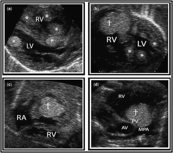
A 1-day-old boy with a maternal history of tuberous sclerosis and prenatal diagnosis of multiple intracardiac rhabdomyomas was transferred to our institution with respiratory distress. Echocardiography showed multiple ventricular mural and septal tumours and one large mobile tumour arising below the pulmonary valve causing right ventricular outflow tract obstruction (Fig 1a–d). Because of its large size, location, and increasing right ventricular outflow tract pressure gradient on serial echocardiograms, the patient underwent surgical resection of the large right ventricular outflow tract cardiac rhabdomyoma on day 11 of life, along with patent ductus arteriosus ligation and suture closure of a small secundum atrial septal defect. The patient’s post-operative course was unremarkable and follow-up echocardiograms at 5 and 11 months of age showed continued regression of cardiac tumours with a decrease in both size and number of lesions.
( a – d ) Echocardiographic still frames in the neonatal period showing multiple ventricular tumours, a large obstructive RVOT tumour, and a clear RA. ( a ) Parasternal long-axis view showing multiple ventricular tumours (*) in the ventricular septum and free wall. ( b ) Subcostal view showing large RVOT cardiac rhabdomyoma (†). Note additional small tumours in the ventricular septum and left ventricular free wall (*). ( c ) Subcostal view to show right ventricular inflow and outflow shows the large obstructive RVOT tumour (†). Note normal RA cavity with absence of atrial tumour. ( d ) Modified parasternal short-axis view showing the large RVOT tumour (†) prolapsing into the pulmonary valve apparatus. AV=aortic valve; LV=left ventricle; MPA=main pulmonary artery; PV=pulmonary valve; RA=right atrium; RV=right ventricle; RVOT=right ventricular outflow tract.

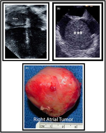
However, at his 2-year follow-up visit, an electrocardiogram showed atrial tachyarrhythmia, and an echocardiogram demonstrated the presence of a new large right atrial tumour, measuring 3.6×3.5 cm, arising from the atrial septum and causing partial obstruction to superior vena cava blood return (Fig 2a–c). The right atrial tumour had never been noted on prior echocardiograms or intraoperatively at the time of right atrial inspection for atrial septal defect closure, suggesting that either a de-novo tumour had arisen or a very small previously unrecognised tumour had suddenly grown in size between 1 and 2 years of age. The patient had developed atrial tachyarrhythmia with impaired ventricular function that was suspected to be related to the dysrhythmia. There was also concern that there may have been sarcomatous change given the rapid growth. These considerations prompted surgical resection. In the operating room, a broad stalk from the atrial septum was noted and the tumour was noted to have effaced the right atrial free wall. Surgical resection of the tumour was successful. Histopathological findings were identical to the previously resected right ventricular outflow tract tumour and consistent with rhabdomyoma. Post-operatively, he remained stable in sinus rhythm and was discharged home 4 days later. At his last follow-up echo at age 4 years, no new cardiac tumours were noted.
( a – c ) Echocardiographic and macroscopic images of the new large right atrial tumour found at 2 years of life. ( a ) Four-chamber view showing the large RA tumour (***). ( b ) Transoesophageal echocardiogram image showing a large homogeneous RA tumour (***). ( c ) Gross macroscopic appearance of the resected RA tumour. RA=right atrium.

Discussion
Cardiac rhabdomyomas are the most common primary cardiac tumours of infancy, and their association with the tuberous sclerosis complex is well established. The natural history of cardiac rhabdomyomas in tuberous sclerosis has been described as single or multiple tumours, most commonly located in the ventricular walls or interventricular septum, detected prenatally or within the first year of life with subsequent stabilisation or regression of tumours.Reference Jóźwiak, Kotulska and Kasprzyk-Obara 1 – Reference Nir, Tajik and Freeman 4 , Reference Smythe, Dyck, Smallhorn and Freedom 6 , Reference Bosi, Lintermans, Pellegrino, Svaluto-Moreolo and Vliers 7 Cardiac rhabdomyomas are managed conservatively; surgery is indicated for patients who have haemodynamic compromise or intractable arrhythmias. Operative management typically involves resection of the intracavitary portion of the tumour without complete excision.
The underlying pathophysiology in the development of all tumours in tuberous sclerosis is thought to be linked to the mammalian target of rapamycin signalling pathway, which controls cell growth and proliferation.Reference Jozwiak, Jozwiak and Wlodarski 8 – Reference Schwartz, Fernández, Kotulska and Jóźwiak 10 The proteins hamartin and tuberin, which are affected by the TSC1 and TSC2 mutations, respectively, in tuberous sclerosis, form a heterodimer, which exerts inhibitory control on mammalian target of rapamycin. Loss of function of either one of these proteins disrupts the heterodimer, leading to increased mammalian target of rapamycin activation. Despite the fact that this explains tumorigenesis in tuberous sclerosis, it does not speak to why cardiac rhabdomyomas in tuberous sclerosis tend towards regression, whereas other tumours including cerebral tumours do not. A possible mechanism is the overexpression of the pro-apoptotic “Bax” protein in cardiac rhabdomyoma cells in patients with tuberous sclerosis when compared with heart cells from normal controls.Reference Kotulska, Larysz-Brysz and Grajkowska 9 More studies are needed to elucidate this further.
After an extensive review of the literature, we found only one case series describing a new-onset cardiac rhabdomyoma or cardiac rhabdomyoma with significant growth after the first year of life. Jóźwiak et alReference Jóźwiak, Kotulska and Kasprzyk-Obara 1 , in a case series of 154 cardiac rhabdomyoma cases, described new-onset cardiac rhabdomyomas or cardiac rhabdomyomas that grew in size after infancy in just six patients in their cohort. They noted some important characteristics of this subset of patients in whom new or growing cardiac rhabdomyomas were identified: every single patient was pubertal or pre-pubertal when the new or growing tumour was identified and were in the age range of 10–15 years; of the six patients, five were female; and mutational analysis was available for four patients and every single one had the TSC2 mutation. Combining these findings with the reported fact that tumour size and number are known to peak in the neonatal period and during infancy with subsequent regression, Jozwiak postulated that sex hormones could be an important contributing factor in tumour development – transplacentally transported maternal hormones in the neonate presenting with multiple tumours, and pubertal hormones in older children presenting with new-onset or growing tumours.
Thus, it is now generally held that cardiac rhabdomyomas peak in incidence within the first year of life following which the course until puberty is uncomplicated. Looked at from this viewpoint, our patient had some important peculiarities. The patient was male, was neither an infant nor pre-pubertal/pubertal, and had developed a tumour in the right atrium, an unusual site. This case illustrates the possibility that cardiac rhabdomyomas can develop de-novo or grow significantly in size even after the high-risk first year of life, and before the child enters puberty. Despite there being no malignant transformation, the tumour proved dangerous by virtue of its size and location, affecting both haemodynamics and electrical conductance. Importantly, the patient was completely asymptomatic at the time of echocardiographic discovery of the tumour, further emphasising the importance of routine surveillance of patients with tuberous sclerosis for cardiac rhabdomyomas even beyond the first year of life.
Conclusion
Routine echocardiographic surveillance is essential in asymptomatic tuberous sclerosis patients with apparently stable or regressing cardiac rhabdomyomas, as new-onset tumours can occur even in the low-risk period of 2–10 years of life.
Acknowledgement
None.
Financial Support
This research received no specific grant from any funding agency, commercial, or not-for-profit sectors.
Conflicts of Interest
None.