Introduction
Ventricular septal defect is the most common CHD, accounting for approximately 30% of all congenital cardiac anomalies. Reference Cohen, Lopez, Allen, Shaddy, Penny, Feltes and Cetta1 Muscular ventricular septal defects constitute 10–15% of all ventricular septal defects and are anatomically classified as mid-muscular, apical, anterior, and posterior. Reference Soto, Becker and Moulaert2,Reference Lopez, Houyel and Colan3 Small muscular ventricular septal defects, especially those detected in infancy, have a high spontaneous closure rate approaching 80%. Reference Cohen, Lopez, Allen, Shaddy, Penny, Feltes and Cetta1 In contrast, large muscular ventricular septal defects are less likely to close and often present with symptoms of congestive heart failure. If left untreated, they pose a risk of pulmonary vascular obstructive disease. Moderate-sized defects lead to left atrial and left ventricular volume overload and pulmonary hypertension. In addition, they predispose one to growth retardation, recurrent lower respiratory tract infections, and endocarditis. Reference Kumar and Kumar4
Surgical closure has traditionally been the standard of care. However, open-heart surgery with cardiopulmonary bypass carries significant morbidity. Because of their favourable anatomy with well-defined rims and sufficient distance from valves and the conduction system, muscular ventricular septal defects are especially amenable to transcatheter closure. Reference Lock, Block and McKay5 This contrasts with peri-membranous ventricular septal defects, where device closure carries a higher risk of atrioventricular block and valvular interference.
Transcatheter closure of ventricular septal defects was first described by Lock et al. in 1987 using the Rashkind double umbrella device. Reference Lock, Block and McKay5 Although various dedicated ventricular septal defect occluders have since been employed, conventional occluders often require large-profile sheaths, limiting their use in infants and small children. Reference Holzer, Balzer and Cao6–Reference Ghosh, Sahai and Agrawal10
The KONAR-MF™ occluder (Lifetech Scientific Co. Ltd., Shenzhen, China) is a novel, medium-profile, double-disc device specifically designed for ventricular septal defect closure. Its bi-directional deployment capability and compatibility with smaller delivery systems make it advantageous in infants and small children.
This retrospective study evaluates the experience with transcatheter muscular ventricular septal defect closure using the KONAR-MF™ occluder across three paediatric cardiac centres in India, focusing on procedural techniques, safety, and outcomes.
Materials and methods
Study design
This was a retrospective, multicentre study conducted between July 2018 and July 2024 at three tertiary paediatric cardiac centres in India. Approval from the institutional ethics committees of all participating centres was obtained.
Patient selection
All patients who underwent transcatheter closure of muscular ventricular septal defects using the KONAR-MF™ occluder during the study period were included. Medical records were reviewed for demographic data, clinical history, chest X-ray, 12-lead electrocardiogram, and transthoracic echocardiography. Significant left-to-right shunting was inferred from left atrium and left ventricle dilatation.
Muscular ventricular septal defects were classified as mid-muscular, apical, anterior, or posterior. Echocardiography was the basic imaging modality. A comprehensive echo for assessment of chamber size, location of defect, presence of associated anomalies, and valve regurgitation was performed. The defect size was measured in two orthogonal views, and the maximum dimension was considered for device selection (Figures 1(a) and (b)). Proximity to valves, relation to the moderator band, and presence of multiple defects were documented. Inclusion criteria:
Transthoracic echocardiographic images of a large muscular ventricular septal defect located below the moderator band. ( a ) Two-dimensional four-chamber view showing the ventricular septal defect (arrow). ( b ) Colour Doppler demonstrating left-to-right shunt across the defect (dotted arrow). ( c ) Apical four-chamber view with device in situ (arrow). ( d ) Colour Doppler showing a small residual shunt (dotted arrow), which resolved on follow-up. RA = right atrium; RV = right ventricle; LV = left ventricle. ( e ) KONAR-MFOTM depicting the dual screws(black arrows), high pressure disc(1) and low pressure disc(2) with inner PTFE membrane.

Patients were eligible if they met at least one of the following indications:
-
1. Clinical signs of congestive heart failure,
-
2. Failure to thrive attributable to the ventricular septal defect,
-
3. Two or more episodes of respiratory infections within the preceding 12 months.
-
4. Evidence of left atrium and left ventricle dilatation.
Adequate rims of atleast 3–4 mm with sufficient distance from adjacent valves were mandatory requirements for device closure.
Device
The KONAR-MF™ occluder is a self-expandable nitinol double-disc device with a central waist and screw attachments at both ends, permitting deployment via either arterial or venous access (Figure 1( e )/supplemental figure S1). It is made of 0.005 mm nitinol wire. Smaller devices (sizes 5/3 mm to 8/6 mm) lack a polytetrafluoroethylene membrane and can be delivered through a 5F Judkin’s right coronary guide catheter. Larger devices (sizes 9/7 mm to 14/12 mm) contain a polytetrafluoroethylene membrane and require 6F–7F delivery sheaths. Device size was typically 1–2 mm larger than the maximum measured defect diameter.
Procedure
Procedures were performed under local or general anaesthesia depending on patient size and access route. Standard arterial and venous access (femoral or jugular) was obtained. Intravenous heparin (100 U/kg IV) was administered, and baseline haemodynamics were recorded. A left ventricle angiogram was done in an appropriate view, left anterior oblique being the most often used (Figure 2( a )) Device delivery was guided by fluoroscopy and echocardiography. Device release was undertaken only after confirming stability, preserved valve function, and absence of significant residual shunt (Figures 1( c ) and ( d )).
Angiographic demonstration of different approaches for muscular VSD closure. ( a ) Left ventriculogram showing muscular VSD (arrow). ( b )Retrograde approach for closure of a mid-muscular VSD with KONAR-MF occluder. ( c ) Jugular venous approach with arteriovenous loop across the defect with buddy wire to provide stability during deployment. ( d ) A KONAR-MF occluder positioned across the defect with LV angiogram showing no residual flow. ( e ) Patent foramen ovale method in a 4.2 kg infant for closure of a muscular VSD using KONAR-MF occluder. Catheter advanced across the PFO into the left atrium, left ventricle and pulmonary artery. ( f ) Successful device deployment across the VSD. (Abbreviation : VSD = ventricular septal defect).

1. Retrograde (Figure 2( b )): Apical and posterior muscular ventricular septal defects were crossed from the left ventricle aspect. Apical defects sometimes have multiple openings on the right ventricle, and trabeculations make it difficult to cross from the right ventricle. A Judkin’s right coronary catheter along with a 0.035 J-tip Terumo wire combination was used. The wire was positioned in the pulmonary artery or the caval vein. The low-pressure disc (Figure 1( e )) was first opened in the right ventricle and then pulled back towards the septum with the waist across the defect. The high-pressure disc (Figure 1( e )) was deployed in the left ventricle under fluoroscopy and transthoracic echo guidance. Defects where the device could be deployed through 5 Fr guide catheters were also closed retrogradely.
In the event of the requirement of larger sheaths, the wire was snared from the venous end to form an arteriovenous loop (Figures 2( c )). The long delivery sheath was passed from the venous end and positioned in the left ventricle. The high-pressure disc was opened in the left ventricle and pulled snugly against the septum, followed by deployment of the low-pressure disc in the right ventricle.
2. Antegrade: This method was most suited for mid-muscular and anterior muscular defects situated above the moderator band. Judkin’s Right Catheter, or Tiger Catheter, was positioned in the left pulmonary artery and connected to the pressure transducer system. The catheter was gradually withdrawn from the left pulmonary artery into the right ventricle, tracing the interventricular septum and monitoring the pressure wave pattern. The pressure waves changed from a pulmonary artery waveform to a right ventricle waveform, and subsequently either a hybrid or left ventricular waveform was seen when the catheter approached the level of the defect and after crossing it. A J-tip Terumo wire was then advanced into the left ventricle and then into the aorta. The Terumo was exchanged for stiff wire over which a delivery sheath was taken from the femoral venous end. The high-pressure retention skirt was first opened in the left ventricle and pulled towards the septum, and then the right ventricle disc was opened and the device deployed.
3. From across the PFO (Figures 2( e ) and ( f )): Two midmuscular defects were closed by this approach. In the presence of a PFO, a wire can be negotiated from the femoral vein into the right atrium across the PFO to the left atrium and then into the left ventricle. The defect crossed from the left ventricle to the right ventricle, eliminating the need for arterial access.
4. Hybrid approach: The perventricular approach was performed in the operating theatre under transesophageal echo guidance in patients with severe hyperkinetic pulmonary hypertension. One patient had two large apical defects requiring two devices and hence posed a significant challenge for direct surgery as well as closure in the catheterisation laboratory. The other child had a single large midmuscular defect of 11 mm, necessitating a big device in a small child weighing 5.5 kg. A lower ministernotomy was done, and the pericardium was opened and retracted. The defect was crossed by direct right ventricle puncture. The device was deployed under transesophageal guidance.
Post-procedural management
All patients were monitored for ≥48 hours. An electrocardiogram and echocardiogram were performed before discharge. Aspirin (3–5 mg/kg/day) was prescribed for six months. In patients with residual shunting, urinalysis was performed to exclude haemolysis.
Follow-up protocol
Patients were followed at 1 week, 3 months, 6 months, 12 months, and annually thereafter. Each visit included clinical evaluation, an electrocardiogram and an echocardiogram to assess device position, residual shunting, ventricular function, and valvular competence.
Statistical analysis
Categorical variables were summarised as frequencies and percentages. Continuous variables were expressed as mean ± standard deviation (SD) or median with interquartile range, as appropriate. Analyses were limited to descriptive statistics.
Results
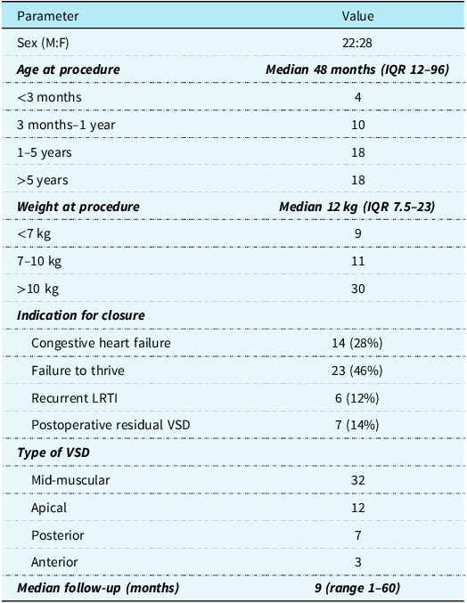
A total of 50 patients underwent transcatheter closure of muscular ventricular septal defects using 54 KONAR-MF™ devices. Baseline characteristics are summarised in Table 1. The median age was 48 months (interquartile range 12–96), and the median weight was 12 kg (interquartile range 7.5–23), with the smallest patient weighing 2.4 kg at 1 month of age. Fourteen procedures were performed in infants; 7 were <6 months old, and 9 weighed <7 kg.
Demographic and clinical characteristics of patients undergoing muscular ventricular septal defect device closure (VSD – ventricular septal defect, LRTI – lower respiratory tract infection)

Clinical indications included failure to thrive in 46% (n = 23), congestive heart failure in 28% (n = 14), recurrent respiratory infections in 12% (n = 6), and postoperative residual ventricular septal defect in 14% (n = 7). Among residual cases, two were closed within 10 days after surgery, while the others were closed between 3 months and 2 years postoperatively. One patient with a history of infective endocarditis also underwent device closure 6 months after complete treatment.
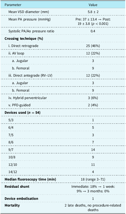
The anatomical distribution of muscular ventricular septal defects was mid-muscular (32), apical (12), posterior (7), and anterior (3). The mean ventricular septal defect diameter was 5.8 ± 2 mm. Mean pulmonary artery pressure decreased from 37 ± 13.4 mmHg to 19 ± 3.8 mmHg (p < 0.001). Eleven patients had pulmonary artery pressure >50% of systemic.
Procedural details are summarised in Table 2. An initial left ventriculogram was done to delineate the defect (Figure 2( a )). Retrograde device deployment (Figure 2( b )) was used in 26 defects (48%), arterio-venous loop with venous deployment (Figures 2( c ) and ( d )) in 14 (26%), direct antegrade right ventricle–left ventricle approach in 9 (16%), hybrid per-ventricular approach in 3 (6%), and patent foramen ovale-guided approach (Figures 2( e) and ( f )) in 2 (4%) (Supplementary Figure S2). The most used devices were sizes 9/7 mm (n = 14) and 12/10 mm (n = 11). The median fluoroscopy time was 18 minutes (range 3–71).
Catheterisation and procedural data (VSD – ventricular septal defect; PA – pulmonary artery; ao – aorta; RV – right ventricle; LV – left ventricle; AV – arteriovenous; PFO – patent foramen ovale)

Outcomes
An immediate residual shunt was observed in 18% of patients (Figure 1( d )), predominantly with non-polytetrafluoroethylene devices; this reduced to 9% at 1 week and resolved completely by 3 months. Mild new tricuspid regurgitation was noted in 14%, which resolved spontaneously on follow-up. No patient developed aortic regurgitation, haemolysis, or atrioventricular block.
Complications and mortality
One instance of device embolisation into the descending aorta occurred due to undersizing; the device was surgically retrieved, and the defect was successfully closed. There were no procedure-related deaths. Two deaths occurred among patients with postoperative residual ventricular septal defects: one was a 4-month-old who died of sepsis 10 days post-procedure, and the other a 2-month-old infant with a single lung who succumbed 2 weeks after the procedure due to sepsis and prolonged ventilation.
The overall survival was 96%. At a median follow-up of 9 months (range 1–60), no conduction disturbances, valvular dysfunction, late embolisation, or recurrent shunt was observed.
Discussion
The technical difficulties of closing large muscular ventricular septal defects in small infants are multifactorial. One major challenge is the requirement for large delivery sheaths, which is overcome by an arteriovenous loop enabling the introduction of sheaths from the venous end. In infants, the creation of an arteriovenous loop with a bulky sheath can exert a “stenting effect” across both atrioventricular and semilunar valves, leading to haemodynamic compromise. Furthermore, kinking of the sheath during looping can hinder smooth device delivery. Reference Carminati, Butera and Chessa7,Reference Elmarsafawy, Hafez and Alsawah8,Reference Diab, Cao and Mora11–Reference Carminati, Butera and Chessa13 The trans-carotid approach by some operators avoids arteriovenous loop formation and provides a direct path Reference Breatnach, Kenny and Linnane14 while jugular venous access may be required for apical or posterior muscular defects. Reference Carminati, Butera and Chessa13 Traditional muscular ventricular septal defect occluders typically required sheaths ranging from 6F to 12F, which limited their applicability in small infants. Reference Carminati, Butera and Chessa7,Reference Carminati, Butera and Chessa13 The retrograde approach is a simpler technique with shorter fluoroscopy and procedure time. Reference Jameel, Arfi and Arif15 As a result, some operators restricted transcatheter closure to defects <6 mm, Reference Alshahrani, Linnane, McCrossan, Oslizlok, McMahon, Walsh and Kenny16 while others opted for perventricular hybrid closure in larger defects. Reference Thakkar, Patel and Shah17
The KONAR-MF™ occluder addresses many of these limitations and has some advantages when compared to other occluders (Supplementary Table S3). This double-disc device with dual-sided screws allows deployment from either the arterial or venous side, reducing the necessity for arteriovenous loop formation. The double-disc system forms a safety feature when occluding defects with pulmonary hypertension. The KONAR-MF™ small range devices can go through 5 Fr guide catheters, and the maximum sheath required is 7 Fr. Reference Elmarsafawy, Hafez and Alsawah8,Reference Walavalkar, Maiya and Pujar9 Conventional muscular ventricular septal defect occluders may need sheaths up to 8F or 9F for delivery. In our multicentre study, retrograde arterial deployment was feasible in 48% of patients, while arteriovenous loop formation was required in only 26%. This represents a major procedural simplification compared with traditional duct and VSD occluders, which mandate venous deployment and require arteriovenous loop creation for larger sizes. Reference Tanidir, Baspinar and Saygi18,Reference Koneti, Azad and Bakhru19
Anterior–superior defects were best approached retrogradely from left ventricle to right ventricle, particularly when located near the outflow tract. Mid-muscular and postero-inferior defects often required jugular access with arteriovenous loop formation to achieve sheath stability. Apical defects were generally approached via jugular or trans-PFO routes. True posterior defects, especially in small infants, were more amenable to perventricular hybrid closure under echocardiographic guidance.
Our findings align with prior studies reporting the safety and efficacy of the KONAR-MF™ device in children <10 kg. Reference Tanidir, Baspinar and Saygi18 Importantly, our series represents the experience of muscular ventricular septal defect closure with KONAR-MF™, including infants as small as 2.5 kg and as young as 1 month. Residual shunting was observed in 18% immediately post-procedure, predominantly with devices lacking polytetrafluoroethylene membranes, but this resolved completely at 3 months, consistent with the natural process of endothelialisation.
Hybrid perventricular closure was employed in three patients, reinforcing its continued value for large or posterior defects with elevated pulmonary pressures. Reference Thakkar, Patel and Shah17 The ability to achieve high procedural success across diverse anatomies and weight ranges underscores the device’s adaptability.
Limitations
Retrospective design, modest cohort size, and non-uniform follow-up are acknowledged limitations. Nonetheless, this is one of the largest real-world multicentre experiences using KONAR-MF™ in muscular ventricular septal defects.
Conclusion
Muscular ventricular septal defect closure using the KONAR-MF™ occluder is safe, feasible, and effective across a wide paediatric age and weight spectrum, including carefully selected infants under 5 kg. The device’s medium-profile configuration, dual-sided deployment, and compatibility with smaller delivery systems provide important procedural flexibility, particularly in anatomically challenging locations.
Our multicentre experience highlights favourable short—and mid-term outcomes, with high procedural success, minimal complications, and no conduction disturbances. Residual shunts observed immediately after the procedure resolved with follow-up, underscoring the reliability of the device.
Supplementary material
The supplementary material for this article can be found at https://doi.org/10.1017/S1047951126111664.
Data availability statement
Clinical deidentified data will be available to the readers on reasonable request.
Financial support
No external funding has been received.
Competing interests
The author, Dr Nageswara Rao Koneti, reports receiving royalty payments for the KONAR-MFTM ventricular septal defect occluder.
Ethical standard
Ethics committee approval has been obtained. Informed consent from patients was waived off, as only the standard of care was practised and patient identity has not been revealed. SJICR/EC/2024/019—Sri Jayadeva Institute of Cardiovascular Sciences and Research, Bangalore. IEC-2024-PEDCARD-035—Amrita Institute of Medical Sciences, Cochin. RCHBH/JUNE/24-cardio-16—Rainbow Children’s Hospital, Hyderabad. The authors assert that all procedures contributing to this work comply with the ethical standards of the relevant national guidelines on human experimentation (Indian Council of Medical Research, 2017) and with the Helsinki Declaration of 1975, as revised in 2008, and have been approved by the institutional ethics committees.