Introduction
Maternal health during gestation is an important factor for fetal development. Around 6% of pregnant women report high levels of psychological stress during pregnancy, which results from several conditions such as depression, panic or domestic violence. Reference Woods, Melville, Guo, Fan and Gavin1 Higher levels of maternal cortisol may cross over the placental barrier promoting long-lasting effects in the regulation of the hypothalamic–pituitary–adrenal (HPA) axis of the offspring, reducing the expression of glucocorticoid receptors (GR) in the brain and exacerbating glucocorticoid secretion. Reference Kapoor, Dunn, Kostaki, Andrews and Matthews2 Thus, stressful events in the prenatal period have been related to hyperactive HPA axis responses, metabolic changes and low birth weight in the offspring in both early and adult life. Reference Kapoor, Dunn, Kostaki, Andrews and Matthews2–Reference Tamashiro and Moran4
The systemic secretion of adrenal glucocorticoids after stress is central for the mobilization of lipids and glucose reserves, increasing the energy demand to help maintain homeostasis. Reference Peckett, Wright and Riddell5 For example, individuals with Cushing syndrome or patients receiving treatment with exogenous glucocorticoid administration present hyperglycemia, insulin resistance and increased adiposity. Reference Geer, Islam and Buettner6 In the liver, the control of triglycerides and gluconeogenesis is mediated by GR. Reference Peckett, Wright and Riddell5 In addition, glucocorticoids act to control the regulation of phosphoenolpyruvate carboxykinase (PEPCK), an essential enzyme in the hepatic production of glucose. Reference Geer, Islam and Buettner6 Peroxisome proliferator-activated receptors (PPARs) α and γ also play a key role in lipid and glucose metabolism, in addition to oxidation and the storage of fatty acids. Reference Grygiel-Gorniak7 Previous studies on rodents have already demonstrated that prenatal stress is associated with long-term alterations in the offspring’s metabolism, with a decreased expression of PPARα and γ in males and an increased expression of GR in prepubertal mice. Reference Maeyama, Hirasawa and Tahara8,Reference Brunton, Sullivan, Kerrigan, Russell, Seckl and Drake9 These changes may be important in lipid metabolism and glucose-insulin control.
It is already known that regular physical activity is an important non-pharmacological tool to prevent the onset of metabolic disorders Reference Zanuso, Jimenez, Pugliese, Corigliano and Balducci10,Reference Pi, Liu and Xi11 and, when performed during pregnancy, may provide benefits to both mother and offspring, both in rodents and humans. Reference Harris, Baer and Stanford12 We have recently shown that pregestational exercise attenuates the effects of prenatal stress in the offspring, in a sex-dependent manner. Reference Luft, Levices and Costa13 However, the long-term effects of exercise before pregnancy and the possible mechanisms involved in the metabolic response are not yet well elucidated. Nevertheless, persistent effects of exercise have been related to cognitive improvement in rats Reference Rezaee, Marandi, Alaei and Esfarjani14 and the attenuation of posttraumatic stress symptoms in humans. Reference Pitts, Whealin and Harpaz-Rotem15 Acute exercise activates the HPA axis and promotes increased gluconeogenic activity. Reference Stranahan, Lee and Mattson16 Physical exercise has also been associated with increased insulin sensitivity in humans with abnormal glucose levels, Reference Herzig, Ahola, Leppaluoto, Jokelainen, Jamsa and Keinanen-Kiukaanniemi17 decreased progression of type 2 diabetes in adults Reference Zanuso, Jimenez, Pugliese, Corigliano and Balducci10 and ameliorated hepatic steatosis in mice. Reference Pi, Liu and Xi11 The relationship between exercise and HPA axis activity involves a series of complex mechanisms, considering that it may be influenced by duration, type and intensity of exercise, as well as by the characteristics of the stressor and the population studied.
The regulation of stress pathways is markedly influenced by sex differences. Reference Sze and Brunton18 Studies with rats have shown that females which are subjected to prenatal stress show greater activation of the HPA axis compared to males. Reference Weinstock19 Across the estrous cycle, female mice present different hormonal levels of progesterone and estrogen, which could act directly in the regulation of the physiology and function of several organs and systems. Reference Viau and Meaney20 Differences between males and females can also be found in metabolic functions and diseases in humans. In general, men have impaired fasting glucose and a greater predisposition to diabetes, while women appear to have a higher incidence of obesity and the accumulation of visceral adiposity. Reference Mauvais-Jarvis21
Considering that fetal exposure to stressful events promote permanent changes in the function of multiple systems and the practice of regular exercise is an important component in the treatment of several diseases, we hypothesized that exercise before pregnancy would contribute to generating beneficial and persistent metabolic effects on the prenatally stressed offspring. Working towards this goal, we investigated the effects of maternal physical exercise before the gestational period on the metabolic changes induced by prenatal stress in adult mice. Body weight, biochemical analyzes, oxidative stress, gene expression of metabolic-related receptors and possible sex differences were evaluated.
Materials and methods
Animals
Male and female Balb/c mice were used to generate the offspring. Animals were kept in individually ventilated boxes, with a light/dark cycle of 12 h (lights on at 07:00 am), at a temperature of 22 ± 2 °C and with free access to water and food. The animals were purchased from the Experimental Biological Models Center (CEMBE) at PUCRS. The experimental protocol was approved by the University Ethics Committee for the Use of Animals (CEUA) under protocol number 15/00446 and all mice were manipulated according to the guidelines of the National Council for the Control of Animal Experimentation (CONCEA).
Experimental design
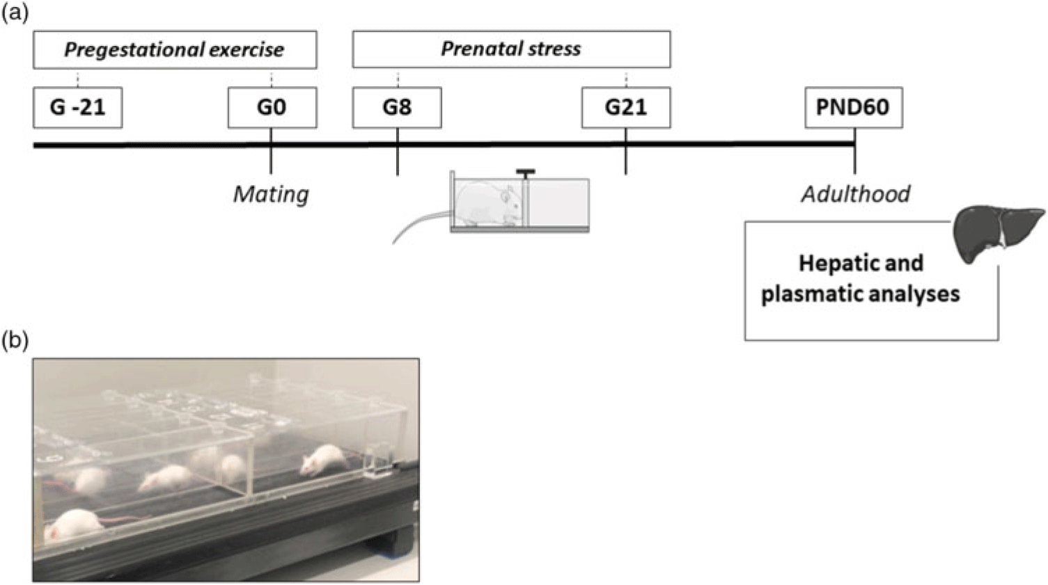
Virgin female Balb/c mice (6–8 weeks) were divided into three experimental groups: control (CON, n = 8 dams), prenatal stress from the second week of pregnancy (PNS, n = 7 dams) and physical exercise 3 weeks before gestation plus prenatal stress from the second week of pregnancy (EX + PNS, n = 8 dams). At first, the females had their estrous cycle monitored through a vaginal smear visualization and, during the fertile period, they were mated with males of the same age and lineage. After confirming the mating (presence of a semen plug), females were separated and placed in individual boxes. Four dams were excluded from the EX + PNS group as they presented difficulties in following the exercise protocol (initial number of animals in the group n = 12; final number of animals in the group n = 8 dams). The cohort characteristics are presented in Table 1. Pregnant females were weighed on days 1, 8 and 15 of the gestation period. The day after birth was considered as postnatal day 1 (PND 1). The litters were randomly adjusted to 4–6 animals per dam on PND 1 and weaned on PND 21. After weaning, males and females were allocated to separate boxes with a maximum of five animals and were maintained until adulthood without any intervention. The offspring were weighed on PND 1, 10 and 60. At 60 days of age (PND 60), female and male mice were euthanized through decapitation, as the use of anesthetics may trigger acute stress responses by activating the HPA axis. The blood was collected for the measurement of plasma glucose, triglycerides and cholesterol concentrations. Moreover, the liver was removed by laparotomy for the evaluation of oxidative stress parameters through the measurement of thiobarbituric acid reactive substances (TBARS), catalase (CAT), reduced glutathione (GSH) and superoxide dismutase (SOD). In addition, glucocorticoid receptor (GR), peroxisome proliferator-activated receptor alfa (PPARα) and peroxisome proliferator-activated receptor gamma (PPARγ) gene expression were evaluated in the liver. The experimental setup is shown in Fig. 1.
Cohort characteristics

Data are shown as absolute number# or mean ± standard deviation (SD)*, as indicated. CON, control; PNS, prenatal stress from the second week of pregnancy; EX + PNS, physical exercise three weeks before gestation and prenatal stress from the second week of pregnancy.
(a) Experimental design of the study and (b) exercise setup. PNS: prenatal stress from the second week of pregnancy; G-21: 21 days before gestational day 0; G0: gestational day 0; G8: gestational day 8; G21: gestational day 21; PND60: post-natal day 60.

Physical exercise
Females of the EX + PNS group were submitted to a protocol of physical exercise on a motorized treadmill, in the last hour of the light cycle, 5 days a week, during the 3 weeks (15 sessions) that preceded the day of mating. The exercise protocol consisted of 60 min of running at a speed of 10 m/min with a 0% slope. Reference Wearick-Silva, Marshall and Viola22 The exercised animals were adapted to the treadmill for 10 min at a speed of 5 m/min three days before the beginning of the exercise protocol. No stressful stimulus was used to motivate the animals to run. After the end of the protocol, animals were returned to their cages with ad libitum access to water and food.
Prenatal stress model
Prenatal restraint stress was applied using the protocol described by Vargas et al. (2016). Reference Vargas, Campos and de Souza23 In brief, the protocol was applied from the eighth day of gestation until the day of birth of the offspring, in a closed cylinder (Insight, Brazil), between 10 am and 12 pm, for 30 min, every other day. PNS females were submitted to 5–6 stressful episodes during pregnancy. This prenatal stress model has been previously proven to be a sufficient stressor to increase corticosterone levels in rodents. Reference Zagron and Weinstock24 Pregnant control mice remained in their home cages without any intervention during the gestational period.
Biochemical analysis
Biochemical analyses were performed on PND 60 on whole blood samples collected in tubes with heparin and centrifuged (1000g) for 10 min. The separated plasma was then frozen at −80°C until the analyses. The plasma levels of glucose, triglyceride, and cholesterol were determined using commercial kits (Labtest Diagnóstica, Brazil), according to the manufacturer’s instructions, in a semi-automatic spectrophotometer (Genesis 8/Spectronic).
Oxidative stress
In order to evaluate the oxidative stress parameters, the liver tissue was homogenized in 10 volumes (1:10, w/v) of sodium phosphate buffer, pH 7.4. The homogenate was centrifuged at 750 g for 10 min at 4°C. The pellet was discarded, and the supernatant was immediately separated and used for the measurements of TBARS, CAT, GSH and SOD. Total protein was measured using the Nanodrop Spectrophotometer (Model 1000, Thermo Scientific).
Thiobarbituric acid reactive substance (TBARS)
The samples were centrifuged, 10 μl of the supernatant was collected and added to 10 µl of sodium dodecyl sulfate (SDS 12.4 mM) and 400 μl of thiobarbituric acid. The mixture was heated for 30 min and then centrifuged at 750 g for 10 min at 25°C. A pink compound was formed and measured spectrophotometrically at 532 nm (Genesis 8/Spectronic). The total content of protein in the sample was measured using the Nanodrop Spectrophotometer (Model 1000, Thermo Scientific). The results were represented as mM TBARS/mg protein. Reference Zanatta, Viegas and Tonin25
Catalase assay (CAT)
For the test, 102 μl of H2O2 30% and 100 μ of Triton X-100 were pipetted with 10 µl of the supernatant from the macerate and read at 240 nm in a semi-automatic spectrophotometer (Genesis 8/Spectronic). One CAT unit is defined as 1 μmol of H2O2 consumed per minute. The total content of protein in the sample was measured using the Nanodrop Spectrophotometer (Model 1000, Thermo Scientific). The results were presented as CAT units/mg protein.
Reduced glutathione (GSH)
For the analyses, 250 μl of metaphosphoric acid was added to 50 µl of liver supernatant and centrifuged. Afterward, 650 μl of Na2HPO4 and 100 μl of the color reagent (5,5′-Dithiobis-2-Nitrobenzoic Acid) were added to 250 μl of the supernatant. A yellow compound was formed and measured in a semi-automatic spectrophotometer at 412 nm (Genesis 8/Spectronic). The total content of protein in the sample was measured using the Nanodrop Spectrophotometer (Model 1000, Thermo Scientific). The results were represented as O.D. 412 µm/g protein. Reference Lima, Krause and da Silva26
Superoxide dismutase (SOD)
The superoxide dismutase was measured with a commercially available kit (RANSOD, Random Labs, UK), according to the manufacturer’s instructions. The total content of protein in the sample was measured using the Nanodrop Spectrophotometer (Model 1000, Thermo Scientific). The results were represented as % inhibition/mg protein.
Quantification of mRNA through real-time PCR
Total cellular RNA was extracted from the liver using the Trizol method (Thermo Scientific) according to the manufacturer’s instructions. The RNA was resuspended in 20 μl of nuclease-free water (Ambion®) and converted to complementary DNA (cDNA) (GoScript™ Reverse Transcription System Protocol – Promega) according to the protocol indicated by the manufacturer. The final concentration of cDNA was analyzed using the fluorometric method (Qubit® – ThermoFisher – Scientific) using a commercial kit (Qubit® dsDNA HS Assay – Thermo Scientific).
Gene expression was performed in real-time quantitative PCR (Step One Plus – Applied Biosystems) using 16 ng of cDNA. The liver samples were prepared in duplicate and the relative expression of messenger RNA (mRNA) was calculated using the Delta-Delta Ct method (ΔΔCt) using the male control group as a reference. Glyceraldehyde-3-phosphate dehydrogenase (GAPDH) was adopted as the reference endogenous gene. A negative control for each primer was used on each plate to check for possible contamination. Amplification reagent measurements were calculated based on the incorporation of the SYBR® Green fluorescence marker (Applied Biosystems) into the double cDNA ribbon for each amplification reaction.
The primers used for real-time PCR were: GAPDH (forward 5′ GGGGAGCCAAAAGGGTCATC 3′; reverse 5′ GACGCCTGCTTCACCACCTTCTTG 3′), GR (forward 5′ GGAATAGGTGCCAAGGGTCT 3′; reverse 5′ GAGCACACCAGGCAGAGTTT 3′), PPARα (forward 5′ TGCAAACTTGGACTTGAACG 3′; reverse 5′ TGATGTCACAGAACGGCTTC 3′) and PPARγ (forward 5′ TGGAATTAGATGACAGTGACTTGG 3′; reverse 5′ CTCTGTGACGATCTGCCTGAG 3′).
Statistical analysis
The normality of data was tested using the Shapiro–Wilk test. Outliers were excluded from analyses. As a normal distribution was found, the data were expressed using mean and standard error of the mean (SEM). Two-way ANOVA with repeated measures was used for the gestational body weight analysis and a one-way ANOVA was used for the offspring body weight analysis. For the evaluation of the interaction between sex (male × female) and the experimental groups (CON × PNS × EX + PNS), a two-way ANOVA was used. Whenever a significant difference was found (p < .05), the LSD post-hoc test for multiple comparisons was used. For biochemical analysis and PCR data, biological replicates were CON n = 8 (n = 10–14 offspring); PNS n = 7 (n = 7–9 offspring); and EX + PNS n = 8 (n = 13–15 offspring). In all cases, the level of significance was set at 5%. Data were analyzed using SPSS version 18.0 (SPSS Inc., USA).
Results
Pregestational exercise increases neonatal body weight
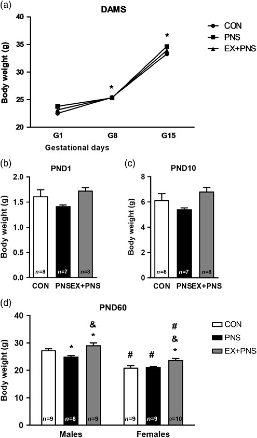
No effects of prenatal stress and pregestational exercise on maternal body weight were found during gestation, although dams showed a significant body weight gain during pregnancy, as was expected (Fig. 2a). A one-way ANOVA showed that there were no significant effects for groups in the assessment of body weight on PND1 (F (2,20) = 3.02; p = .071) and PND10 (F (2,20) = 3.33; p = .056). Results are shown in Fig. 2b and 2c, respectively.
Maternal and offspring body weight. (a) Maternal body weight gain during pregnancy was evaluated during the gestational period. Groups were compared by a two-way ANOVA with repeated measures followed by the LSD post-hoc test. Data are shown as mean ± SEM, CON = 8, PNS = 7 and EX + PNS = 8 animals per group. *p < 0.05 denotes significant differences between G8 and G15 compared to G1. The offspring body weight was evaluated at (b) PND1 and (c) PND10. Groups were compared by one-way ANOVA. Data are shown as mean ± SEM, n = 7–8 animals per group (exact numbers are within the bars). Adult mice’s body weight was evaluated at (d) PND60. Groups were compared by a two-way ANOVA followed by the LSD post-hoc test. Data are shown as mean ± SEM, n = 8–10 animals per group (exact numbers are within the bars). *p < 0.05 denotes significant differences compared to the CON group in the same sex; & p < 0.05 denotes significant differences compared to the PNS group; # p < 0.05 denotes significant sex differences within the same group. CON, control; PNS, prenatal stress from the second week of pregnancy; EX + PNS, physical exercise 3 weeks before gestation and prenatal stress from the second week of pregnancy.

Pregestational exercise and prenatal stress alter body weight in adult life in a sex-dependent manner
A two-way ANOVA revealed significant main effects for group (F (2,48) = 17.91; p = .000002) and sex (F (1,48) = 118.00; p = .000001). Post-hoc analyses showed that PNS males had a significantly lower body weight when compared to CON (p = .009) and EX + PNS (p = .000011) groups. Also, at PND 60, EX + PNS males showed a heavier body weight when compared to the CON group (p = .029). On the other hand, prenatal stress did not influence female body weight in adult life. However, exercise before pregnancy induced a body weight increase in females when compared to the CON (p = .001) and PNS (p = .002) groups. As expected, in the comparisons between the sexes, females showed a lower body weight when compared to the males of their respective groups (p < .0001). Results are shown in Fig. 2d.
Prenatal stress and pregestational exercise influence the offspring’s liver metabolism in a sex-dependent manner
We evaluated the ability of pregestational exercise in preventing the effects of prenatal stress on liver-related biochemical markers. No main effects for group, sex and interaction were found for glucose. For the triglyceride, we detected a significant effect for sex (F (1,46) = 4.12; p = .048). Cholesterol analysis revealed a significant effect for group (F (2,46) = 4.75; p = .013) and sex (F (1,46) = 18.42; p = .00009). Fig. 3c shows no significant differences (p = .057) between EX + PNS and CON group for plasma cholesterol in males. However, there was a significant increase in the plasma cholesterol in PNS females when compared to CON (p = .008). This increase found in the PNS group was prevented by exercising before pregnancy (p = .021). When sex effects were analyzed, EX + PNS females had an increased triglyceride secretion (p = .021) when compared to the males of the same group (Fig. 3b). Sex differences have also been demonstrated for cholesterol, as CON (p = .014) and EX + PNS (p = .001) females showed lower cholesterol levels compared to the males from their respective experimental groups (Fig. 3c).
Biochemical parameters of liver metabolism. Plasma concentrations of (a) glucose, (b) triglyceride and (c) cholesterol were evaluated at PND 60 in males and females. Groups were compared by a two-way ANOVA followed by the LSD post-hoc test. Data are shown as mean ± SEM, n = 5–11 animals per group (exact numbers are within the bars). *p < 0.05 denotes significant differences compared to the CON group in the same sex; & p < 0.05 denotes significant differences compared to the PNS group; #p < 0.05 denotes significant sex differences within the same group. CON, control; PNS, prenatal stress from the second week of pregnancy; EX + PNS, physical exercise 3 weeks before gestation and prenatal stress from the second week of pregnancy.

Pregestational exercise does not prevent the effects of prenatal stress on female liver catalase
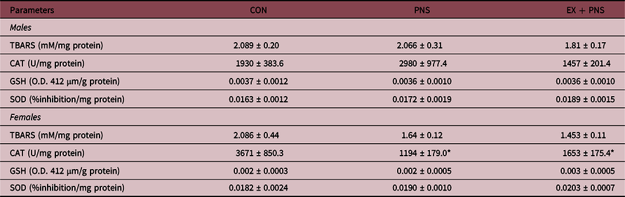
We also investigated whether pregestational exercise would be capable of preventing possible effects of prenatal stress on oxidative responses in the liver. The liver enzymatic antioxidant defense was evaluated through the activities of CAT, SOD and GSH, besides the production of TBARS. No significant main effects for group, sex and interaction were observed for the TBARS, GSH and SOD analysis. A significant interaction between group and sex (F (5,34) = 3.68; p = .036) was shown for catalase and post-hoc analysis revealed, in the females, a significant decrease in both PNS (p = .013) and EX + PNS (p = .039) groups when compared to the CON (Table 2).
Hepatic oxidative stress markers

Groups were compared by two-way ANOVA followed by the LSD post hoc test. Data are shown as mean ± SEM, n = 5–9 animals per group. TBARS, thiobarbituric acid reactive substances; CAT, catalase; GSH, glutathione; SOD, superoxide dismutase; CON, control; PNS, prenatal stress from the second week of pregnancy; EX + PNS, physical exercise three weeks before gestation and prenatal stress from the second week of pregnancy.
* p < 0.05 denotes significant differences compared to the CON group in the same sex.
Pregestational exercise alters liver gene expression in a sex-dependent manner
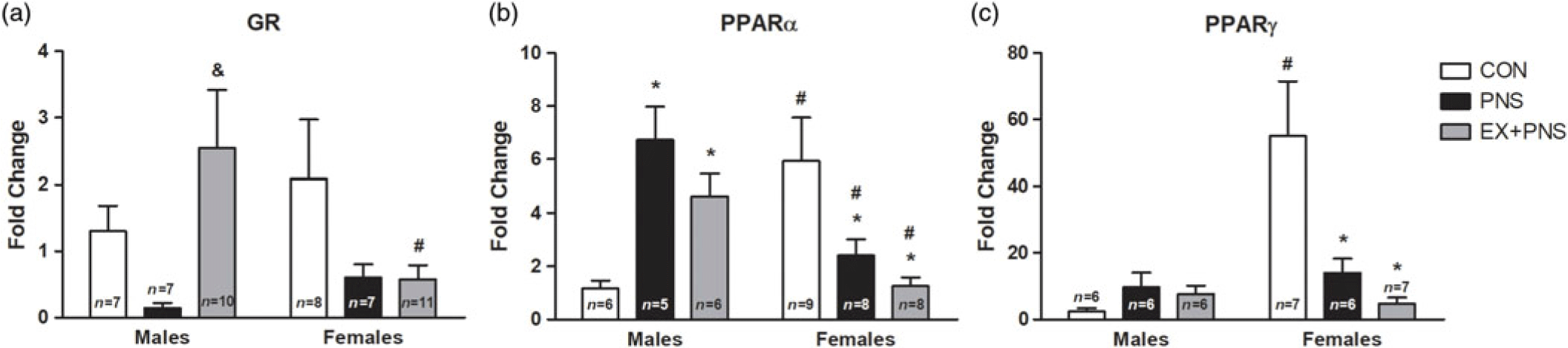
Considering the previous results presented, we evaluated GR, PPARα and PPARγ mRNA expression in the liver. A significant interaction between group and sex was demonstrated for both GR (F (5,44) = 3.56; p = .037) and PPARα (F (5,36) = 11.37; p = .0001). As for PPARγ, a significant main effect for group (F (2,32) = 4.52; p = .019), sex (F (1,32) = 6.95; p = .013) and interaction (F (5,32) = 7.51; p = .002) was found. Post-hoc analysis showed no significant differences in GR expression between CON and PNS male groups, although a significant GR mRNA expression increase (p = .006) was seen when EX + PNS was compared to the PNS group (Fig. 4a). No significant differences were observed for female GR gene expression (Fig. 4a). There was a significant increase in the expression of PPARα in the liver of PNS (p = .002) and EX + PNS (p = .036) males in comparison to the CON group (Fig. 4b). On the other hand, the PPARα expression significantly decreased in both the PNS (p = .012) and EX + PNS (p = .001) females when compared to the CON group (Fig. 4b). Post-hoc analysis showed a significant decrease in PPARγ expression in PNS (p = .001) and EX + PNS (p = .000049) females compared to the CON group (Fig. 4c). Both EX + PNS and PNS did not change the liver PPARγ expression in males when compared to the CON group (Fig. 4c). When sex effects were analyzed, a significant decrease in the GR mRNA expression (p = .011) in EX + PNS females (Fig. 4a) and the PPARα mRNA expression in both the PNS (p = .009) and EX + PNS (p = .029) females (Fig. 4b) was observed when compared to the males from the same group. On the other hand, the CON females showed a higher mRNA expression for PPARα (p = .002) and PPARγ (p = .000046) when compared to the CON males (Fig. 4b and 4c, respectively).
Hepatic gene expression. (a) Tissue glucocorticoid receptor, (b) peroxisome proliferator-activated receptors α and (c) γ mRNA were measured in males and females. Groups were compared by a two-way ANOVA followed by the LSD post-hoc test. Data is shown as mean ± SEM, n = 5–11 animals per group (exact numbers are within the bars). *p < 0.05 denotes significant differences compared to the CON group in the same sex; & p < 0.05 denotes significant differences compared to the PNS group; # p < 0.05 denotes significant sex differences within the same group. GAPDH, glyceraldehyde-3-phosphate dehydrogenase; GR, glucocorticoid receptor; PPARα, peroxisome proliferator-activated receptor α; PPARγ, peroxisome proliferator-activated receptor γ; CON, control; PNS, prenatal stress from the second week of pregnancy; EX + PNS, physical exercise three weeks before gestation and prenatal stress from the second week of pregnancy.

Discussion
An adverse environment in utero has the ability to affect fetal development and growth, promoting long-term changes in the offspring’s metabolism. However, the long-term effects of physical exercise before the gestational period on prenatally stressed offspring, as well as the involvement of sex differences in these responses, have not yet been elucidated. The present study has shown that exercise before pregnancy was able to prevent some of the prenatal stress effects on the metabolic parameters of the offspring in a sex-dependent manner.
Stressful events during the gestational period may disrupt the regulation of metabolic homeostasis. Reference Brunton, Sullivan, Kerrigan, Russell, Seckl and Drake9 Both repeated variable prenatal stress and restraint prenatal stress are well-established protocols used in several studies. Reference Maeyama, Hirasawa and Tahara8,Reference Luft, Levices and Costa13,Reference Kinnunen, Koenig and Bilbe27,Reference Markham, Mullins and Koenig28 Although differences regarding the time and frequency of the repeated restraint stress exposure exist, several studies have demonstrated that its use during pregnancy leads to impaired spatial learning and memory, anxiety, increased corticosterone levels, among others. Reference Su, Zhang and Dang29,Reference Palacios-Garcia, Lara-Vasquez and Montiel30 Moreover, it is well established that exposure to adverse events during the prenatal period is considered to generate alterations in both body weight and composition. Reference Pankevich, Mueller, Brockel and Bale31–Reference Mueller and Bale33 Our data showed no effect of stress and pregestational exercise on the weight gain of mothers during gestation. Similarly, Klein et al. demonstrated that swimming before gestation does not affect the body weight of pregnant female rats. Reference Klein, Dos Santos Rodrigues and Hozer34 Although swimming has been used as a stressor in repeated variable stress paradigms, Reference Sickmann, Skoven and Bastlund35,Reference Sickmann, Arentzen, Dyrby, Plath and Kristensen36 studies have also shown its use as a form of exercise, demonstrating positive neural adaptations, in addition to decreasing corticosterone levels. Reference Liu, Xu, Lu, Zhang, Sheng and Ni37,Reference Song, Kim, Kim, Park and Lee38 There is also evidence that mothers who perform physical activities from the beginning of the gestation period and then decrease the exercise levels closer to birth generate significantly heavier children, probably due to the increase in the body fat percentage. Reference Finken, van der Voorn, Heijboer, de Waard, van Goudoever and Rotteveel39 Using the same stress model during the last 2 weeks of gestation, Vargas et al. found no significant differences in the mice’s birth weight, corroborating the present findings. Reference Vargas, Campos and de Souza23 Nevertheless, in adulthood, our study showed that prenatal stress decreased body weight only in males in an effect prevented by maternal exercise. Adult females from exercised mothers also presented greater weight compared to the other groups. It is already known that physical activity influences the maintenance of body weight and obesity in rodents. Reference Mifune, Tajiri and Nishi40 In a study using rats, treadmill exercise before and during gestation promoted a decrease in the offspring weight at weaning. Reference Quiclet, Dubouchaud and Berthon41 In mice, the same period of exercise generated an increase in the percentage of lean mass and a decrease of fat mass only in the male adult offspring. Reference Carter, Lewis and Wilkerson42 Some differences found between studies may be related to the species used and the type of exercise employed. Moreover, the effects of gestational exercise may be related to changes in body composition, as observed in a study with rats showing sex-dependent effects. Reference Rosa, Blair and Vickers43
Considering the body weight results found, we sought to investigate possible liver regulatory mechanisms, since it is a key organ for the control of several metabolic functions. Our data showed that maternal stress caused sex-dependent effects on cholesterol, which is a regulator of lipid organization. Clinical and experimental studies have already demonstrated the beneficial effects of physical exercise on diseases associated with lipid and hepatic alterations, Reference van der Windt, Sud, Zhang, Tsung and Huang44 just as the treadmill running reduced cholesterol in obese and dyslipidemic mice. Reference Cho, Kim, Jang, Kim and Kang45,Reference Ferreira, Pinto and Iborra46 Increased plasma cholesterol in PNS females had not yet been reported, although maternal stress has already been implicated in the increased lipid accumulation in the liver of prepubertal mice. Reference Maeyama, Hirasawa and Tahara8 It is possible that the beneficial effect of physical exercise prior to gestation in prenatally stressed mice could be associated with both the cholesterol metabolism and sex-dependent mechanisms. Surprisingly, our data showed that males from the exercise group had increased plasma cholesterol, indicating a possible influence of sex hormones since estrogens are already known to be regulators of lipid metabolism. Reference Palmisano, Zhu, Eckel and Stafford47 Although the relationship between exercise and cholesterol levels is well established, Reference Zhang, Zhao, Xu, Chen and Li48–Reference Wen, Jadhav, Williamson and Rideout50 we speculate that the association between the withdrawal of exercise before pregnancy and stress may have led to this negative metabolic outcome.
Regarding oxidative stress, our study showed that prenatal stress promotes changes in the antioxidant capacity, observed by a decreased hepatic CAT only in females. At the same time, pregestational exercise was not able to influence this effect. CAT is the enzyme responsible for the decomposition of hydrogen peroxide into water and oxygen and it is important for protecting cells from oxidative stress. Reference Jones, Eklow, Thor and Orrenius51 To date, there have been no studies evaluating the effects of maternal stress on the activity of CAT in the liver of rodents. However, chronic restraint stress is a classic model used to induce alterations in the activity of antioxidant enzymes. It is already known that early-life stress is able to decrease both antioxidant enzymes CAT and SOD levels and thereby increase the production of brain reactive oxygen species in male and female juvenile rats. Reference Jia, Sun, Su, Dang and Chen52 Our data demonstrated an effect of prenatal stress in the regulation of oxidative stress only in females, indicating a possible contribution of sex hormones in the antioxidant system response. The type and intensity of physical exercise are also important factors for the production of oxidative stress. A previous study with male mice demonstrated an efficient regulation of antioxidant enzymes and a reduction of oxidative damage in the liver of rodents which were submitted to treadmill exercise compared to sedentary ones. Reference da Silva, Pinho, Rocha, Tuon, Silveira and Pinho53 Maternal exercise has also been associated with the reversal of deleterious effects of prenatal stress in mice. Reference Bustamante, Henriquez, Medina, Reinoso, Vargas and Pascual54 Corroborating our findings, a study on rats using maternal swimming before and during gestation did not prevent oxidative changes in the brain of the offspring with neonatal hypoxia-ischemia. Reference Marcelino, de Lemos Rodrigues, Miguel, Netto, Pereira Silva and Matte55
Glucocorticoids are steroid hormones that act predominantly through GR, which are expressed in the hepatic tissue and are involved in the regulation of several energy control processes, besides being associated with the excessive production of oxidative stress. Reference Sato, Takahashi, Sumitani, Takatsu and Urano56 In addition, it has already been observed that hepatic GR can participate in the regulation of genes involved with cholesterol. Reference Lemke, Krones-Herzig and Berriel Diaz57,Reference Zollner and Trauner58 In females, our data showed no differences between the groups in the GR gene expression. Similarly, in a murine prenatal model, repeated social stress has been associated with long-term alterations in liver markers of metabolic activity in males and females, although with no changes on the hepatic GR. Reference Brunton, Sullivan, Kerrigan, Russell, Seckl and Drake9 Altered GR expression may suggest that exposure to maternal in utero glucocorticoids can program the physiology and promote greater susceptibility to metabolic diseases in a sex-dependent manner. The interaction between glucocorticoids and GR is fundamental for the liver homeostatic control, and the decreased expression of these receptors in the tissue has been associated with disorders such as fatty liver and hypercortisolism. Reference John, Marino, Sanchez and Hinds59 In contrast to the females, pregestational exercise increased the hepatic GR mRNA in males. It has already been observed in rodents that physical exercise induces the increase of circulating glucocorticoid levels, besides increasing the GR expression in the adipose tissue and then inducing lipolysis. Reference Campbell, Fediuc, Hawke and Riddell60 Although the effects of this type of exercise on the offspring’s liver have not been addressed, it is possible that our pregestational exercise protocol has acted as a positive modulator of liver GR expression in a sex-dependent manner. Exercising on a treadmill has also been previously considered to be a stressor, although evidence using a similar protocol has already demonstrated a decreased level of glucocorticoids (corticosterone) in exercised mice. Reference Li, Liang, Guan, Fan, Chi and Yang61
Females showed a decreased expression of PPARα and γ, whereas males showed an increased expression of PPARα when subjected to stress during pregnancy. In addition, exercise was not able to prevent these effects in both sexes. A study on rats reported that prenatal stress promotes a trend to decrease hepatic mRNA expression of PPARα and γ only in males. Reference Brunton, Sullivan, Kerrigan, Russell, Seckl and Drake9 Moreover, the study has shown a decreased peroxisome proliferator-activated receptor coactivator 1a (Pgc1a) and transcriptional coactivator (that interacts with PPARγ) gene expression in females. Reference Brunton, Sullivan, Kerrigan, Russell, Seckl and Drake9 Sex differences seem to be an important variable in determining the effects of early life stress on these markers. Reference Brunton, Sullivan, Kerrigan, Russell, Seckl and Drake9,Reference Paternain, de la Garza, Batlle, Milagro, Martinez and Campion62 PPARs play a key role in the metabolism of carbohydrates, lipids and proteins. Reference Monsalve, Pyarasani, Delgado-Lopez and Moore-Carrasco63 For example, liver disruption of PPARγ in mice is associated with increased adiposity and hyperlipidemia, Reference Gavrilova, Haluzik and Matsusue64 and the lack of PPARα contributes to the development of steatosis. Reference Kersten, Seydoux, Peters, Gonzalez, Desvergne and Wahli65 The long-term exercise was already related to the increased liver PPARα in male rats in association with decreased triglyceride and cholesterol. Reference Zhang, Liu and Li66 It is unclear what leads to the increased PPARα gene expression in PNS males, but the response in the EX + PNS group may be related to hepatic GR levels, since the interaction between these two receptors has already been observed in an inflammatory model. Reference Bougarne, Paumelle and Caron67 Very little data are available on mice, related to PPARs gene expression sex-differences. There are studies showing that PPAR-alfa and estrogen receptors share cofactors, suggesting that there is an important role of sex hormones in this response. Reference Rando and Wahli68 In lymphocytes, higher basal levels in PPARα mRNA and protein in male mice compared to females was demonstrated. Reference Park, Park, Lee, Bothwell and Choi69 Similarly, a study on rats showed that the expression of PPAR-alfa mRNA and protein levels are higher in male than in female rat livers. Reference Jalouli, Carlsson and Ameen70 To the best of our knowledge, there are no studies showing sex differences in the expression of PPAR-gamma in mice.
Our study has some limitations, including the lack of a corticosterone assessment in the mothers, which prevented us from investigating if the exercise protocol used was able to alter corticosterone levels. Although it is well-known that both restraint stress has been associated with increased glucocorticoid levels during pregnancy in rodent models, Reference Maghsoudi, Ghasemi, Ghaempanah, Ardekani, Nooshinfar and Tahzibi71,Reference Lim, Fedulov and Kobzik72 as well as moderate chronic exercise decreases stress-related hormone levels, Reference Inoue, Okamoto and Shibato73,Reference Patki, Li and Allam74 to the best of our knowledge, there are no studies that have measured corticosterone levels using a similar protocol. Additionally, in order to avoid the use of stressful stimuli to keep mice running, few animals were removed from the study, as they did not adapt to the treadmill. Aiming to minimize the possibility of exercise acting as a stressful stimuli per se, all sessions were visually monitored to identify animals with possible difficulties and/or visual signs of distress. Exercising in the last hour of the light cycle may have also influenced the results, as animals are more active during the nocturnal period. PCR was performed using a single reference gene, which would also be considered as a limitation, although GAPDH is stably expressed between groups. Also, direct analysis of the proteins resulting from the mRNA changes detected is also a limitation of the present study, as in mice, the gene expression represents 40% of the variation in protein expression. Reference Schwanhausser, Busse and Li75 Thus, although we did not find significant differences in mRNA expression, different levels of protein could be observed in the liver.
In conclusion, physical exercise before the gestational period prevents the effects of prenatal stress on adult body weight and cholesterol in a sex-dependent manner. However, despite the beneficial metabolic effects found, it was not possible to observe significant differences in the expression of important genes related to the hepatic regulation of this response, such as GR and PPARs. The sex-dependent effects found for stress and exercise suggests the influence of sex steroid hormones, such as estradiol, progesterone and testosterone, although the precise mechanisms still require further investigations.
Acknowledgements
The authors would like to thank CNPq, CAPES and PUCRS for the concession of scholarships.
Financial Support
This study was financed in part by the Coordenação de Aperfeiçoamento de Pessoal de Nivel Superior – Brasil (CAPES) – Finance Code 001; and Conselho Nacional de Desenvolvimento Científico e Tecnológico (CNPQ) – Process Number 401178/2016-1.
Conflicts of Interest
None.
Ethical standards
The authors assert that all procedures contributing to this work complied with the ethical standards of the Ethical Committee of Experimental Animals of the Pontifícia Universidade Católica do Rio Grande do Sul and were approved by the institutional committee from this institution.