Introduction
Post-traumatic stress disorder (PTSD) is classified as an anxiety disorder resulting from experiencing or witnessing an extreme traumatic stressor, which implies potential loss of life or serious injury to the self or others. Its three main symptoms clusters include intrusions (i.e. flashbacks or nightmares), avoidance of trauma-related stimuli and hyperarousal (APA, 1994).
Over the past decade, it has become increasingly clear that several brain structures play a key role in the generation of PTSD symptoms. Positron emission tomography and single photon emission computed tomography (SPECT) studies have found regional cerebral blood flow (rCBF) changes during trauma recall in PTSD patients, with reports of either increased or decreased rCBF (depending on the paradigm and on the subject sample) mainly within hippocampus, amygdala and the medial prefrontal cortex (mPFC) (for recent reviews, see Nutt & Malizia, Reference Nutt and Malizia2004; Francati et al. Reference Francati, Vermetten and Bremner2007; Karl et al. Reference Karl, Schaefer, Malta, Dörfel, Rohleder and Werner2006; Liberzon & Sripada, Reference Liberzon and Sripada2008). However, other structures have been consistently found to be involved in PTSD, i.e. anterior and posterior insular cortex (Osuch et al. Reference Osuch, Benson, Geraci, Podell, Herscovitch, McCann and Post2001; Liberzon et al. Reference Liberzon, Britton and Phan2003; Lindauer et al. Reference Lindauer, Booij, Habraken, van Meijel, Uylings, Olff, Carlier, den Heeten, van Eck-Smit and Gersons2008), posterior cingulate cortex (PCC) (Bremner et al. Reference Bremner, Narayan, Staib, Southwick, McGlashan and Charney1999a, Reference Bremner, Staib, Kaloupek, Southwick, Soufer and Charneyb; Sachinvala et al. Reference Sachinvala, Kling, Suffin, Lake and Cohen2000), as well as definite portions of the parietal lobe such as inferior parietal lobule (IPL)/supramarginal gyrus (Bremner et al. Reference Bremner, Narayan, Staib, Southwick, McGlashan and Charney1999a, Reference Bremner, Staib, Kaloupek, Southwick, Soufer and Charneyb, Reference Bremner, Vythilingam, Vermetten, Southwick, McGlashan, Staib, Soufer and Charney2003; Shaw et al. Reference Shaw, Strother, McFarlane, Morris, Anderson, Clark and Egan2002) and precuneus (PCN) (Molina et al. Reference Molina, Isoardi, Prado and Bentolila2010).
Psychological questionnaires, including self-rating scales (SRS), are useful tools for the assessment of psychiatric disorders, as well as symptom severity and follow-up treatment outcomes (Högberg et al. Reference Högberg, Pagani, Sundin, Soares, Åberg-Wistedt, Tärnell and Hällström2008). Such behavioural measures have also been used in studies investigating correlations with brain perfusion. For instance, rCBF in PTSD patients was correlated with symptom severity (Lucey et al. Reference Lucey, Costa, Adshead, Deahl, Busatto, Gacinovic, Travis, Pilowsky, Ell, Marks and Kerwin1997; Kim et al. Reference Kim, Lyoo, Lee, Kim, Sim, Bae, Kim, Lee and Jeong2007), flashback intensity (Osuch et al. Reference Osuch, Benson, Geraci, Podell, Herscovitch, McCann and Post2001), anxiety and depression measures (Lucey et al. Reference Lucey, Costa, Adshead, Deahl, Busatto, Gacinovic, Travis, Pilowsky, Ell, Marks and Kerwin1997; Bonne et al. Reference Bonne, Gilboa, Louzoun, Brandes, Yona, Lester, Barkai, Freedman, Chisin and Shalev2003) and clinician-administered PTSD scale (Bonne et al. Reference Bonne, Gilboa, Louzoun, Brandes, Yona, Lester, Barkai, Freedman, Chisin and Shalev2003; Peres et al. Reference Peres, Newberg, Mercante, Simão, Albuquerque, Peres and Nasello2007). Recently, our group has used a series of SRS in order to test some secondary outcome variables in PTSD subjects before and after Eye Movement Desensitization and Reprocessing treatment, showing that such instruments are sensitive and capable of discriminating between symptomatic and non-symptomatic subjects (Högberg et al. Reference Högberg, Pagani, Sundin, Soares, Åberg-Wistedt, Tärnell and Hällström2008).
The present study was designed to investigate rCBF alterations in PTSD patients and to seek possible correlations between brain perfusion and PTSD-related SRS in order to cross-check their diagnostic value and to look for their neural correlates. On the basis of the reviewed literature, we predict that functional changes between symptomatic and non-symptomatic subjects will be found in limbic structures, comprising insular cortex and posterior cingulate. On the other hand, the literature on the neural correlates of SRS, especially those employed in the present study, is lacking. Hence, a second aim of this study was to investigate to what extent the correlations between rCBF and SRS were consistent with the rCBF changes related to PTSD.
Method
Subjects
The present study investigates drivers of underground and long-distance trains in Stockholm and Sweden registered by the company as having either once or several times experienced a ‘person under train’ accident or having been assaulted at work (Pagani et al. Reference Pagani, Högberg, Salmaso, Tärnell, Sanchez-Crespo, Soares, Åberg-Wistedt, Jacobsson, Hällström, Larsson and Sundin2005, Reference Pagani, Högberg, Salmaso, Nardo, Sundin, Jonsson, Soares, Aberg-Wistedt, Jacobsson, Larsson and Hällström2007; Högberg et al. Reference Högberg, Pagani, Sundin, Soares, Åberg-Wistedt, Tärnell and Hällström2008), in which a set of SRS was administered.
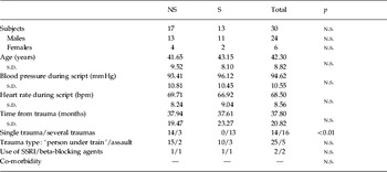
Altogether, 33 subjects were initially recruited for the present study, 16 of whom developed PTSD (S) and 17 who did not (NS). However, three subjects had to be removed because an inspection of their magnetic resonance imaging (MRI) structural images (collected at the same time as SPECT) revealed a very high degree of dilatation in the anterior and posterior horns of the lateral ventricles. This was further confirmed by the fact that these subjects showed outlying values in the ‘cerebrospinal fluid/intra-cranial volume’ ratio (i.e. 0.294, 0.285, 0.273, respectively, being mean+2×s.d.=0.204+2×0.033=0.270). Given that such an abnormality would have affected our analyses, we removed these subjects from our sample and performed all statistical analyses on 30 subjects (whose demographic and clinical variables are summarized in Table 1).
Demographic and physiological data in patients with post-traumatic stress disorder (PTSD; S) and in traumatized non-PTSD controls (NS)

bpm, Beats per min; SSRI, selective serotonin reuptake inhibitor.
In total, 13 subjects with current PTSD (S) were compared with 17 subjects who had experienced these traumatic events but had never developed PTSD (NS). When subjects took part in the study, a period of time varying between 3 months and 6 years had elapsed since the traumatic event. Symptoms had to be present for at least 1 month and occurring >3 months earlier. Only a full PTSD diagnosis was accepted, whereas exclusion criteria were other psychiatric conditions, drug or alcohol abuse or dependency, significant medical condition, neurological illness or a history of head injury.
Before entering the study, written informed consent was obtained. The study was approved by the Local Ethics and Radiation Safety Committees.
Diagnosis and SRS
The diagnosis of PTSD was established according to the DSM-IV criteria (APA, 1994). The SCID-I (First et al. Reference First, Spitzer, Gibbon and Williams1997) was carried out by a psychiatrist not otherwise engaged in the study. Further diagnostic interviews were performed by the same psychiatrist in order to evaluate ratings of symptoms and functional assessment using the following scales: the GAF (APA, 1994); the HAMA (Hamilton, Reference Hamilton1969); the HAMD (Hamilton, Reference Hamilton1960).
Participants were also asked to perform several SRS, administered on a computer immediately before the interview and described as follows: Beck Anxiety Inventory (BAI; Beck et al. Reference Beck, Epstein, Brown and Steer1988), inquiring about how the subject has been feeling in the last week in regard to subjective somatic or panic-related symptoms; the Impact of Event Scale (IES), evaluating the amount of intrusions and avoidance during the last week related to a past stressful event (Horowitz et al. Reference Horowitz, Wilmer and Alvarez1979) and considered a measure of the current subjective distress (Joseph, Reference Joseph2000); the Social Disability Index (SDI), focusing on everyday functions, relations and work during the preceding month (Ormel et al. Reference Ormel, Vonkorff, Oldehinkel, Simon, Tiemens and Üstün1999); World Health Organization (10) Well-Being Index (WHO-10), focusing on overall subjective well-being over 1 week without regard to diagnosis (Bech et al. Reference Bech, Gudex and Johansen1996), covering symptoms of depression, anxiety and vitality (i.e. emotions, four items) and concerning various aspects of coping skills and adjustment to life (i.e. cognitive evaluations, six items).
Experimental design and symptom provocation procedure
Subjects were investigated by SPECT, performed following the radiotracer administration during an autobiographical script-driven symptoms provocation (Pagani et al. Reference Pagani, Högberg, Salmaso, Tärnell, Sanchez-Crespo, Soares, Åberg-Wistedt, Jacobsson, Hällström, Larsson and Sundin2005), whereas SRS were administered independently from SPECT within ±15 days from it.
To study the reactivity of the subjects upon exposure to a trigger, a symptom provocation procedure was introduced according to the method described by Lang et al. (Reference Lang, Levin, Miller and Kozak1983). The script was read by a research assistant and recorded on tape. For each participant, an individualized trauma script was obtained. It was presented in the present tense and second person, focusing on perceptual details and physiological responses in order to evoke the traumatic memory as effectively as possible.
The subjects were admitted to a quiet neutral room and were positioned on a couch. They were then monitored for blood pressure and heart rate and an intravenous (i.v.) line was inserted into the right cubital vein. The previously recorded script was then presented to each subject, by using earphones. After the tape had run for 15 s, the radiopharmaceutical was injected in bolus into the i.v. line. The script duration was 1 min and 30 s and, subsequently, the subjects were asked to recall the event in their mind for one more minute. Subjects were brought to the SPECT camera 20 min later.
SPECT image acquisition
1000 MBq (27.0 mCi) of 99mTc-d,l-hexamethylpropylene amine oxime (99mTc-HMPAO, Ceretec®; Amersham International plc, UK) was injected by i.v. within 20 min from reconstitution. SPECT brain imaging was performed using a three-headed γ camera (TRIAD XLT20; Trionix Research Laboratory Inc., USA) equipped with low-energy, ultra-high resolution collimators. The projection data were acquired for 15 s per projection at 90 equal angles of a complete revolution (0–360°). Before reconstruction, the projection data were pre-processed using a 2D Hamming filter with a cut-off frequency of 2.25 cycles/cm. Sectional images were reconstructed by filtered back projection using a Ramp filter with a cut-off frequency of 0.6 cycles/cm. During pre-processing, correction for attenuation was made. No scatter correction was applied. Both acquisition and reconstruction were performed in 128×128 matrices with a pixel size of 2.22×2.22 mm.
Image pre-processing
Data were analysed with SPM2 (Wellcome Department of Cognitive Neurology, UK) as implemented in Matlab 6.5.1. All images were normalized to a predefined SPECT template based on the Montreal Neurological Institute reference brain by a bilinear interpolation method into a common anatomical space and smoothed with a Gaussian kernel filter of 12 mm (FWHM) to account for the inter-subject normal variations and to increase the signal:noise ratio. Grey matter threshold was set at 0.8 and normalization of global CBF to 50 was performed by proportional scaling.
Statistical analysis
The voxel-based analyses were performed using SPM2 with a ‘one scan per subject, analysis of covariance’ design model, for the contrasts between groups. Correlation analyses between rCBF and SRS were carried out with the ‘single-subjects covariates only’ design model. In both analyses, age and months to SPECT were introduced as nuisance variables. The ratio ‘grey matter/intra-cranial volume’ (the latter calculated as the sum of grey matter, white matter and cerebro-spinal fluid) was introduced as covariate because a contingent reduced volume may cause an apparent change in the rCBF, which is not related to brain activity.
For both t tests and correlation analyses, height thresholds of p<0.001 were set, with p<0.05 corrected for multiple comparisons at cluster level and p<0.001 uncorrected at voxel level. Only those clusters containing more than 400 contiguous voxels were accepted as significant, based on the calculation of the partial volume effect resulting from the spatial resolution.
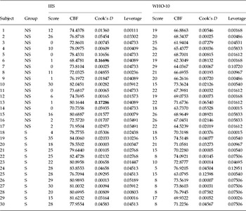
The following contrasts were formed: S >NS; NS >S. Moreover, the four SRS were submitted to statistical parametric mapping (SPM) analysis, seeking possible positive or negative correlations with CBF data in the group of subjects as a whole (i.e. irrespective of PTSD diagnosis, n=30). In order to evaluate the robustness of any significant correlations found, we calculated Cook's D (Cook, Reference Cook1977) and leverage statistics (see Table 2), whose critical values depend on the sample size (4/n=4/30=0,1333). This was done by extracting CBF values from SPM and by entering them into SPSS 13.0 (SPSS Inc., USA).
Cook's D and leverage statistics to evaluate the robustness of significant correlations (critical values: 4/n=4/30=0.1333)

IES, Impact of Event Scale; WHO-10, World Health Organization (10) Well-Being Index; CBF, cerebral blood flow; NS, traumatized non-post-traumatic stress disorder (PTSD) controls; S, patients with post-traumatic stress disorder.
Items above critical values are shown in bold.
SPM2 co-registers the individual SPECT to the 152 brains average of the Montreal Neurological Institute (http://www.bic.mni.mcgill.ca). Because its template does not completely match the Talairach brain, it is necessary to correct the SPM coordinates. This was achieved by using the subroutine implemented by Matthew Brett (http://imaging.mrc-cbu.cam.ac.uk/imaging/CbuImaging), which gives the correspondence between SPM coordinates and Talairach coordinates. Brodmann areas were then identified after importing the corrected coordinates, by means of the Talairach Deamon Database (http://www.talairach.org/).
Results
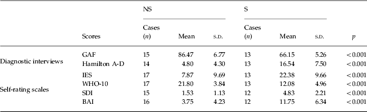
Both diagnostic interviews and SRS showed highly significant scores differences (as determined by t tests) between groups, PTSD subjects having higher IES, SDI and BAI scores and lower WHO-10 scores (see Table 3). IES scores showed high positive correlations with BAI (Pearson's r=0.66) and SDI (0.78); whereas WHO-10 showed high negative correlations with IES (−0.73), SDI (−0.81), and BAI (−0.55).
Psychometric ratings of study groups and statistical differences
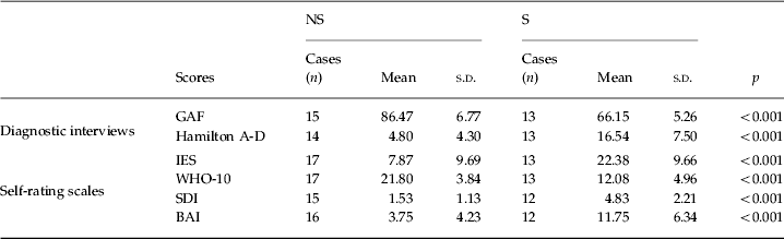
S, Post-traumatic stress disorder patients; NS, traumatized control subjects; GAF, Global Assessment of Functioning; IES, Impact of Event Scale; WHO-10, World Health Organization (10) Well-Being Index; SDI, Social Disability Index; BAI, Beck Anxiety Inventory.
Comparison between PTSD patients and controls
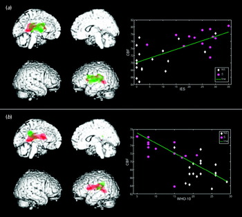
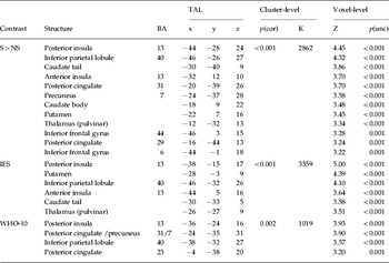
The comparison between S and NS (S >NS) showed a significantly higher tracer uptake in the left hemisphere in posterior and anterior insula, posterior cingulate, inferior parietal lobule, precuneus, nucleus caudatus, putamen, thalamus and inferior frontal gyrus (see Table 4 and structures displayed in red in Fig. 1). Conversely, the comparison between NS and S (NS >S) did not show any significant result.
(a) Left side: regions showing a higher regional cerebral blood flow (rCBF) in post-traumatic stress disorder (PTSD) as compared with non-PTSD subjects (S >NS; in red), regions showing a positive correlation between CBF and Impact of Event Scale (IES) scores (in green), and overlap between the two contrasts (in yellow); right side: scatter plot showing the strength of the correlation between CBF and IES scores (magenta circles, PTSD subjects; white diamonds, traumatized controls; green line, regression line); (b) left side: regions showing a higher rCBF in PTSD as compared with non-PTSD subjects (S >NS; in red), regions showing a negative correlation between CBF and World Health Organization (10) Well-Being Index (WHO-10) scores (in green), and overlap between the two contrasts (in yellow); right side: scatter plot showing the strength of the correlation between CBF and WHO-10 scores (magenta circles, PTSD subjects; white diamonds, traumatized controls; green line, regression line).

Brain regions showing a higher regional cerebral blood flow (rCBF) in S compared with NS (top); a positive correlation between CBF and IES scores (mid); a negative correlation between CBF and WHO-10 scores (bottom)

S, Post-traumatic stress disorder patients; NS, traumatized control subjects; IES, Impact of Event Scale; WHO-10, World Health Organization (10) Well-Being Index; BA, Brodmann area; TAL, Talairach coordinates; p(cor), statistical significance at cluster-level (whole-brain corrected); K, cluster size; Z, z-scores; p(unc), statistical significance at voxel-level (uncorrected).
Correlation analyses
IES scores positively correlated with tracer uptake (r=0.68) in the left hemisphere in posterior and anterior insula, IPL, nucleus caudatus, putamen and thalamus (see Table 4 and structures displayed in green in Fig. 1a).
WHO-10 scores negatively correlated with tracer uptake (r=−0.70) in the left hemisphere in posterior insula, posterior cingulate, IPL and PCN (see Table 4 and structures displayed in green in Fig. 1b).
SDI and BAI scores did not show any significant correlations with CBF at the chosen statistical threshold.
The clusters stemming from all three analyses substantially overlapped in posterior insula and IPL, whereas the clusters stemming from the comparison S >NS and the IES correlation also considerably overlapped along anterior insula and the basal ganglia.
Discussion
In the present study, correlation analyses were performed in order to detect brain regions whose activity is linked to symptom-related (BAI, IES) or more general (SDI, WHO-10) SRS scores. Our most relevant result was that IES scores positively correlated and WHO-10 scores negatively correlated with rCBF in a cohort of both PTSD and non-PTSD subjects, i.e. irrespective of PTSD diagnosis. To the best of our knowledge, this is the first time that such a finding is reported. Furthermore, it was shown that these correlations occurred to a great extent in the same regions in which rCBF significantly differed between symptomatic and non-symptomatic subjects during an autobiographical trauma script, i.e. anterior and posterior insula, PCC, IPL, PCN and the basal ganglia in the left hemisphere.
The IES is a measure of current subjective distress, which assesses the frequency of intrusive and avoidant phenomena associated with the experience of a particular event (Horowitz et al. Reference Horowitz, Wilmer and Alvarez1979). This instrument was shown to be capable of differentiating between PTSD and non-PTSD subjects (Bryant & Harvey, Reference Bryant and Harvey1996) and was successfully used in several studies on either pharmacological or psychotherapeutic treatments (see Joseph, Reference Joseph2000). IES scores have been found to be able to predict greater subsequent distress and other PTSD symptoms (McFarlane, Reference McFarlane1992; Joseph et al. Reference Joseph, Dalgleish, Thrasher, Yule, Williams and Hodgkinson1996).
The WHO-10 is a measure of the current subjective well-being and focuses on the personal evaluative reactions to one's own life in terms of both emotions and cognitive evaluations (Bech et al. Reference Bech, Gudex and Johansen1996). Subjective well-being is a transversal integrative construct, which has been much developed in the last decades (Diener et al. Reference Diener, Eunkook, Lucas and Smith1999) and widely adopted in different fields not strictly related to PTSD. However, subjective well-being is interesting at least for two reasons. First, it represents a criterion of mental health from the individual's own perspective (Diener et al. Reference Diener, Suh and Oishi1997). Second, it has proved to be a sensitive tool for the assessment of remission from PTSD (i.e. disappearance of symptoms) both immediately and several months after therapy (Högberg et al. Reference Högberg, Pagani, Sundin, Soares, Åberg-Wistedt, Tärnell and Hällström2008).
To sum up, our results indicate that a higher rCBF in some PTSD-sensitive brain regions is related to a higher subjective distress and to a lower subjective well-being. Such evidence has at least two implications. First, SPECT supports the validity of SRS by showing a common neural substrate between PTSD and some psychological constructs. Second, the structures identified with the correlation analyses should be implicated in the neural representation and/or processing of intrusions and avoidance (IES), as well as subjective well-being in terms of emotions, coping skills and life satisfaction (WHO-10).
Insular cortex
Several functional and structural studies have consistently shown the involvement of anterior (Liberzon et al. Reference Liberzon, Britton and Phan2003; Chen et al. Reference Chen, Xia, Li, Liu, He, Zhang, Yan, Zhang and Hu2006; Geuze et al. Reference Geuze, Westenberg, Jochims, de Kloet, Bohus, Vermetten and Schmahl2007; Lindauer et al. Reference Lindauer, Booij, Habraken, van Meijel, Uylings, Olff, Carlier, den Heeten, van Eck-Smit and Gersons2008) or posterior insula (Corbo et al. Reference Corbo, Clément, Armony, Pruessner and Brunet2005; King et al. Reference King, Abelson, Britton, Phan, Taylor and Liberzon2009), or both (Felmingham et al. Reference Felmingham, Kemp, Williams, Falconer, Olivieri, Peduto and Bryant2008; Whalley et al. Reference Whalley, Rugg, Smith, Dolan and Brewina2009) in PTSD pathophysiology. Studies on PTSD using a correlational approach have demonstrated that activity in anterior insular cortex is associated with flashback intensity (Osuch et al. Reference Osuch, Benson, Geraci, Podell, Herscovitch, McCann and Post2001), re-experiencing symptom severity (Hopper et al. Reference Hopper, Frewen, van der Kolk and Lanius2007) and avoidance symptoms (Strigo et al. Reference Strigo, Simmons, Matthews, Grimes, Allard, Reinhardt, Paulus and Stein2010) and that grey matter density reduction in this region directly correlates with trauma load (Nardo et al. Reference Nardo, Högberg, Looi, Larsson, Hällström and Pagani2010). In addition, it was shown that both anterior and posterior insular activity correlated with alexithymia scores (Frewen et al. Reference Frewen, Lanius, Dozois, Neufeld, Pain, Hopper, Densmore and Stevens2008).
On the other hand, alterations in insular activity have also been observed in healthy subjects, anteriorly during induced anxiety (Chua et al. Reference Chua, Krams, Toni, Passingham and Dolan1999), both anteriorly and posteriorly when processing negative emotions (Phan et al. Reference Phan, Wager, Taylor and Liberzon2002), and also in patients with various anxiety disorders or symptoms (Osuch et al. Reference Osuch, Ketter, Kimbrell, George, Benson, Willis, Herscovitch and Post2000; Stein et al. Reference Stein, Simmons, Feinstein and Paulus2007). Finally, some functional MRI (fMRI) studies have established altered patterns of functional connectivity among either anterior or posterior insula and other limbic regions in PTSD subjects (Lanius et al. Reference Lanius, Williamson, Densmore, Boksman, Neufeld, Gati and Menon2004, Reference Lanius, Williamson, Bluhm, Densmore, Boksman, Neufeld, Gati and Menon2005; Simmons et al. Reference Simmons, Paulus, Thorp, Matthews, Norman and Stein2008; Fonzo et al. Reference Fonzo, Simmons, Thorp, Norman, Paulus and Stein2010).
Insular cortex is highly regarded as an integrative interoceptive centre (Craig, Reference Craig2010) crucially involved in autonomic and limbic functions, body representation and subjective emotional response (Wiens, Reference Wiens2005), pain perception (Nagai et al. Reference Nagai, Kishi and Kato2007) and subjective experience of self-relevant aversive stimuli (Paulus & Stein, Reference Paulus and Stein2010). The insula is also considered to play a pivotal role in several neuropsychiatric diseases, including anxiety disorders such as PTSD (Etkin & Wager, Reference Etkin and Wager2007; Nagai et al. Reference Nagai, Kishi and Kato2007).
As regards its functional anatomy, the insular cortex seems to be organized according to a posterior-to-anterior gradient of integrative representations (Craig, Reference Craig2010). In fact, posterior insula contains a somatotopically organized ‘primary interoceptive centre’, which represents all affective feelings from the body (sensory aspects of emotions) and subserves maintenance and well-being of the body. Conversely, anterior insula is part of a network that engenders emotional awareness and self-consciousness, instantiating all subjectively perceived feelings within a unified and conscious representation (subjective feeling state).
Thus, a functional dichotomy seems to exist between a ‘posterior centre of interoception’ related to corporeal awareness (i.e. perception and evaluation of bodily signals) and an ‘anterior centre of emotional awareness’, possibly also playing executive and monitoring roles (Simmons et al. Reference Simmons, Paulus, Thorp, Matthews, Norman and Stein2008; Craig, Reference Craig2010). Both processes can be altered in PTSD and the different findings in terms of anterior/posterior insula reported in various studies might be related to the differential involvement of the two processes depending on the samples, type of controls and tasks employed.
It has been suggested that, in PTSD, traumas could be stored in the form of somatic memories and expressed as alterations in physiological stress response (van der Kolk, Reference van der Kolk1994). Accordingly, a failure of the declarative memory may lead to a reorganization of the trauma on a somatosensory level (physical sensations). This interpretation is supported by a study demonstrating that patients affected by dissociative identity disorder initially recall their trauma not as a narrative, but as somatosensory re-experiencing (van der Hart et al. Reference van der Hart, Bolt and van der Kolk2005). Posterior insula seems to be a good candidate to underlie a somatosensory reorganization of trauma-related memories. Consistently, an fMRI study on dissociative PTSD patients has shown that enhanced activity in this region is related to non-conscious (but not to conscious) fear processing (Felmingham et al. Reference Felmingham, Kemp, Williams, Falconer, Olivieri, Peduto and Bryant2008). Thus, a hyperactivity of posterior insula could be related to the activation of somatic representations of the traumatic experience (dissociated memories), which remain non-integrated and fail to reach the declarative memory. This interpretation is in line with a view of PTSD as a memory disorder characterized by the reliving of non-integrated traumatic memories (van der Kolk et al. Reference van der Kolk, Burbridge and Suzuki1997).
There are several explanations for an altered activity in anterior insula in PTSD. It has been argued that an unbalanced coordination between left and right anterior insulae might underlie dysfunctional feelings and/or awareness (Craig, Reference Craig2010). Increased activity in this region could be related to the processing of fear or other negative valenced emotional responses to symptom-provoking stimuli, suggesting the existence of a dysfunctional emotional regulation system (Etkin & Wager, Reference Etkin and Wager2007). Insular cortex seems to play a general role in several anxiety disorders (Paulus & Stein, Reference Paulus and Stein2010). In fact, the insula is considered critical for the evaluation of the potential impact of stimuli on the body, i.e. the detection of emotionally salient stimuli and the generation/regulation of affective responses. Its enhanced activity might result from an increased anticipatory response to potential aversive consequences, even if not actually present. It has been hypothesized that anxious subjects have reduced signal:noise ratio of interoceptive afferents, so they cannot easily differentiate between body signals associated with potential aversive stimuli and those that are not (Paulus & Stein, Reference Paulus and Stein2010).
In the present study, rCBF in both posterior and anterior insular cortex was found to be increased in PTSD subjects as compared with traumatized controls by using a symptom provocation paradigm. Moreover, irrespective of symptom provocation and PTSD diagnosis, activity in both portions of the insula increased with higher subjective distress scores, whereas only activity in posterior insula increased with lower subjective well-being scores. Thus, our findings suggest that trauma reliving, intrusions and avoidant behaviours imply altered activity in both posterior and anterior insula, i.e. a supposed dysregulation of both interoception (dissociated somatic memories) and (aversive/negative) emotional awareness, whereas subjective well-being seems to be associated only with interoceptive disturbance. In other words, feeling one's own body properly would be a critical component of well-being, as experiencing alteration in this process would be a critical component of PTSD. Conversely, an altered emotional awareness, or an enhanced negatively valenced emotional processing, would be associated with PTSD, but does not seem to affect subjective well-being.
Default mode network
Our results show that trauma reliving in PTSD subjects as well as higher subjective distress and lower well-being scores are all associated with higher activity in the IPL, whereas only trauma reliving and lower subjective well-being scores are associated with higher activity in the PCC and PCN.
The IPL has been found to be implicated in PTSD in a series of former symptom provocation studies (Bremner et al. Reference Bremner, Narayan, Staib, Southwick, McGlashan and Charney1999a, Reference Bremner, Staib, Kaloupek, Southwick, Soufer and Charneyb; Lanius et al. Reference Lanius, Williamson, Densmore, Boksman, Neufeld, Gati and Menon2004; Morey et al. Reference Morey, Dolcos, Petty, Cooper, Hayes, LaBar and McCarthy2009), as well as in activation studies investigating emotional valence (Bremner et al. Reference Bremner, Vythilingam, Vermetten, Southwick, McGlashan, Staib, Soufer and Charney2003) and working memory (Shaw et al. Reference Shaw, Strother, McFarlane, Morris, Anderson, Clark and Egan2002, Reference Shaw, Moores, Clark, McFarlane, Strother, Bryant, Brown and Taylor2009; Morey et al. Reference Morey, Dolcos, Petty, Cooper, Hayes, LaBar and McCarthy2009). The IPL is a broad multi-sensory integrative region and part of the dorsal executive network implicated in working memory. It takes part in several higher cognitive functions such as detection of salient stimuli (Husain & Nachev, Reference Husain and Nachev2006), perceptual awareness (Milner & Goodale, Reference Milner and Goodale1995), consciousness (Taylor, Reference Taylor2001), body image and the concept of ‘self’ (Torrey, Reference Torrey2007).
The PCC has been found to be more active in PTSD as compared with controls in previous symptom provocation and resting state studies (Sachinvala et al. Reference Sachinvala, Kling, Suffin, Lake and Cohen2000; Bremner et al. Reference Bremner, Narayan, Staib, Southwick, McGlashan and Charney1999a, Reference Bremner, Staib, Kaloupek, Southwick, Soufer and Charneyb, Reference Bremner, Vythilingam, Vermetten, Southwick, McGlashan, Staib, Soufer and Charney2003). Moreover, it has been suggested that a hypometabolism in this structure could play a role in dissociative symptoms (Lange et al. Reference Lange, Kracht, Herholz, Sachsse and Irle2005) and a very recent volumetric study by our group has shown significantly lower grey matter concentrations in the PCC of PTSD subjects as compared with traumatized controls (Nardo et al. Reference Nardo, Högberg, Looi, Larsson, Hällström and Pagani2010). The PCC is involved in a number of cognitive functions closely related to PTSD, such as episodic and autobiographical memory retrieval (Desgranges et al. Reference Desgranges, Baron and Eustache1998), pain perception (Chételat et al. Reference Chételat, Desgranges, de la Sayette, Viader, Berkouk, Landeau, Lalevée, Le Doze, Dupuy, Hannequin, Baron and Eustache2003; Nielsen et al. Reference Nielsen, Balslev and Hansenb2005), consciousness and self-centred reflection (Vogt & Laureys, Reference Vogt and Laureys2005; Bluhm et al. Reference Bluhm, Williamson, Osuch, Frewen, Stevens, Boksman, Neufeld, Théberge and Lanius2009) and processing of distressing information (Fischer et al. Reference Fischer, Wik and Fredrikson1996).
The PCN has also been found in PTSD studies, often in functional coupling with the PCC. Some fMRI studies have found that the PCN is associated with imagery or recall of traumatic memories in PTSD subjects, especially those exhibiting dissociative symptoms (Lanius et al. Reference Lanius, Williamson, Densmore, Boksman, Neufeld, Gati and Menon2004, Reference Lanius, Williamson, Bluhm, Densmore, Boksman, Neufeld, Gati and Menon2005). Moreover, other studies have identified hypometabolism in this region at rest in PTSD subjects as compared with controls (Lange et al. Reference Lange, Kracht, Herholz, Sachsse and Irle2005; Molina et al. Reference Molina, Isoardi, Prado and Bentolila2010). The PCN has been related to conscious visuospatial representation and mental imagery, the so-called ‘mind's eye’ (Fletcher et al. Reference Fletcher, Shallice, Frith, Frackowiak and Dolan1996; Cavanna, Reference Cavanna2007). Functionally, the PCN is strongly related to the PCC and is hence implicated in consciousness, self-reflection and episodic/autobiographical memory retrieval (Cavanna, Reference Cavanna2007). Thus, alterations in the normal functioning of the PCN might be related to impairments in the declarative memory system and conscious self-processing.
Interestingly, our results converge in showing that trauma reliving, subjective distress and well-being are all associated with a subset of regions belonging to the so-called ‘default mode network’ (DMN), a widespread brain system preferentially active when individuals are not focused on the external environment, but rather engaged in internally focused tasks such as autobiographical remembering, which require mental simulation or imagining scenes (Buckner et al. Reference Buckner, Andrews-Hanna and Schacter2008).
Importantly, DMN does not include primary sensory or motor areas, rather it operates in opposition to other brain systems involved in focusing external attention and sensory processing. The whole network comprises the mPFC, PCC, PCN, IPL and various portions of the temporal cortex (including the parahippocampal gyrus). The PCC has reciprocal connections with the IPL and the temporal cortex (Kobayashi & Amaral, Reference Kobayashi and Amaral2003, Reference Kobayashi and Amaral2007), which makes it a core region for declarative memory retrieval. The PCN also has connections with occipital areas (involved in visual processing) and with the PFC (Cavada & Goldman-Rakic, Reference Cavada and Goldman-Rakic1989; Leichnetz, Reference Leichnetz2001), rendering it strategic for visual imagery and self-related envisioning.
It has been well established that all nodes within the system show a strong intrinsic connectivity in resting conditions, i.e. the so-called ‘spontaneous cognition’ (Greicius et al. Reference Greicius, Krasnow, Reiss and Menon2003; Fox et al. Reference Fox, Snyder, Vincent, Corbetta, van Essen and Raichle2005). However, within the DMN two subsystems have been identified: an anterior node (mPFC), apparently more involved in self-relevant mental simulations; various posterior nodes (PCC, PCN, IPL and temporal cortex), implicated in successful retrieval of information from memory (Buckner et al. Reference Buckner, Andrews-Hanna and Schacter2008).
Two recent fMRI studies have purposely investigated intrinsic connectivity (i.e. activity in resting state) within the DMN in PTSD subjects. They found that spontaneous activity between PCC/PCN and other DMN nodes (and also with amygdala and hippocampus/parahippocampal gyrus) was reduced in PTSD subjects as compared with controls (Bluhm et al. Reference Bluhm, Williamson, Osuch, Frewen, Stevens, Boksman, Neufeld, Théberge and Lanius2009), and that connectivity of the PCC with the anterior cingulate and the amygdala was associated with current PTSD symptoms (Lanius et al. Reference Lanius, Bluhm, Coupland, Hegadoren, Rowe, Théberge, Neufeld, Williamson and Brimson2010).
In the present study, we found that increased activity in the IPL is associated with the processing of trauma-related stimuli, as well as with more frequent intrusions and avoidant behaviours, and with a lower subjective well-being. This is consistent with the impairments in conscious recollection and emotional processing, which typically occurs in PTSD, and possibly with any change in self-processing.
On the other hand, PCC and PCN result to be involved in trauma-related reliving and subjective well-being, but not in the experiencing of intrusions and avoidant behaviours. As centres of self-awareness, these regions might not be implicated in the automaticity of these symptoms, which could be more likely ascribed to the insula and the basal ganglia (whose activity directly correlates with IES scores). PCC and PCN are rather supposedly implicated in the altered declarative memory retrieval. In particular, a dysfunction of the PCC/PCN (in combination with the above-mentioned insular dysfunction) could contribute to the unsuccessful integration of the somatic memories into a conscious, coherent experience available to the declarative system. However, PCC/PCN are also likely involved in the conscious aspects of self-reflection, mental imagery and (aversive) emotional experience related to the traumatic event, whose alterations would be associated with the worsened emotional and cognitive life-related evaluations assessed with the WHO-10.
Finally, we should acknowledge the major limitations of the present study, i.e. the lack of a baseline condition to compare brain activity elicited by the symptom provocation procedure, and the relative small sample size, especially in regard to the PTSD subsample (n=13). However, we should also highlight the novel features of this study: the use of SRS, the correlational approach, the consistency between results of different analytic approaches (t tests and correlations).
Conclusions
This study revealed higher rCBF in PTSD as compared with non-PTSD subjects in limbic and parietal regions and in the basal ganglia. In most of these regions, CBF was also found to increase with higher IES and lower WHO-10 scores, irrespective of the PTSD diagnosis. Our results suggest a view of PTSD characterized by memory disturbances (including dissociation) and dysfunctional emotional regulation. They also seem to provide a cross-validation for IES and WHO-10 scales by means of SPECT, supporting their diagnostic validity and promoting their use in future works. Finally, the SRS correlations that were found suggest the existence of a continuous (i.e. not an ‘all-or-none’) representation of subjective well-being and distress in PTSD-sensitive structures and the involvement of such structures in the processing of these psychological constructs.
Acknowledgements
The study was funded by the Stockholm Public Transport Authority, Connex Sverige AB, Swedish State Railways, Stockholm County Council, The Vårdal Foundation, LJ Boethius Foundation, the Gadelius Memorial Foundation, and the Söderström-Königska Foundation, the Dipartimento per i Rapporti Internazionali, National Research Council (CNR), Italy, the Medical Research Council (MFR), Sweden. We thank Berit Tärnell for her valuable assistance during the study.
Declaration of Interest
None.