Introduction
There has been a growing interest in the development of flexible, portable, solution processable, low-cost, and thin-film photovoltaics.[Reference Rogers, Someya and Huang1] To achieve a cost competitiveness and widespread commercial adaptation of the solar cell technology, small amounts of materials and low-cost manufacturing technologies are necessary. More specifically, the development of liquid-phase deposition of materials on flexible substrates would bring a paradigm shift in the photovoltaic research.
ZnO has emerged as a cost-effective alternative to the most commonly used conducting electrode material, indium tin oxide (ITO). ZnO has many desirable properties, such as its wide band gap (~3.37 eV), visible light transparency, piezoelectricity, photocatalytic activity, biocompatibility, infrared absorption, and ease of production.[Reference Baruah and Dutta2] Unlike TiO2, ZnO can be molded into various nanostructures by simply controlling the synthesis parameters. ZnO nanorods and nanowires provide high surface area and direct conduction pathway for the rapid collection of photogenerated electrons. Liquid-phase synthesis of ZnO nanostructures is preferred due to the energy-efficient, inexpensive, and straightforward nature of the process. The hydrothermal growth process requires two steps of seeding and growth treatments carried out in liquids, which result in epitaxial crystal growth of ZnO nanorods. First, seeding is achieved by depositing ZnO nanoparticles onto substrates for forming nucleation sites which lower the thermodynamic barrier and hence enable further growth of high aspect ratio ZnO nanorods. Second, high aspect ratio ZnO nanorods were grown in a solution of zinc acetate and triethylamine under relatively mild conditions.
ZnO nanorods have mostly been grown on rigid substrates, such as ITO-glass electrodes, for photovoltaics and sensing applications.[Reference Ko, Lee, Kang, Nam and Yeo3, Reference Cheng, Chiu, Lee, Tsai and Hsieh4] However, growing solution-based, uniform, dense, and well-aligned ZnO nanorods on flexible substrates are more desirable. Recently, there have been several reports on the surface modification of flexible substrates, such as cotton and nylon fabrics, paper, and polymer films, by ZnO nanorods for various applications.[Reference Hsieh, Lin and Yang5–Reference Mao, Shi, Zhang and Cao7]
Electrospinning has become one of the most powerful tools for fabricating highly porous and interconnected network of nanofibers with high surface area that can house a large number of functional groups, making this technique particularly attractive for the development of multifunctional materials containing aligned metal-oxide nanostructures.[Reference Sahay, Kumar, Sridhar, Sundaramurthy, Venugopal, Mhaisalkar and Ramakrishna8, Reference Miao, Miyauchi, Simmons, Dordick and Linhardt9] ZnO nanorods have been grown on electrospun cellulosic polymers and titanium dioxide, for photocatalysis, sensing, and photovoltaic applications.[Reference Kanjwal, Barakat, Sheikh, Park and Kim10, Reference Kanjwal, Sheikh, Barakat, Li, Kim and Chronakis11] Polyamide nanofibers produced by electrospinning exhibit excellent mechanical properties, such as toughness and high tensile strength, and dimensional stability, and are resistant to most solvents. Highly oriented polymer chains and small fiber diameters can be easily obtained using polyamides which are crucial for the formation of transparent materials. Recently, we reported the growth of well-aligned, highly dense ZnO nanorods on the electrospun cellulosic nanofibers.[Reference Athauda, Butt, Harikumar and Ozer12] However, to date, there has been no report on the use of electrospun polyamide nanofibers as the 3D scaffold for the growth of ZnO nanorods.
Here, we report for the first time, the hydrothermal growth of branched ZnO nanorods on electrospun polyamide nanofibers as the first step toward the development of transparent, flexible, low-cost, scalable photovoltaic materials. Both methods are applicable for large-scale synthesis. Electrospun polyamide nanofibers have been used as plating template to deposit Pd catalyst before.[Reference Su, Jia, Schempf and Ding13] Since it is feasible to have polyamide nanofibers with diameters significantly smaller than the wavelength of the visible light and their T g (glass transition temperature) is lower than most other common polymers, they are the ideal candidates for self-supporting transparent electrode materials. Although we used nylon 6/6 as a model polyamide in this study, the reported method will be applicable to the other polyamides.
Experimental
Electrospinning procedure
20 wt % nylon 6/6 stock solution (M w ~ 262.35 g/mol, Sigma Aldrich) in formic acid (puriss. 98%, density: 1.22 g/mL) was prepared by overnight stirring. An aliquot of the freshly prepared nylon 6/6 solution was loaded into a 10 mL plastic syringe that was equipped with a blunt 18-gauge stainless-steel needle. A Gamma High Voltage Research power supply that can generate up to 30 kV DC voltage was attached to the tip of the needle, which was placed in a syringe pump. The aluminum collector was placed 18 cm away from the tip of the needle and was attached to the grounding electrode. The electrospun fibers were collected onto glass slides. The electrospinning parameters were as follows: applied voltage = 12 kV; flow rate = 6 µL/min; electrospinning duration = 2 h.
Preparation of ZnO seed solution
0.1 M zinc acetate dihydrate (ACS reagent, ≥98%) (1.0975 g) was dissolved in 50 mL of i-propanol (anhydrous, 99.5%) and the solution was vigorously stirred at 85 °C for 15 min. Then, 0.1 M triethylamine (≥99.5%) was added dropwise, which resulted in a clear solution. The reaction was maintained at that temperature for an additional 10 min and then aged at room temperature for 3 h (pH of the seed solution = 7.01). The average particle size of the ZnO nanocrystals seed solution (0.1 M) was ~31.5 ± 10.0 nm, as measured by a particle size analyzer.
Preparation of ZnO growth solution
Equimolar aqueous solutions of zinc nitrate hexahydrate (reagent grade 98%) and hexamethylenetetramine (ACS reagent, ≥99.0%) were used to grow the ZnO nanorods. First, 0.025 M hexamethylenetetramine (1.928 g) was prepared in 550 mL of deionized water. Once completely dissolved, 0.025 M (4.090 g) of zinc nitrate hexahydrate was added to this solution, which was followed by stirring for 24 h (pH of the growth solution = 6.11).
Growth of ZnO nanorods on electrospun nylon 6/6 nanofibers
The electrospun nanofibers were first dip-coated with the ZnO seed solution for 8 min, rinsed with ethanol, heat treated at 120 °C for 1 h, and then dried in air for 24 h. The electrospun fibers were then immersed in the growth solution, which was subsequently transferred into a closed, capped glass vial and heated at 95 °C for 8 h. The container was removed from the oven at the end of the reaction period and allowed to reach equilibrium at room temperature for approximately 10–12 h. The treated samples were thoroughly rinsed with DI water and dried in air.
Hydrophobic modification of the electrospun samples
The samples were treated with 10 mM 1-dodecanethiol in ethanol (ACS reagent, ≥99.5%, 200 proof, absolute) for 24 h. The samples were then washed in ethanol to remove excess reagents and allowed to dry in air.
Measurements and characterization
The surface morphology of the samples was investigated using a JEOL JSM 6060 LV scanning electron microscope, in which the samples were coated with a 15 nm layer of gold before observation. The crystal structure of the ZnO nanorods was analyzed using a Shimadzu XRD-6100 x-ray diffractometer (XRD) (Cu Kα (0.15406 nm) radiation), employing a scanning rate of 0.02°/s and 2θ ranges from 10° to 70° for ZnO. Thermogravimetric analysis (TGA) was performed using a Mettler Toledo 851 with a TSO 801RO robotic arm. The samples were heated from 40 to 600 °C at a rate of 10 °C/min under a nitrogen atmosphere at a flow rate of 40 mL/min. A small amount of the electrospun samples was scraped off the glass surface and analyzed.
The static water contact angles (WCAs) were measured using an AST Products VCA goniometer, which uses a computer-controlled syringe to dispose a 5 µL DI water droplet onto the substrates while calculating the contact angles at room temperature. Hydrophobicity is indicated by the larger WCAs. All contact angles were reported by taking the average of measurements of five random points on each sample surface. Light absorbencies were measured using a Varian Cary 50 UV-Vis spectrophotometer in a wavelength range of 280–480 nm and a scan rate of 300 nm/min.
Results and discussion
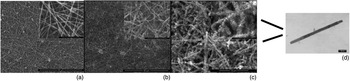
Figure 1(a) reveals the SEM images of electrospun nylon 6/6 nanofibers with average diameters of 100 nm. The electrospun nanofibers have uniform diameters, smooth surfaces, and are randomly oriented on the glass slides. The density of the nanofiber networks can be controlled by varying the electrospinning duration. ZnO nanocrystals adsorbed on the surface of the electrospun nanofibers after the seeding step are clearly seen in Fig. 1(b). ZnO nanorods, with the average diameters of 50 nm and the length of 900–1000 nm, were grown successfully on the electrospun nylon 6/6 nanofibers that penetrate the fiber network and cover the entire length of the nanofibers [Figs. 1(c) and (d)].
SEM images of (a) the nylon 6/6 nanofibers, (b) the seeded nylon 6/6 nanofibers, and (c) the nylon 6/6 nanofibers with ZnO nanorods (insets depict images at higher magnification), and (d) TEM image of a ZnO nanorod.

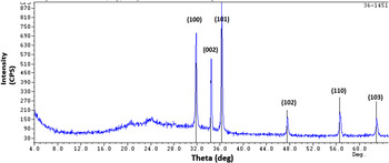
Figure 2 presents XRD pattern of the ZnO nanorods grown on electrospun nylon 6/6 naonfibers. Six reflection peaks appeared at 2θ = 31.9° (100), 34.6° (002), 36.5° (101), 47.7° (102), 56.8° (110), and 63.1° (103), respectively, and all the diffraction peaks could be indexed as the hexagonal wurtzite structure of ZnO with the lattice constants a = 0.3249 nm, c = 0.5206 nm, which were consistent with the values in the standard card (JCPDS 36-1451). No diffraction peaks from any other impurity phases are found, confirming that the only single-phase hexagonal ZnO is present. The enhanced (002) diffraction peak implies that the ZnO nanorods are grown along the c-axis orientation. The sharp diffraction peaks demonstrate that the as-synthesized ZnO nanorods have high crystallinity.
XRD patterns of ZnO nanorods grown on electrospun nylon 6/6 nanofibers.

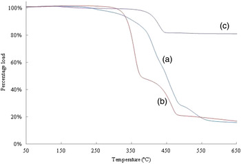
The amount of ZnO nanorods grown on electrospun nylon 6/6 nanofibers was determined by TGA. Figure 3 reveals the TGA curves of nylon 6/6, the seeded, and the ZnO nanorods-grown nanofibers. The loading extent of ZnO nanorods was calculated by the weight loss of the sample at the end of the 600 °C heating cycle by using the TGA instrument, and that was found to be 66 wt% which is significantly higher than those of ZnO grown on cotton (38%) and nylon fabrics (31%) previously prepared in our lab (unpublished data). The significantly higher amount of the ZnO loading can be attributed to the high surface area of the electrospun nanofibers and/or high amount of seeding that promote the growth of ZnO nanorods. This surprising result warrants further investigation.
TGA of (a) nylon 6/6 nanofibers, (b) seeded nylon 6/6 nanofibers, and (c) nanofibers with ZnO nanorods.

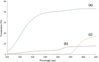
Figure 4 displays UV transmittance spectra of nylon 6/6 nanofibers with and without ZnO nanorods over the wavelength range of 280–420 nm. Untreated nylon 6/6 nanofibers absorbed approximately 21% of the incoming radiation between 320 and 380 nm. However, a 100% absorption of incoming light below 380 nm was observed with nanofibers containing ZnO nanorods. Excellent UV absorption feature of the ZnO nanorods/electrospun nylon 6/6 materials can find many potential applications in UV detectors, UV blocking, and energetic materials.
UV transmission spectra of (a) nylon 6/6 nanofibers, (b) seeded nylon 6/6 nanofibers, and (c) nanofibers with ZnO nanorods.

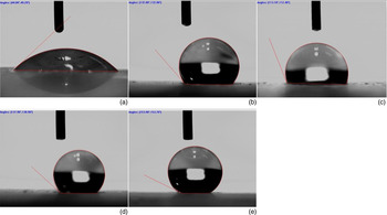
The wettability of a surface can be quantified by determining the contact angle between the liquid droplet and the surface. The WCAs of the samples are depicted in Fig. 5. The electrospun nylon 6/6 nanofibers have a very low hydrophobicity (44.0°). After 1-dodecanethiol treatment, however, WCA of nylon 6/6 nanofibers increased to 132.0°. The seeding process caused an increase in the WCA of the electrospun nylon 6/6 fibers from its original value of 44.0° to 113.1°. With ZnO nanorods grown on nylon 6/6 nanofibers, WCA was found to be 137.9°, which, after 1-dodecanethiol treatment, increased to 153.9°. The increase of the WCA with ZnO growth can be attributed to the increasing roughness of the nanofiber surfaces and the large hydrophobic surfaces of the treated nanorods.
WCA images of electrospun (a) nylon 6/6 nanofibers, (b) nylon 6/6 nanofibers treated with 1-dodecanethiol, (c) seeded nylon 6/6 nanofibers, (d) nylon 6/6 nanofibers with ZnO nanorods, and (e) nylon 6/6 nanofibers with ZnO nanorods treated with 1-dodecanethiol.

Conclusions
We developed a simple and facile method to grow ZnO nanorods on electrospun polyamide nanofibers with high WCAs and UV absorption by the combination of solution-based hydrothermal growth and electrospinning technique. Highly crystalline hexagonal wurtzite ZnO nanorods were obtained. The method described here can be easily extended to any other metal oxide–polyamide combination. Moreover, as the two techniques used are low cost, the method described here can easily be adapted for a large-scale fabrication. Hydrophobicity brings about self-cleaning properties, which can be highly beneficial for the protection of the functional devices against harsh environmental conditions.
Acknowledgments
We would like to thank the Department of Chemistry & Biochemistry at The University of Tulsa for its support. We greatly appreciate the financial support from The University of Tulsa Institute of Nanotechnology, TU Student Research Grants Program, and the Faculty Development Summer Fellowship and Research Programs for financial support. We would also like to thank Mr Rick Portman and Mr Winton Cornell for their help with SEM and XRD analysis, respectively.