Isolated left ventricular noncompaction is an important cause of childhood cardiomyopathy. According to recent reports, this anomaly accounts for almost one-tenth of all types of cardiomyopathy identified in an Australian epidemiological study of children,1 and similarly accounted for almost one-tenth of cases of cardiomyopathy diagnosed at the Texas Children's Hospital.2 Among adults undergoing echocardiography, its prevalence has been estimated to be 0.05%.3 Better availability, and increased technical sophistication, of cardiac imaging has led to increased recognition of the abnormality4, 5, 6 in people of all ages, including those without symptoms.
Although left ventricular noncompaction was originally described in case reports of children with other cardiac malformations,7, 8 the diagnosis may be missed in many children because of limited awareness of the condition.9 To the best of our knowledge, the prevalence of noncompaction of the left ventricle has not previously been described in children with additional congenital cardiac malformations. Moreover, the influence of any ventricular noncompaction on the prognosis for these children is unknown. In an effort to gain more information, we examined the prevalence, as identified through angiography, of left ventricular noncompaction in children diagnosed with congenital cardiac disease, proceeding to establish the presenting features and outcomes for these children.
Methods
Design of the study
This was an institution-based study of a cohort of patients with congenital cardiac disease treated in the Cardiology Department at the Royal Children's Hospital, Melbourne. This is the sole paediatric cardiac unit in Victoria, and serves a population of 5.5 million people.
Selection of patients
Our population comprised all patients less than 18 years of age undergoing left ventricular angiography between January, 1994, and December, 2000. To avoid any bias in selection, and to permit accurate documentation of outcome, we did not include children referred from overseas, or those referred solely for assessment for cardiac transplantation.
Diagnostic criterions for left ventricular noncompaction
Patients with left ventricular noncompaction were identified during a systematic review of all left ventricular angiograms performed during the period of study, using standard morphologic criterions.10, 11 The morphologically left ventricle was defined as a thick-walled, essentially ellipsoid ventricle; with a predominantly circular shape in short axis, and a morphologically mitral valve supported by paired papillary muscles. Children with functionally univentricular hearts in which the dominant ventricle was of right morphology, as in those with hypopla-sia of the left heart, or those with indeterminate ventricular morphology, were excluded from the study. Thus, in this study, a functionally single left ventricle was defined on the basis of absence or hypoplasia of the morphologically right ventricle sufficient to preclude a conventional biventricular surgical repair.
Patients with isolated atrial septal defects within the oval fossa, and those with patency of the arterial duct, were also excluded from consideration, as a minority of patients with these diagnoses underwent left ventricular angiography prior to definitive therapy.
The left ventricle was divided into 5 segments, based on common angiographic views, although variations of these views were used in some anatomically complex cases. The right anterior oblique view usually demonstrated the anterior and inferior segments of the left ventricular wall, and the left anterior oblique view usually demonstrated the septal, lateral, and apical segments.
Left ventricular noncompaction was diagnosed when any angiographic view clearly demonstrated a two-layered structure of the endocardium, with numerous and excessively prominent endocardial trabeculations and deep intertrabecular recesses, which filled with dye from the left ventricular cavity. For the purposes of quantifying the noncompacted and compacted layers, the epicardial contour was distinguishable as a soft tissue silhouette against lung tissue on the angiographic image (Fig. 1). To maintain concordance with diagnostic criterions used in previous studies,2, 3, 10 the measured thickness of the noncompacted layer in the most severely affected region was required to equal or exceed the thickness of the compacted epicardial layer at end-systole.

Figure 1. Angiogram showing a left anterior oblique view of the left and right ventricles in systole, from the thirty-first patient listed in Table 2, with a muscular ventricular septal defect and pulmonary valvar stenosis. The silhouette of the compacted lateral left ventricular myocardium can be easily visualised in the lower right corner of the angiographic view, thus enabling assessment of the ratio of noncompacted to compacted myocardium in systole.
The medical charts of all children diagnosed with left ventricular noncompaction were reviewed for information regarding the nature and timing of presenting symptoms, and the outcome in terms of the latest assessment of left ventricular function and clinical state. Survival for children with left ventricular noncompaction was compared with the survival of children from the same population, with the same principal congenital cardiac malformations, that is the same morphological diagnostic groups, who underwent left ventricular angiography, but did not have angiographic evidence of left ventricular noncompaction. The outcomes assessed were death or transplantation. Survival was analysed as time from diagnosis until death, transplantation, or 1st April 2003, when the vital state of all patients was ascertained. Follow-up was arranged for any patient not seen within the preceding 12 months.
Statistical analysis
Proportions were computed with 95% confidence intervals using exact mid-p-value intervals. Fisher's exact test was used to assess the association between two categorical variables. Crude survival was estimated using the Kaplan-Meier method. Survival was modelled using a proportional hazards model, with time since diagnosis as underlying timescale. Estimates of rate-ratios and survival curves were derived from that. The model was analysed using three explanatory variables in the same analysis, specifically the presence of left ventricular noncompaction, a functionally single left ventricle, and the age at diagnosis. A p-value less than 0.05 was considered significant.
Results
Between 1994 and 2000, we performed a total of 1653 angiograms of the morphologically left ventricle among local patients with congenital cardiac disease. Of these, 1628 (98.5% of angiograms) from 1535 patients were available for review. Angiographic features of left ventricular noncompaction were present in 63 angiograms from 31 patients, and for a majority of these children, left ventricular noncompaction comprised a new diagnosis. Allowing for the exclusion of 20 patients with isolated atrial septal defect within the oval fossa, or patency of the arterial duct, the prevalence of left ventricular noncompaction was 2.0% (95% CI: 1.4%–2.9%) in this population of children with congenital cardiac disease.
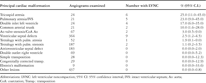
In Table 1, we show the range of principal associated cardiac malformations among the children undergoing angiography during the period of study, and the corresponding prevalence of left ventricular noncompaction within each diagnostic group.
Table 1. Incidence of left ventricular noncompaction among children with congenital cardiac disease undergoing left ventricular angiography.

The median number of affected segments was 3, with a range from 2 to 5. The number of noncompacted segments did not differ between children with a balanced ventricular arrangement and those with a functionally single left ventricle (p equals 0.42).
Almost half (48%) of children with left ventricular noncompaction had varying forms of a functionally univentricular heart with a hypoplastic and incomplete right ventricle, precluding biventricular repair. In a further assessment, 15 of 69 (22%, 13%–33%) children with a functionally single left ventricle were found to have left ventricular noncompaction, compared to 16 of 1446 (1.1%, 0.7%–1.8%) children with balanced ventricular arrangements (p less than 0.0001).
In Figures 1–3, we illustrate features of left ventricular noncompaction from patients in our study, showing the findings in the setting of different cardiac malformations. There was a complete spectrum of the extent of noncompacted myocardium in terms of segments involved, and also in the ratio of noncompacted to compacted myocardium. In some patients, the right ventricle also showed a remarkable appearance of noncompaction (Fig. 1), but it was not our purpose in this study formally to assess noncompaction involving the right ventricle.

Figure 2. Angiogram from the same patient as shown in Figure 3, that is the sixth patient listed in Table 2, showing a right anterior oblique view of the dominant left ventricle in systole.

Figure 3. Angiogram showing a left anterior oblique view of the left ventricle in systole, from the sixth patient listed in Table 2, with noncompaction of the dominant left ventricle in the setting of double inlet left ventricle and combined sub-valvar and valvar pulmonary stenosis.
Presenting features
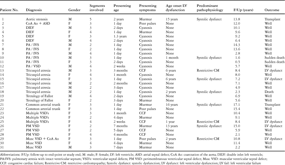
In Table 2, we show the presenting features and outcomes of individual children with left ventricular noncompaction. The majority of children with noncompaction presented in the neonatal period with symptoms of their principal cardiac malformation. None of the children with noncompaction had been diagnosed with neurological disorders, and none of the children were known to have had cerebrovascular events or embolizations.
Table 2. Clinical details of individual patients with left ventricular noncompaction associated with additional cardiac malformations.

Outcomes
Of the patients with noncompaction, 2 died suddenly, and 3 died or underwent cardiac transplantation after experiencing progressive left ventricular dysfunction. The median age at death or transplant was 2.3 years, with a range from 1.6 to 15.9 years.
Survival was assessed and compared only in the 760 children from the principal diagnostic groups in which we found patients with left ventricular noncompaction (Fig. 4). Age at diagnosis showed a non-significant negative trend with mortality, p equals 0.17. In this analysis, the proportional hazards model showed a significant effect of both the presence of left ventricular noncompaction and the presence of a functionally single left ventricle when entered separately into the model. In a model with both variables, the estimated effects of left ventricular noncompaction and a functionally single left ventricle were both in the vicinity of 2. That is, the presence of either condition doubled mortality, the rate ratio estimate of left ventricular noncompaction being 2.34, with 95% confidence intervals from 0.79 to 6.94, the rate ratio estimate of functionally single left ventricle being 2.04, with 95% intervals from 0.79 to 5.30, and an effect of 2.04 × 2.34, or 4.7, when both conditions were present. The p-values for left ventricular noncompaction and single ventricle were 0.127 and 0.142 respectively, but removing either variable from the model rendered the other significant. The p value for left ventricular noncompaction alone was 0.011, and that for functionally single ventricle alone was 0.021, so in this light we found it appropriate to report the non-significant effects of the two variables separately, as they both have a clear effect, even though non-significant when both are in the model.

Figure 4. Estimated survival curves for the 760 children shown in Table 1 with the principal congenital cardiac malformations in which we observed left ventricular noncompaction. Assuming an age at diagnosis of 1 year, survival is compared for four mutually exclusive subgroups within this group: those with and without left ventricular noncompaction, and those with and without a functionally single ventricle. The median age at diagnosis for the cohort is 0.97 years. The vertical lines represent 95% confidence intervals for the survival at 0.6, 1, and 3 years from diagnosis. Abbreviations: CHD: congenital heart disease; LVNC: left ventricular non-compaction; SV: functionally single ventricle.
The estimated survival curves for patients with and without noncompaction, and with and without a functionally single left ventricle are shown in Figure 4. At 5 years from diagnosis, survival among the 16 children with a balanced ventricular arrangement and evidence of noncompaction was 92%, with confidence intervals from 77% to 97%, compared to 96%, with intervals from 95% to 98%, among the 675 children with the same principal cardiac malformations but without evidence of noncompaction (Fig. 4).
Among the 31 children with left ventricular noncompaction, we did not find a significant difference in mortality between those with a functionally single left ventricle and those with a biventricular arrangement, although all the 3 early deaths were among children with a functionally single left ventricle.
Freedom from death or transplantation was unrelated to the number of noncompacted left ventricular segments. We did not assess the effect on survival of the ratio of noncompacted to compacted myocardium in any patient.
At latest follow-up, left ventricular dysfunction had developed in 5 of 26 surviving patients with noncompaction, and one of these patients had been listed for cardiac transplantation. Left ventricular dysfunction occurred in those with and without a functionally univentricular morphological arrangement (Table 2). Of these patients, 2 have predominantly impaired left ventricular systolic function, with echocardiographic left ventricular ejection fractions of 35% and 41%, respectively, while 3 patients have predominantly restrictive cardiomyopathy, with left ventricular end-diastolic pressures greater than 18 millimetres of mercury at cardiac catheterization (Table 2).
Discussion
Our study provides novel information about the prevalence of left ventricular noncompaction in children with a wide variety of cardiac malformations. Left ventricular noncompaction was detected in 2% of children with congenital cardiac disease under-going left ventricular angiography; with a prevalence varying from 1% of children with a balanced ventricular arrangement, to 22% of those having a functionally single left ventricle.
A number of factors may have contributed to this unexpectedly high prevalence. It is possible that the sensitivity of angiography is greater than echocardiography in detecting the myocardial features of noncompaction in this population of children with abnormal cardiac anatomy. This issue was not addressed in this study. Also, in recent years the proposed criteria for the diagnosis of left ventricular noncompaction in structurally normal hearts have become more stringent. The particularly high prevalence in children with functionally single left ventricle may be due to abnormal pressure or volume loading beginning during fetal life, or a common genetic factor resulting in both the functionally univentricular arrangement and ventricular noncompaction.
For many of our patients, the diagnosis of left ventricular noncompaction had not previously been made. This suggests that, with greater awareness of the clinical entity, the true prevalence and the clinical spectrum of left ventricular noncompaction may be greater than previously recognized.2, 4, 9, 12 Better imaging of the myocardium will also improve the accuracy of diagnosis. As the sophistication of magnetic resonance imaging improves, detailed static and cine images of the ventricular myocardium may further delineate the spectrum of left ventricular noncompaction, and give improved morphological and functional information.5, 6
Aetiology of left ventricular noncompaction
Left ventricular noncompaction is characterized by its genotypic and phenotypic heterogeneity.10, 13, 14 The myocardial abnormality was initially described among several infants with obstruction of the left ventricular outflow tract,7 in whom noncompaction was thought to represent an arrest of normal transition between fetal and adult myocardium, due to elevated left ventricular pressures. More recently an underlying genetic basis has been further elucidated, with both familial and sporadic cases being described.15, 16 Mutations in the G4.5 gene have been demonstrated in some individuals with isolated left ventricular noncompaction, including those without a typical phenotype of Barth syndrome.13 A genetic basis has also been suggested for subjects with left ventricular noncompaction and pulmonary valvar stenosis.13
An increasing body of evidence supports the notion that pathological ventricular noncompaction is the result of failure of a developmental process within the myocardium.12 During normal embryologic development, the trabecular layer of the maturing ventricular wall compacts progressively from base to apex.17 Pathological ventricular noncompaction may be a result of varying degrees of failure of this process. The pathophysiologic mechanisms for this are not fully understood, but may explain the heterogeneity of the condition.6, 12
The morphological spectrum of left ventricular noncompaction ranges from mildly increased trabeculation in a single cardiac segment, to significant trabeculation involving the apex, septal and free walls of the left ventricle.12 It is not clear whether the clinical implications of ventricular noncompaction correlate with the proportion of affected myocardium, but it is clear that the range of clinical scenes is broad. The presentation includes onset of congestive cardiac failure from infancy1 to adult life,15, 18 with a variable combination of systolic dysfunction and restrictive physiology.
Recent calls for the recognition and classification of left ventricular noncompaction as a distinct cardiomyopathy11 acknowledge the need for a greater understanding of its epidemiology and pathophysiology.
Our findings question a prevailing concept that left ventricular noncompaction is predominantly an isolated disorder that can only be diagnosed in the absence of other congenital cardiac malformations. The traditional criterions for its definition should be reconsidered with respect to children with congenital cardiac disease.
Outcomes
To our knowledge, there are few other studies reporting the outcome of a large group of patients with left ventricular noncompaction associated with additional congenital cardiac abnormalities. Our clinical findings are in keeping with those found in a small cohort of children from Saudi Arabia.19 Importantly, we found that the rate of death of patients with evidence of noncompaction was double that of those with comparable cardiac structural abnormalities without noncompaction. There was an apparent cumulative effect in subjects with a functionally single left ventricle. Moreover, nearly one-fifth of surviving subjects with left ventricular noncompaction developed persisting cardiac dysfunction, which sometimes became manifest many years after their initial presentation. These findings suggest the added impact of left ventricular noncompaction on the outcome of patients with congenital cardiac disease. We speculate that the presence of noncompaction may further impair myocardial performance among these patients. Microcirculatory coronary dysfunction has been described in subjects with isolated left ventricular noncompaction.20 This may result in both ventricular dysfunction and cardiac arrhythmias, and may account for the late myocardial dysfunction in the children included in our study. Ventricular noncompaction might also explain the increased incidence of late myocardial dysfunction among children with functionally single ventricles.
Limitations of the study
Our retrospective study has several limitations. Patients with symptoms, and those with more complex cardiac malformations, are more likely to undergo cardiac catheterization. For this reason, the outcomes for children with congenital heart disease, and those with left ventricular noncompaction, may not be truly representative of all children with these conditions.
Diagnostic consistency in this study was maintained by the use of pre-defined criterions applied to a single form of cardiac imaging. While left ventricular noncompaction can be readily identified from a variety of imaging modalities, including echocardiography,2, 4, 9 a systematic review of all echocardiograms undertaken on our patients with congenital heart disease was beyond our present scope. Moreover, we found that echocardiograms on children with congenital heart disease varied greatly in their technical quality. They were often performed in an opportunistic manner, varying with the condition of the child, their behaviour and the principle or presenting abnormalities being investigated. For these reasons, subjects in this study were recruited using angiography as the primary modality for imaging.
The limitations of angiography in diagnosing noncompaction are also significant. Though varying in thickness and location, the noncompacted endocardial layer of myocardium is easily identifiable, but the extent of the compacted layer is more difficult to visualize. In our study, we identified the margin of the compacted layer in all cases as a soft tissue silhouette against the lung tissue.
Our sample size was relatively small. The wide confidence intervals indicate that the ratios for rate of death should be interpreted with caution. Although our results cannot be used to predict the survival of any future population, they do provide a basis for further investigation of the issues.
Conclusions
Left ventricular noncompaction was found in 2% of our studied children with congenital heart disease undergoing left ventricular angiography, and is more common in children with functionally single ventricles. We suggest that our identification of late cardiac dysfunction, and the trend towards diminished long-term survival, justify additional scrutiny for ventricular noncompaction in children with congenital cardiac disease.
Financial Support
For the duration of this work Marina Hughes was supported by a KPMG Foundation Fellowship in Cardiovascular Disease Research, awarded by the Royal Australasian College of Physicians.