Introduction
Inclusion of hepatic venous flow is essential to prevent Pulmonary arteriovenous Malformations (PAVMs) after Kawashima surgery in heterotaxy patients. Reference Srivastava, Preminger and Lock1 Typically, Y-graft was used to incorporate hepatic veins into the Fontan circuit, which can be technically demanding and prone to kinking. However, in patients with bilateral hepatic venous drainage, this becomes even more complex. The use of transcatheter Fontan techniques offers an alternative for high-risk surgical cases. Our case report describes hepatic venous inclusion via a catheter-based intervention in a post-Kawashima patient with complex bilateral hepatic venous drainage.
Case report
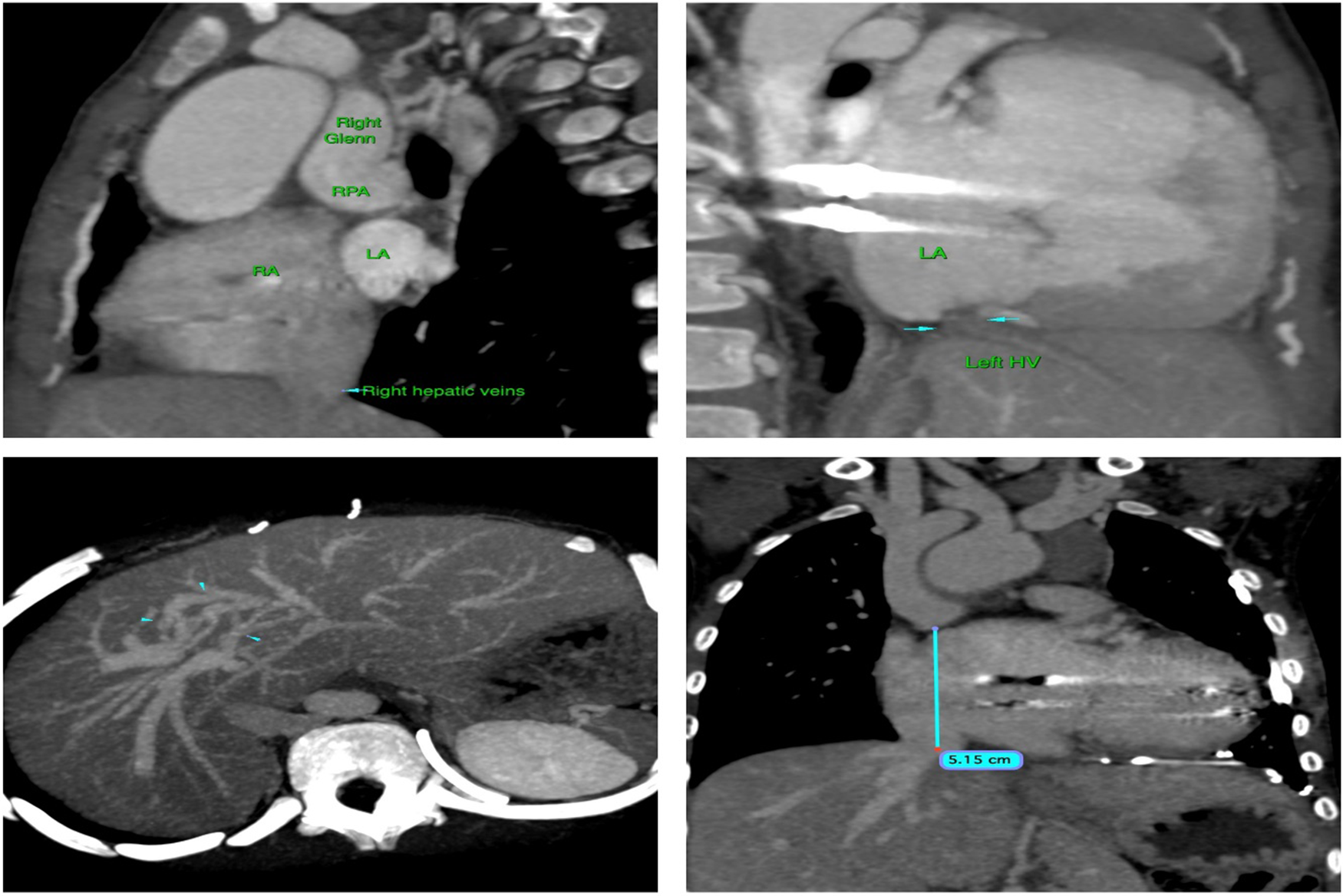
A 6-year-old girl from Africa, diagnosed with heterotaxy, left isomerism, unbalanced atrioventricular septal defect, severe pulmonary stenosis, and interrupted IVC, underwent Kawashima surgery at 1 year of age. At 4 years, she developed complete heart block and elevated mean pulmonary artery pressures (17 mmHg), complicating her pre-Fontan evaluation. Due to the ventricular dysfunction with elevated pulmonary artery pressures, Fontan completion was deferred. Instead, the patient underwent pacemaker implantation and left hepatic vein ligation as it was deemed non routable by surgery and to simplify future hepatic vein inclusion to complete Fontan. One year later, she returned for the hepatic vein diversion procedure. Clinical examination revealed hypoxaemia with a saturation of 78% and mild ventricular dysfunction. A computerised tomographic pulmonary angiogram showed good-sized branch pulmonary arteries with venous collaterals between the left and right hepatic veins (Figure 1). The liver function test showed normal values, and ultrasound liver showed normal hepatic and portal vein velocity with no evidence of portal hypertension except for few areas of hypertrophy noted in the right lobe of liver. Cardiac catheterisation confirmed elevated pulmonary artery pressures (mean-18 mmHg), but due to multiple previous sternotomies along with PAVMS and considering the complex heterotaxy syndrome, a catheter-based Fontan completion was preferred as we gained experience in performing transcatheter Fontan in unprepared surgical cases. Reference Ganesan, Muthukumaran, Viswanathan and Lakhani2,Reference Sivaprakasam, Ganesan and Ramkishore3
Computerized tomographic pulmonary angiogram images showing realtion of Glenn anastomosis with the common atrium and right hepatic veins and there is a ligated left hepatic vein (Blue arrow) without flows into the common atrium with well developed collaterals between right and left heaptic vein(Blue arrow heads).

Procedure
After obtaining consent from the parents, under general anaesthesia, intravenous antibiotics were administered, and right femoral venous and right internal jugular venous access was established using 6 Fr sheaths. Pre-procedure computerised tomographic pulmonary angiogram showed well-established collaterals between the left and right hepatic veins, guiding the site for puncture between the pulmonary artery and atrium. We planned to create connection between the pulmonary artery and the atrium, followed by placement of bare metal stents at the hepatic vein-right atrium (RA) junction, and a covered stent from the pulmonary artery to the hepatic vein-RA junction.
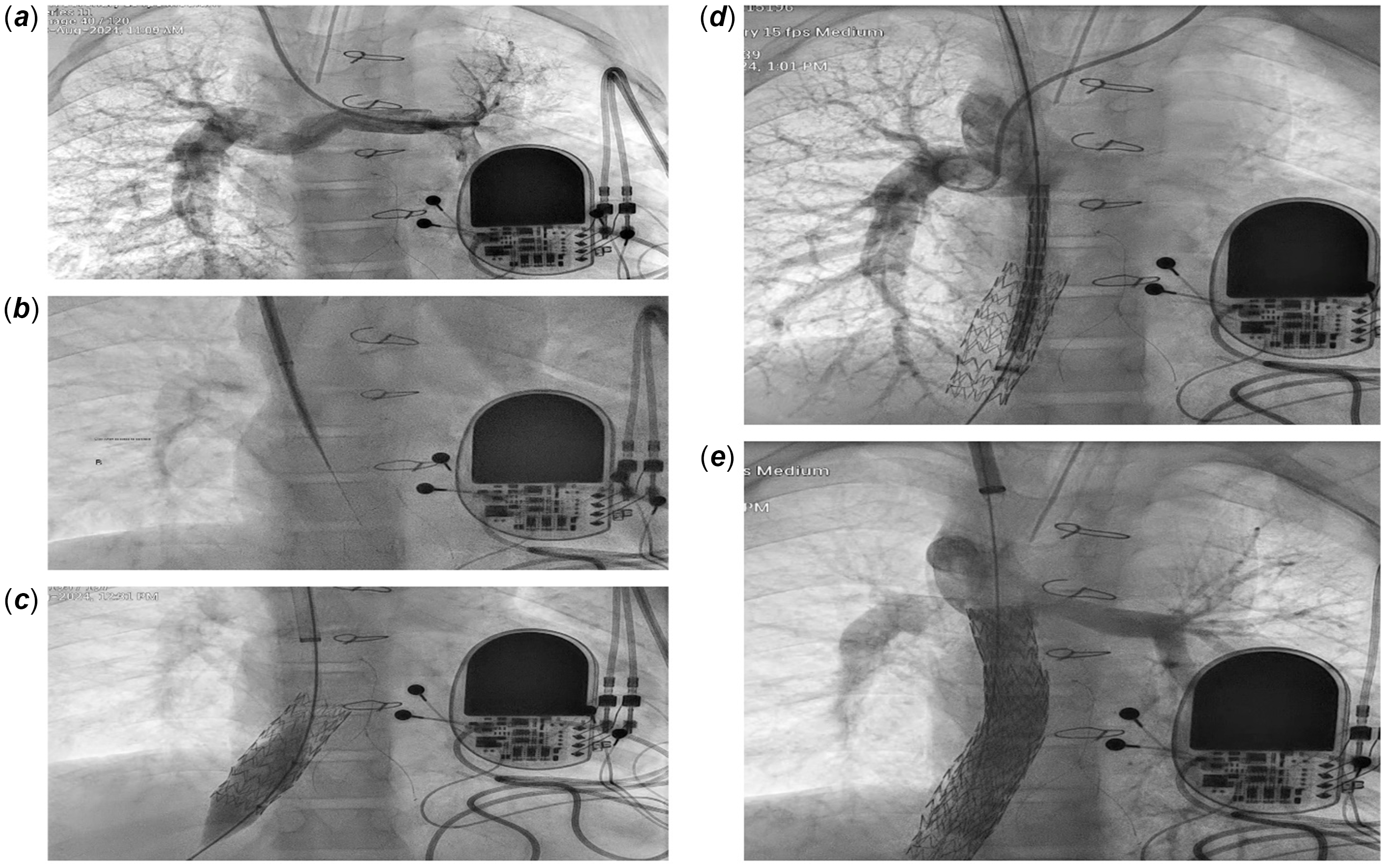
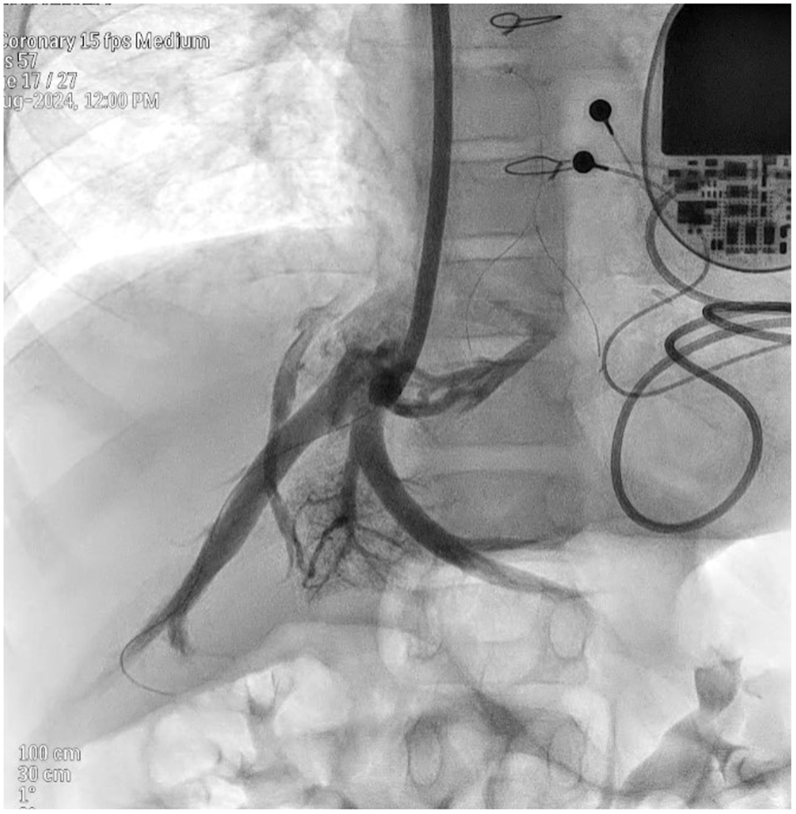
Through the right internal jugular venous approach using 8 Fr Swartz TM (St. Jude medical, Plymouth, USA), transeptal guiding introducer along with the dilator the PA over the common atrium was tented using hand angiograms in frontal and lateral projections. Using the hard end of 0.014 Asahi Gaia third wire (ASAHI INTECC, Aichi, Japan) along with the cautery, we perforated the pulmonary artery and atrial roof and entered the common atrial chamber (Figure 2 B). Check angiograms of pulmonary artery, and echocardiogram was performed to rule out pericardial and pleural collections before advancing the dilator of Swartz sheath into the common artium. After creating communication between pulmonary artery and atrium, the Gaia third wire was exchanged with exchange Terumo wire and placed deep inside the hepatic vein which was later replaced with Lunderquist wire (Cook medical, Denmark) using a Judkins right catheter. After upgrading the sheath to 14 Fr in the right internal jugular venous, pigtail angiograms were done to delineate the hepatic venous anatomy, and it showed good unobstructed drainage of left hepatic veins through collaterals into right and middle hepatic vein (Figure 3/ Video 1). Measurements were made that showed hepatic vein diameter of 14 mm, anchoring uncovered stent length of 44 mm and a covered stent length from base of pulmonary artery to hepatic vein of 55 mm was measured. A 44 mm Zephyr XL stent (SLTL, Gujarat, India) was crimped over 14 X 45 mm BIB balloon (NuMED Inc., NY, USA) and deployed in right hepatic vein with the stent extending into the mid atrial cavity followed by post dilatation with 16 x 20 mm Atlas Gold balloon (Bard Inc.,AZ, USA) (Figure 2 C). A 59 mm Zephyr covered stent was crimped over 12 X 80 Mustang balloon (Boston scientific, Marlborough, MA, USA) and deployed from the pulmonary artery end to approximate the IVC-RA junction (Figure 2D). There was migration of the stent near the pulmonary artery end hence another 39 mm Zephyr covered stent was deployed to cover the pulmonary artery end with post dilatation using 14- and 16-mm diameter balloons. Mean pulmonary artery pressure was 18 mmHg post-procedure, and the oxygen saturation improved to 88–90% in room air with good flow across the Fontan circuit (Figure 2E). No procedure-related complications occurred, and she was extubated and monitored overnight.
Angiographic still images showing Glenn injection (A) followed by creation of communication between pulmonary artery(PA) and atrium(B) with uncovered stent deployment in right hepatic vein followed by covered stent deployment from PA to heaptic vein -atrium junction (D).Fontan completion showing good flow across the circuit & branch pulmonary arteries(E).

Hepatic venous angiogram showing drainage of the left sided ligated hepatic veins into the other hepatic viens through collaterals.

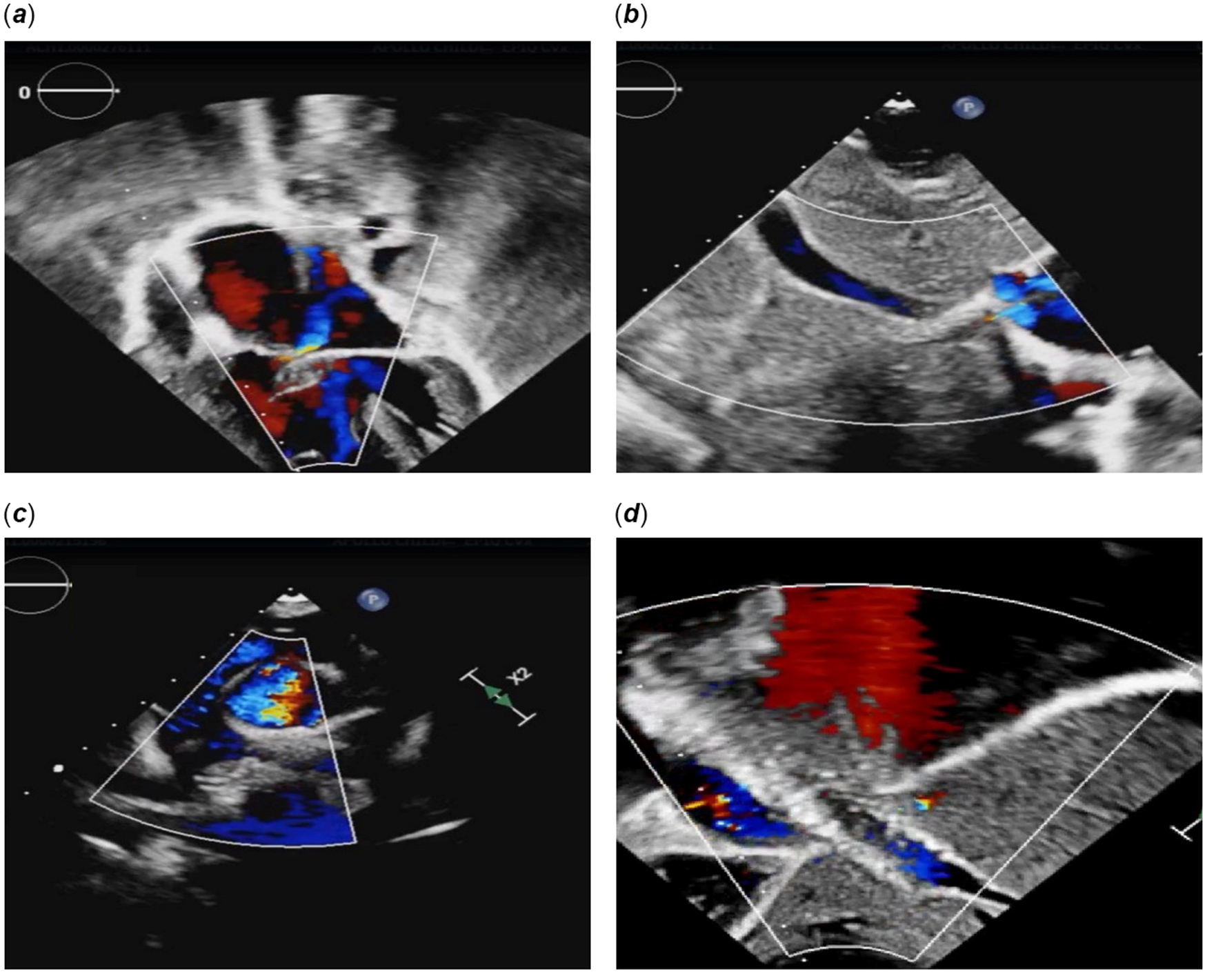
The patient was discharged on anticoagulation therapy with warfarin, aspirin, and sildenafil. One week later, her follow-up echocardiogram demonstrated good flow in the branch pulmonary arteries and unobstructed hepatic venous flow into the Fontan circuit (Figure 4).
Echocardiographic images showing unbalanced atrioventricular septal defect(A)with hepatic vein draining into common atrium(B).Post procedure showing well flowing Glenn anastomosis(C) with uncovered stent in the hepatic vein (D).

Discussion
Surgical techniques, including hepato-azygous connections,4 have been described in literature for complex hepatic venous anatomy inclusion. There were also techniques of surgical preparation during Kawashima surgery to complete transcatheter Fontan later, Reference Lugones, Junco, Biancolini, Martínez and Damsky Barbosa5,Reference Prabhu, Anderson, Ward, Karl and Alphonso6 but these may not always be feasible in cases with elevated pulmonary pressures or complex venous anatomy. In this case, due to the high risk associated with surgical Fontan, a transcatheter approach was chosen. The presence of well-developed venous collaterals between the hepatic veins allowed for a successful percutaneous hepatic vein diversion. Ligation of the left hepatic vein, combined with the development of venous collaterals, enabled us to achieve a redirection of hepatic venous flow without surgical intervention.
This is the first reported case of transcatheter Fontan completion in a post-Kawashima patient following hepatic vein ligation. It demonstrates that ligation of a separately draining hepatic vein can result in collateral development and still allow for successful transcatheter redirection with completion of Fontan can be done without complications.
Conclusion
For heterotaxy patients at high surgical risk, transcatheter hepatic vein diversion offers a safe and effective alternative to surgical Fontan completion.