Introduction
Unilateral vocal fold palsies can be caused by a wide range of pathologies, which can affect the recurrent laryngeal nerve along its course. Early detection of the underlying pathology is important, to rule out neoplastic lesions.
Radiographic investigations such as ultrasonography,Reference Wang, Chen, Lou, Yang, Hu and Shieh1 chest radiographyReference Altman and Benninger2 and computed tomography (CT)Reference Kang, Roh, Lee, Jung, Choi and Nam3,Reference Paddle, Mansor, Song and Franco4 scans have been utilised in the assessment of unilateral vocal fold palsies that remain unexplained after thorough history-taking and physical examination. In our institution, most clinicians routinely request CT scans of the skull base to arch of aorta as an initial investigation for patients with a clinical diagnosis of idiopathic unilateral vocal fold palsy.
A review of the literature showed that although the yield of routine CT imaging in the initial assessment of idiopathic vocal fold paralysis has been well studied, the role of CT imaging in cases of vocal fold hypomobility or paresis is not as well established. The paucity of studies investigating the diagnostic yield of CT in vocal fold paresis and the lack of Asian-based data prompted us to perform this study.
The primary aim of our study was to investigate the diagnostic yield of CT in the initial evaluation of patients with a clinical diagnosis of idiopathic unilateral vocal fold palsy. Our secondary aims were to determine the various pathologies detected on positive CT scans and to investigate the predictive factors for positive CT findings.
Materials and methods
Institutional board review approval was obtained for this retrospective study. The study included all adult patients seen at Tan Tock Seng Hospital, Singapore, from 2010 to 2018, with a clinical diagnosis of idiopathic unilateral vocal fold palsy, who underwent contrast-enhanced CT of the neck and chest.
We performed an initial search of cases using relevant diagnosis codes keyed into our electronic database. We then went through clinical data to obtain our final study subjects, who were patients with documented findings of unilateral vocal fold palsy with no obvious cause identified after thorough history-taking and physical examination, and who subsequently underwent CT evaluation. Subjects with a known cause of vocal fold palsy, neurological conditions (e.g. stroke, parkinsonism), primary laryngeal or pharyngeal neoplasm, or with a history of prolonged endotracheal intubation (more than 7 days), prior neck or chest surgery, rheumatoid arthritis, or medical conditions that could result in joint hypomobility, were excluded from the chart review.
Statistical analysis was performed using SPSS® software version 26. Data were analysed primarily using quantitative analysis. We also ran univariate analysis (chi-square test) to determine whether the patient or disease factors were predictive of positive CT findings.
Results
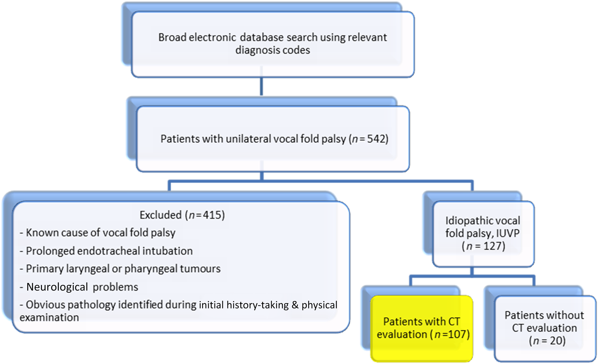
Of the 107 patients included in our study (Figure 1), 22 had positive CT findings that identified the cause of the vocal fold palsy. This equated to a diagnostic yield of 21 per cent. Patients with vocal fold paresis had a lower positive CT scan yield, of 11.5 per cent, than those with vocal fold paralysis (a yield of 29.1 per cent).
Flow chart showing the patient selection process leading to the recruitment of 107 subjects into the study. CT = computed tomography

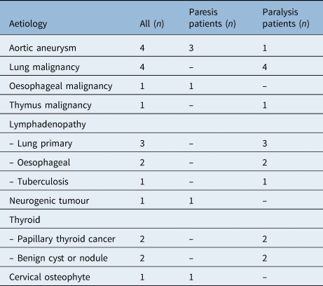
Malignancies (i.e. lung, thyroid, oesophagus, thymus, metastatic lymphadenopathy and neurogenic) accounted for 63.6 per cent (n = 14) of the positive findings (Table 1). Five out of the six cases of lymphadenopathy detected on CT were subsequently investigated and found to be secondary to primary lung (n = 3) and oesophagus (n = 2) malignancies. Lung malignancy (n = 7) was the most common cause of idiopathic unilateral vocal fold paralysis, accounting for 31.8 per cent of the positive CT findings (lung primary n = 4; metastatic lymph node from lung metastasis n = 3). Malignancy accounted for 75 per cent and 33 per cent of the positive CT findings in vocal fold paralysis and paresis cases respectively. Other identified causes of the idiopathic unilateral vocal fold palsy included aortic aneurysm (n = 4), benign thyroid nodule or cyst (n = 2), tuberculosis lymphadenopathy (n = 1), and cervical osteophyte (n = 1).
Breakdown of various aetiologies of vocal fold palsy detected on CT

CT = computed tomography
Nine of the 22 patients with positive CT findings had a chest radiography performed within six months of the CT scan; 3 patients showed aortic aneurysm and 6 showed a pulmonary or hilar mass.
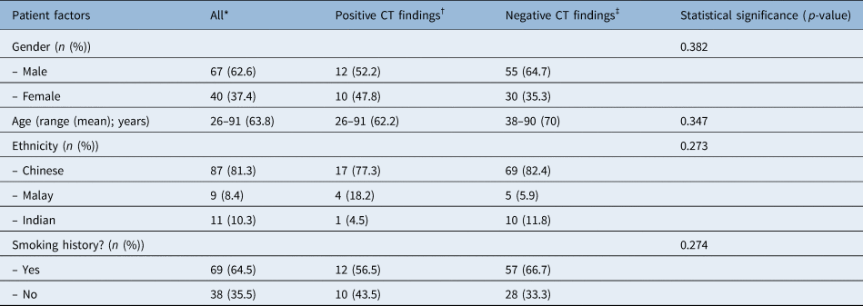
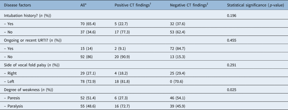
Analysis of patient and disease factors did not reveal any significant predictors of positive CT findings, except for degree of weakness (Tables 2 and 3). Patient characteristics such as gender, age, history of smoking and ethnicity were not predictors of positive CT findings. Patients with vocal fold paralysis had a clinically significant higher chance of having a positive CT finding compared to those with vocal fold paresis (p = 0.025).
Statistical analysis of effect of various patient factors on diagnostic CT yield

*n = 107; †n = 22; ‡n = 85. CT = computed tomography
Statistical analysis of effect of various disease factors on diagnostic CT yield

*n = 107; †n = 22; ‡n = 85. CT = computed tomography; UTRI = upper respiratory tract infection
During the follow-up period (mean duration of 14.3 months), none of the remaining 85 patients with negative CT findings were diagnosed with disease that accounted for their idiopathic unilateral vocal fold palsy.
Discussion
Studies have shown variation in CT diagnostic yield in the evaluation of idiopathic unilateral vocal fold palsy. Paddle et al.Reference Paddle, Mansor, Song and Franco4 reported a diagnostic yield of 2.9 per cent and an incidental yield of 27.6 per cent for idiopathic unilateral vocal fold palsy. Badia et al.Reference Badia, Hillel, Shah, Johns and Klein5 reported a 0 per cent diagnostic yield. In our centre, CT has high diagnostic yield, of 20.6 per cent. We also found a positive correlation between the degree of vocal fold hypomobility and the incidence of positive CT findings (29.1 per cent in patients with vocal fold paralysis vs 11.5 per cent in patients with vocal fold paresis, p = 0.025).
Of note, malignancy accounted for more than half (63.6 per cent) of the causative diagnoses detected on CT in our series, notably higher than in other studies,Reference MacGregor, Roberts, Howard and Phelps6,Reference Yumoto, Minoda, Hyodo and Yamagata7 which report rates of 13–33 per cent. Hence, we recommend routine CT imaging in the investigation of idiopathic unilateral vocal fold palsy. This is particularly so for vocal fold paralysis cases, where malignancy accounted for three-quarters of the positive diagnostic CT findings.
Altman and BenningerReference Altman and Benninger2 suggested chest radiography as the first-line investigation to evaluate for any compressive aortic aneurysm, or intrathoracic pulmonary or nodal diseases. Chen et al.Reference Chen, Young, Donovan and Ongkasuwan8 recommended initial evaluation with thyroid ultrasound and chest radiography. Proponents of initial ultrasound of the thyroid and X-rays of the chest have cited the higher radiation risks and healthcare costs associated with CT.
In our series, 36 per cent of the positive CT findings were secondary to pulmonary neoplasms and aortic aneurysms, and 18 per cent were secondary to thyroid neoplasms and nodules. These pathologies could potentially have been detected on chest X-ray and thyroid ultrasound. However, these studies would have missed the other 46 per cent of positive diagnoses, including neoplastic lesions. Moreover, patients may still have to undergo CT for further detailed evaluation of the lung and mediastinum pathology, resulting in more investigations and higher healthcare costs. Further prospective studies on the cost-effectiveness and diagnostic rate of initial chest X-ray or ultrasound thyroid evaluation versus CT imaging would be useful.
The role of repeat interval CT imaging for patients with initial negative CT findings is a subject of controversy. Noel et al.Reference Noel, Jeffery and Damrose9 reported that 8 out of 207 of those with initial negative CT scans went on to develop alternative causative diagnoses, with a mean time to diagnosis of 27 months. The authors therefore recommended routine repeat imaging within two years after the initial negative CT scan. The eight patients detected in the Noel et al. study underwent repeat imaging because of the development of new clinical findings or symptoms, rather than it being a routine repeat interval scan. In our series, patients with initial negative CT findings had a low likelihood of missed occult pathology, and none of the 85 patients subsequently developed disease that accounted for their idiopathic unilateral vocal fold palsy, during a mean follow-up period of 14.3 months. Hence, we concur with Tsikoudas and colleagues’Reference Tsikoudas, Paleri, El-Badawey and Zammit-Maempel10 recommendation that there is no added benefit of repeat interval CT evaluation for clinically asymptomatic patients with initial negative CT findings. We therefore recommend a clinical-based approach with repeat imaging only if the patient develops new symptoms or clinical findings.
• Unilateral vocal fold palsy can be caused by a range of pathologies that can affect the recurrent laryngeal nerve along its course
• Only degree of vocal fold hypomobility was a significant predictor of diagnostic computed tomography (CT)
• Malignancy accounted for 63.6 per cent of positive CT findings in idiopathic unilateral vocal fold palsy, with lung malignancy being the most common
• Unilateral vocal fold palsy and change in voice can be the only presentations in patients with underlying malignancy and aortic aneurysm
• Routine use of CT is recommended initially for investigating idiopathic unilateral vocal fold palsy, especially for paralysis
• Regular interval CT is not necessary if initial scan findings were negative, with no new clinical findings or symptoms
Limitations to this study include its retrospective nature and the small sample size; causative associations between the vocal fold palsy and the aetiology detected can only be assumed. The documentation of vocal fold mobility status as paresis and paralysis is subject to the attending physician's observational bias. Electromyography or examination under anaesthesia were not performed in this study; hence, a mechanical cause of vocal fold fixation is not entirely ruled out before labelling as idiopathic vocal fold paralysis or paresis.
Conclusion
Malignancy is a common aetiology for idiopathic unilateral vocal fold palsy. Routine use of CT is recommended in the initial investigation of idiopathic unilateral vocal fold palsy, as the risk of missing a potential malignancy outweighs the risk of radiation exposure. In circumstances where CT is not readily available, alternative investigations with thyroid ultrasound and chest X-ray can be considered. However, the patient needs to be informed that a negative ultrasound or chest X-ray does not fully preclude the presence of a causative diagnosis or malignancy.
Further long-term prospective studies are required to validate the usefulness of routine interval repeat CT evaluation for earlier detection of potential causative pathologies (e.g. neoplasm) that may not be clinically apparent at the early stages. Further prospective study will also be useful to ascertain the cost-effectiveness and diagnostic rate of initial chest X-ray or thyroid ultrasound scanning versus CT imaging.
Competing interests
None declared