Introduction
The trilobite suborder Phacopina is partially defined based on the fusion of the facial sutures during development, which prevents the librigenae being separated from the cranidium (Jaanusson Reference Jaanusson1975). This means the sutural gape mode of molting, where cephalic sutures (facial, rostral, and hypostomal) are opened to create an anterior exuvial gape, is impossible (Drage et al. Reference Drage, Holmes, García-Bellido and Daley2018). Instead the individual must disarticulate the entire cephalon from the trunk, called Salter’s mode of molting (see Daley and Drage Reference Daley and Drage2016; Drage et al. Reference Drage, Holmes, García-Bellido and Daley2018). The developmental timing of facial suture fusion varies interspecifically within Phacopina and has so far been described from several species within the family Phacopidae (superfamily Phacopoidea) (Crônier et al. Reference Crônier, Auffray and Courville2005; Crônier and Fortey Reference Crônier and Fortey2006). Postembryonic development is also comparatively well known for the Phacopidae (e.g., Osmólska Reference Osmólska1963; Chatterton Reference Chatterton1971, Reference Chatterton1980; Chatterton and Speyer Reference Chatterton and Speyer1997; Crônier et al. Reference Crônier, Bartzsch, Weyer and Feist1999; Crônier Reference Crônier2007), but this group represents a more derived lineage within the Phacopina (Jaanusson Reference Jaanusson1975). On the contrary, the superfamily Dalmanitoidea likely represents an earlier divergence of the phacopines, originating in the Lower Ordovician (Jaanusson Reference Jaanusson1975). Examining notable morphological and behavioral changes such as facial suture fusion in Dalmanitoidea is therefore crucial for understanding the evolution of development within Phacopina. Considering the hypothesized decline in morphological variability and polymorphism in more derived members of various trilobite clades (e.g., Ramsköld Reference Ramsköld1991; Webster Reference Webster2007), which has also been reported for the order Phacopida (Webster Reference Webster2007: Fig. 3E,F), we might expect that such changes in behavior and morphology were less developmentally constrained in the earlier lineages of this group. To explore this notion, and to trace the origins of facial suture fusion in the Phacopina, we here examine the postembryonic development of Dalmanitina (Dalmanitoidea: Dalmanitidae; following the taxonomy of Fortey [Reference Fortey1997]).
To date, the development of Dalmanitoidea is known only from the family Dalmanitidae. In particular, several species of Dalmanitina and closely related taxa have seen descriptions of small numbers of juvenile specimens. These include the original description of D. proaeva elfrida from Bohemia (see Barrande [Reference Barrande1852], also partially refigured by Whittington [Reference Whittington1956] and Šnajdr [Reference Šnajdr1990]), D. olini from Sweden and Wales (Temple Reference Temple1952, Reference Temple1957), D. nanchengensis from China (Lu and Wu Reference Lu and Wu1983), Dalmanites puticulifrons from North America (Whittington and Campbell Reference Whittington and Campbell1967), and Mucronaspis grandis from Bohemia (Budil Reference Budil1996). These works lack description of the full sequence of morphological change during ontogeny, as most studies comprise either too few specimens or disarticulated material. For example, the material of D. nanchengensis, while numerous and permitting a description of the protaspid period, was inadequate for providing a full post-protaspid sequence, because only a single articulated meraspid was identified.
This study presents the postembryonic development of two species of Dalmanitina (Reed, Reference Reed1905) from the Sandbian and Katian strata of the Prague Basin and is the first comprehensive description of developmental patterns in Dalmanitoidea. While Dalmanitina is a more derived genus within the dalmanitoid lineage, which originated in the Lower Ordovician (compared with the Middle Ordovician origins of Dalmanitina; Šnajdr Reference Šnajdr1982), Dalmanitina represents a comparatively large data set of early-diverging phacopine trilobites in the context of the greater body of work on ontogeny of the Phacopoidea. This data set was gathered by examining postembryonic stages of D. proaeva elfrida and D. socialis from the National Museum Prague (NMP) and Czech Geological Survey (CGS) and has allowed for detailed biometric analysis of their development. In particular, this developmental sequence is significant due to its exemplification of the timing of facial suture fusion events, traceable by the noteworthy preservation of a large number of specimens.
Geological Setting
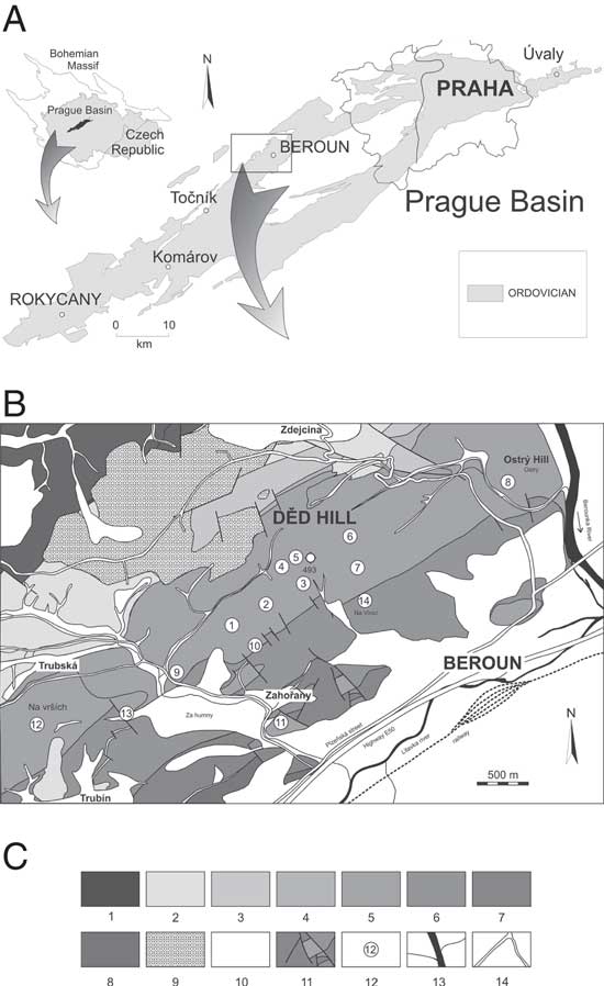
In the Prague Basin (Barrandian area, Czech Republic; see Havlíček Reference Havlíček1981, Reference Havlíček1982), all representatives of Dalmanitina occur exclusively in the strata representing the Berounian regional stage (corresponding approximately to the Sandbian and lower part of the Katian global stages; see Havlíček and Marek Reference Havlíček and Marek1973; Havlíček and Fatka Reference Havlíček and Fatka1992; Havlíček et al. Reference Havlíček, Vanĕk and Fatka1994; Fatka et al. Reference Fatka, Kraft, Kraft, Mergl, Mikulas and Štorch1995; Havlíček Reference Havlíček1998; Fatka and Mergl Reference Fatka and Mergl2009; Gutiérrez-Marco et al. Reference Gutiérrez-Marco, Sá, García-Bellido and Rábano2017). The Berounian comprises in the Prague Basin the Libeň, Letná, Vinice, Zahořany, and Bohdalec Formations. Their constituent fauna are broadly comparable despite their different lithologies. Juveniles of Dalmanitina have historically been documented or collected at several localities for each of the Letná, Vinice, and Zahořany Formations in the Prague Basin. They are rare and poorly preserved, or absent, from all of the remaining stratigraphic levels of the Berounian. This study therefore focuses on the many well-preserved specimens in the historical collections at the NMP and CGS from the Letná, Vinice, and Zahořany Formations (obtained from locations discussed below; see Fig. 1), with an emphasis on the particularly rich material of D. proaeva elfrida occurring in the Vinice Formation.
A, The Ordovician of the Prague Basin (after Manda Reference Manda2008; Fatka et al. Reference Fatka, Budil and David2015). B, Simplified geological map of the northwest neighborhood of Beroun (after Chlupáč Reference Chlupáč1965, 1993, Reference Chlupáč2002; Chlupáč et al. Reference Chlupáč, Brunnerová, Havlíček, Kovanda, Kříž, Šalanský, Štych and Zelenka1989; Havlíček et al. Reference Havlíček, Brunnerová, Holub, Hrkal, Cháb, Chlupáč, Kovanda, Rudolský, Šalanský, Štorch and Volšan1993). C, Key to the geological map of the Prague Basin: 1, Neoproterozoic, Kralupy-Zbraslav Group; 2, Floian, volcanites of Komárov Volcanic Complex (Floian to Sandbian); 3, Libeň Formation, Řevnice Quartzite facies (Sandbian); 4, Libeň Formation, Libeň Shale facies (Sandbian); 5, Letná Formation (Sandbian); 6, Vinice Formation (Sandbian–Katian); 7, Zahořany Formation (Katian); 8, Bohdalec Formation (Katian); 9, Carboniferous; 10, Quaternary undivided; 11, faults; 12, position of localities; 13, rivers and brooks; 14, roads and highways. Key to the numbered localities in B (see Chlupáč Reference Chlupáč1965): 1–7, small pits and outcrops at Děd Hill cumulatively named as “Drabov” in the nineteenth century (most museum samples have this name); 8, Ostrý Hill; 9, Čížovka; 10, Zahořany north of the village; 11, Zahořany, Háj; 12, fields between Trubín and Trubská Villages; 13, Trubská rokle Gorge (cumulative name); 14, Vinice Farm, stratotype of Vinice Formation (cumulative name). 1–8 and 12, Letná Formation localities; 10 and 11, Letná and Zahořany; 13–14 Letná and Vinice.

Juveniles of D. socialis were collected from multiple localities in the Letná Formation. Several small pits were created by fossil collectors at Děd Hill northwest of the town of Beroun in the nineteenth century (Fig. 1, localities 1–7), and cumulatively designated as from “Drabov.” These pits correspond to several fossiliferous intervals that can be difficult to distinguish (Chlupáč Reference Chlupáč1965, 1993, Reference Chlupáč2002). The exact geographic and stratigraphic source of NMP material of D. socialis from the locality Veselá is similarly uncertain (an old farm northeast of the town of Beroun, near the Vráž Village, also corresponds with localities 1–8; see Chlupáč Reference Chlupáč2002; Vokáč et al. Reference Vokáč, Hartl, David, Pavlovič, Doubrava, Kozák and Grigar2015: p. 150).
Juveniles of D. proaeva elfrida are very common in the Vinice Formation. The rich material stored in the NMP collections (particularly the Barrande collection, representing many of the described specimens) comes mainly from Trubín. The exact geographic position of this locality is poorly known. As for the Drabov localities in the Letná Formation, Trubín is likely represented by a series of recently overgrown natural outcrops and small collectors’ pits exploited north of the village Trubín in the nineteenth century (Fig. 1, locality 12; see Chlupáč Reference Chlupáč2002). This cumulative locality name may also overlap with the better-known outcrop at Trubská rokle Gorge, north of Trubín (Fig. 1, locality 13), which provided material of D. socialis and D. proaeva elfrida from the Letná and Vinice Formations, respectively. Some of the studied specimens of D. proaeva elfrida also come from Vinice, the type locality of the Vinice Formation. This was formerly exposed at the farm on the southeastern slope of Děd Hill (now mostly overgrown collection pits; see Fig. 1, locality 14). Studied specimens of D. proaeva accessioned at the CGS had been collected from several localities in the Zahořany Formation (see localities 10 and 11 in Fig. 1 and Supplementary Material 1).
The Letná Formation (Sandbian age) is characterized by alternation of coarser and finer sediments in beds several centimeters to decimeters thick (see Havlíček Reference Havlíček1998), all with strong bioturbation and diverse ichnofossils (see Mikuláš Reference Mikuláš1998a, Reference Mikulášb). Fine-grained sandstones to graywackes, sandy siltstones, clayey siltstones, and clayey sandstones dominate, with rare clayey shales. The thickness of the formation varies between 650 and 40 m (Havlíček Reference Havlíček1998). Dalmanitina socialis and Deanaspis goldfussi dominate among trilobite species (with many other genera present); non-trilobite arthropods, bivalves, gastropods, conulariids, brachiopods, and echinoderms are also present. These are characteristic of the inshore quartzose sandstones, while a poor atheloptic trilobite association is typical for the offshore slope settings. Rare graptolites, trilobites of the Cyclopygid Biofacies, and brachiopods of the Paterula Community occur in poorly oxygenated black shales in central parts of the basin (for a summary, see Fatka et al. Reference Fatka, Lerosey-Aubril, Budil and Rak2013).
The Vinice Formation (uppermost Sandbian to Katian) is characterized by a marine transgression over the stable segments bounding the Prague Basin (see Havlíček Reference Havlíček1998). Black shales are the dominant lithofacies. Due to anoxic or dysoxic near-bottom conditions, the benthic fauna of the Vinice Formation is reasonably monotonous and consists mostly of bivalves, gastropods, and hyolithids, accompanied by a few trilobite species (D. proaeva elfrida and Deanaspis senftenbergi in particular) and brachiopods (see Havlíček and Vaněk Reference Havlíček and Vaněk1966). Ichnofossil diversity is much lower than in the underlying Letná Formation. The benthic fauna typical of the Vinice Formation supposedly inhabited a poorly aerated, soft-bottom environment. Rare graptolites, trilobites of the Cyclopygid Biofacies, and the Paterula brachiopod community represent the pelagic and epibenthic forms. Diverse echinoderms, accompanied by trilobites, brachiopods, cephalopods, and conulariids are confined to the oolitic ironstones in the uppermost parts of the formation.
The Zahořany Formation (Katian) is composed of siltstones and rare intercalations of shales and silty sandstones, with a thickness of between 70 and 400 m across the Prague Basin (Havlíček Reference Havlíček1998). Storm sediments with hummocky cross-stratification are common within the formation at the thinner extremities, and bioturbation is frequent (Mikuláš Reference Mikuláš1998a). The fauna includes abundant brachiopods, gastropods, bivalves, hyolithids, trilobites (D. proaeva and Marrolithus ornatus in particular), and echinoderms. In the deeper parts of the formation, brachiopods gradually decrease in number, and the benthic fauna becomes poorly diversified (Havlíček Reference Havlíček1998).
Materials and Methods
Materials
The material of D. proaeva elfrida accessioned at the NMP that comes from the Vinice Formation is preserved as internal molds (usually covered by a thin film of iron hydroxides) in dark gray (almost black) shales. Specimens of D. socialis accessioned at the NMP from the Letná Formation are preserved in gray to brown siltstones to fine-grained sandstones. The specimens of D. proaeva from the Zahořany Formation accessioned at the CGS are preserved in gray siltstones. These have not been conclusively described to subspecies level (they are mostly represented by disarticulated cephala/cranidia), but are assigned to D. proaeva and are very closely related (almost indistinguishable) to D. proaeva elfrida (see Šnajdr Reference Šnajdr1982: p. 181). Generally, the material from the Vinice Formation is more dorsoventrally flattened, but rarely affected tectonically and preserves more exoskeletal detail. By contrast, the material from the Letná Formation is less flattened, but in most cases poorly preserved due to the coarsely grained sediment. Sometimes, however, the specimens from the Letná Formation seem to preserve partially decalcified remains of exoskeleton with surface sculpture (see “Results”). The material from the Zahořany Formation is more three-dimensional and generally well preserved, but contains only holaspides.
Methods
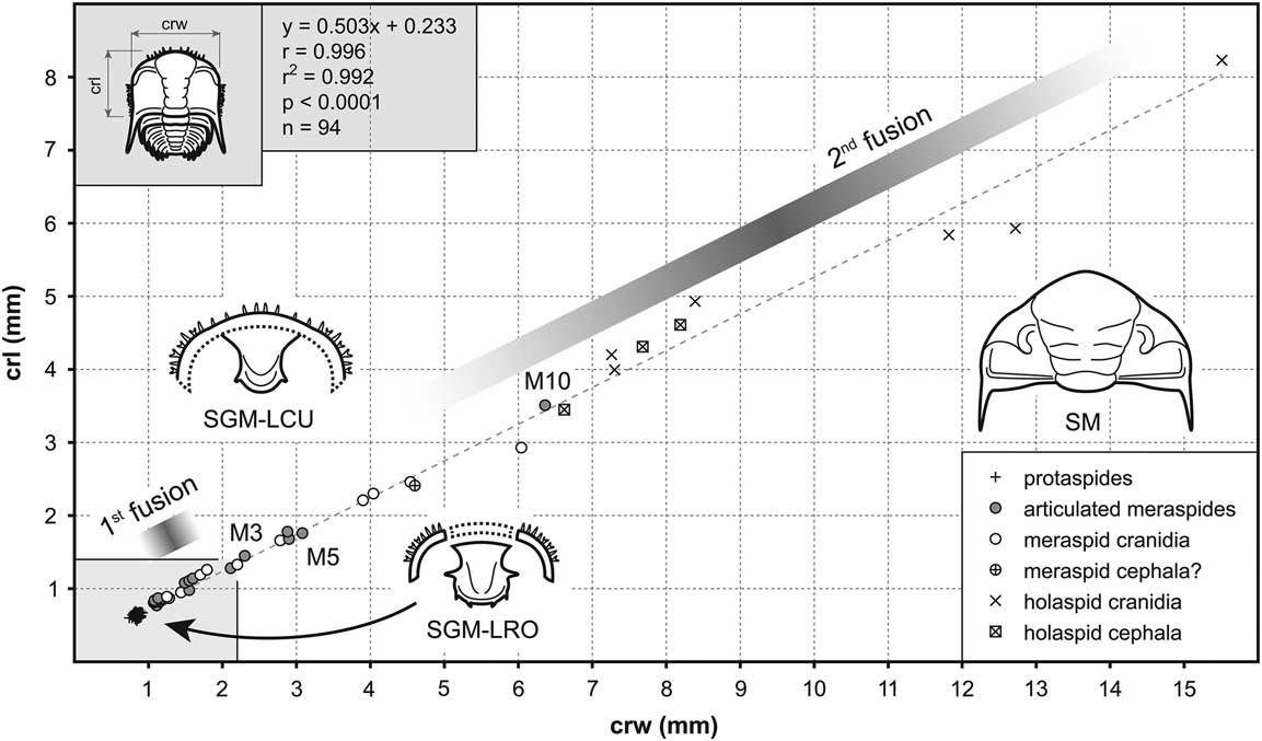
All measured specimens of D. proaeva elfrida (n=98), D. socialis (n=31), and D. proaeva (n=8) from the NMP and CGS (see Supplementary Material 1 and 2) were coated with ammonium chloride sublimate and photographed using the digital microscope Keyence VHX-2000 and/or DSLR camera Canon EOS 6D. The material was subsequently measured using the optical image analyzer TpsDig2 (Rohlf 2006). In all of these specimens, two parameters were measured: (1) sagittal length of the cranidium/cephalon (crl) and (2) transverse width of the cranidium/cephalon across the occipital ring (crw; see Fig. 2). In the earliest (P1 to M2) complete individuals, five parameters were measured in total: (1) sagittal length of the exoskeleton, excluding spines (exl); (2) transverse width of the cranidium across the occipital ring (crw); (3) sagittal length of the cranidium (crl); (4) sagittal length of the trunk, excluding spines (trl); and width of the trunk (trw; see Fig. 3). Width and length measurements were displayed on scatter plots in PAST (v. 2.13; Hammer et al. Reference Hammer, Harper and Ryan2001) to examine developmental trajectory (using reduced major axis linear regression) and to distinguish individual developmental stages. Growth-related developmental traits (growth rate [GR] and index of conformity to Dyar’s rule [IDC]) were calculated according Fusco et al. (Reference Fusco, Garland, Hunt and Hughes2012) from arithmetic means of each cluster or stage.
Scatter plot of cranidial or cephalic width (crw) vs. cranidial or cephalic length (crl) for Dalmanitina proaeva elfrida from the Vinice Formation (Sandbian to Katian) of the Prague Basin. Abbreviations: M3, meraspid degree 3; M5, meraspid degree 5; M10, meraspid degree 10; SGM-LRO, sutural gape mode of molting separating librigenae (and doublure) and hypostome; SGM-LCU, sutural gape mode of molting with a joined lower cephalic unit; SM, Salter’s mode of molting. A number of holaspid specimens detailed in Supplementary Material 1 and 2, both with fused and unfused sutures, are greater than 15 mm in length.

Scatter plots of cranidial width (crw) vs. exoskeletal length (exl), cranidial width (crw) vs. cranidial length (crl), and width of the trunk (trw) vs. length of the trunk (trl) for Dalmanitina proaeva elfrida from the Vinice Formation (Sandbian to Katian) of the Prague Basin. Abbreviations: P1, first protaspid stage; P2, second protaspid stage; M0, meraspid degree 0; M1, meraspid degree 1; M2, meraspid degree 2.

Repositories and Institutional Abbreviations
The studied material is housed in the collections of the National Museum, Prague, Czech Republic (prefix NMP), the Museum of Comparative Zoology, Cambridge, Massachusetts (prefix MCZ), and the Czech Geological Survey, Prague, Czech Republic (prefix CGS).
Results
Postembryonic Developmental Sequence of D. proaeva elfrida
The postembryonic development of trilobites is traditionally divided into protaspid, meraspid, and holaspid periods (cf. Chatterton and Speyer Reference Chatterton and Speyer1997; Hughes et al. Reference Hughes, Minelli and Fusco2006). The studied material of D. proaeva elfrida contains individuals belonging to all of these periods, ranging from 0.78 to 61.20 mm in sagittal exoskeletal length and 0.74 to 37.65 mm in maximum exoskeletal width. The size distribution of the individual cranidia and cephala during postembryonic development can be described by reduced major axis linear regression (Fig. 2). Specimen measurements show size clustering, at least for the earliest developmental stages up to meraspid degree 1 (M1; Fig. 2). Protaspid stage 2 (P2) and meraspid degree 0 (M0) overlap considerably in size (forming the second cluster; Fig. 3) but have distinctly different morphologies (Figs. 4 and 5). M0 specimens show an articulation structure and longer trunk with five axial rings (Fig. 5A,B), whereas P2 specimens show no articulation structure and have a shorter trunk with only four axial rings (Fig. 4H,I).
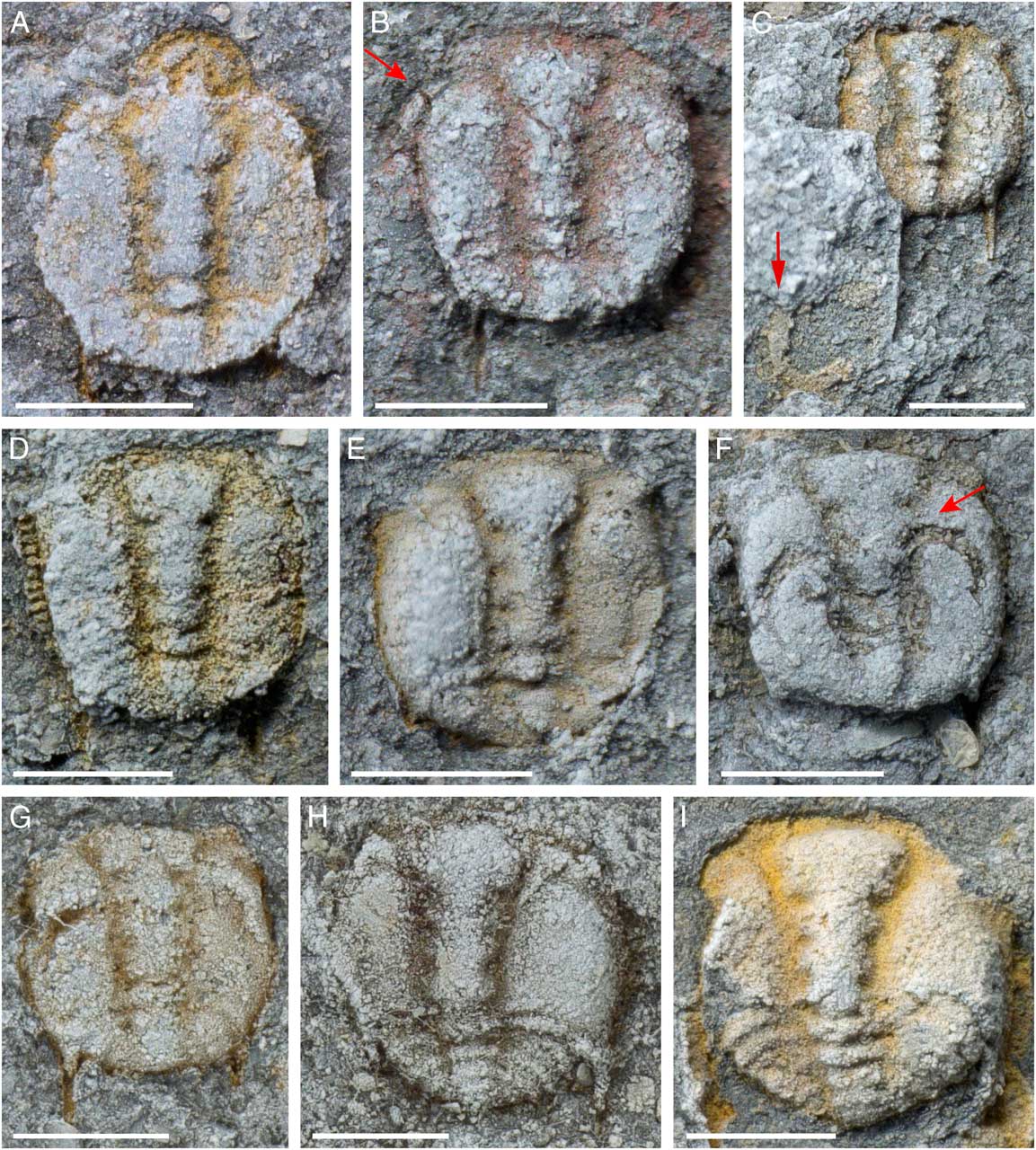
Protaspides of Dalmanitina proaeva elfrida from the Vinice Formation (Sandbian to Katian) of the Prague Basin. Red arrows in B and F point to locations of separated librigenae/doublure, and in C to the hypostome. A–C show these structures as independent (similar to the Nutcracker molt configuration; Drage et al. Reference Drage, Holmes, García-Bellido and Daley2018), while F and G are akin to Maksimova’s molt configuration (Henningsmoen Reference Henningsmoen1975). A–G, protaspid stage 1; H, I, protaspid stage 2. A, NMP L46704; B, NMP L15057; C, NMP L15046; D, NMP L46778; E, NMP L15056; F, NMP L46688; G, NMP L46713; H, NMP L46661; I, NMP L15050. All illustrated specimens are from Trubín (see Fig. 1, localities 12–14). Scale bars, 0.5 mm.

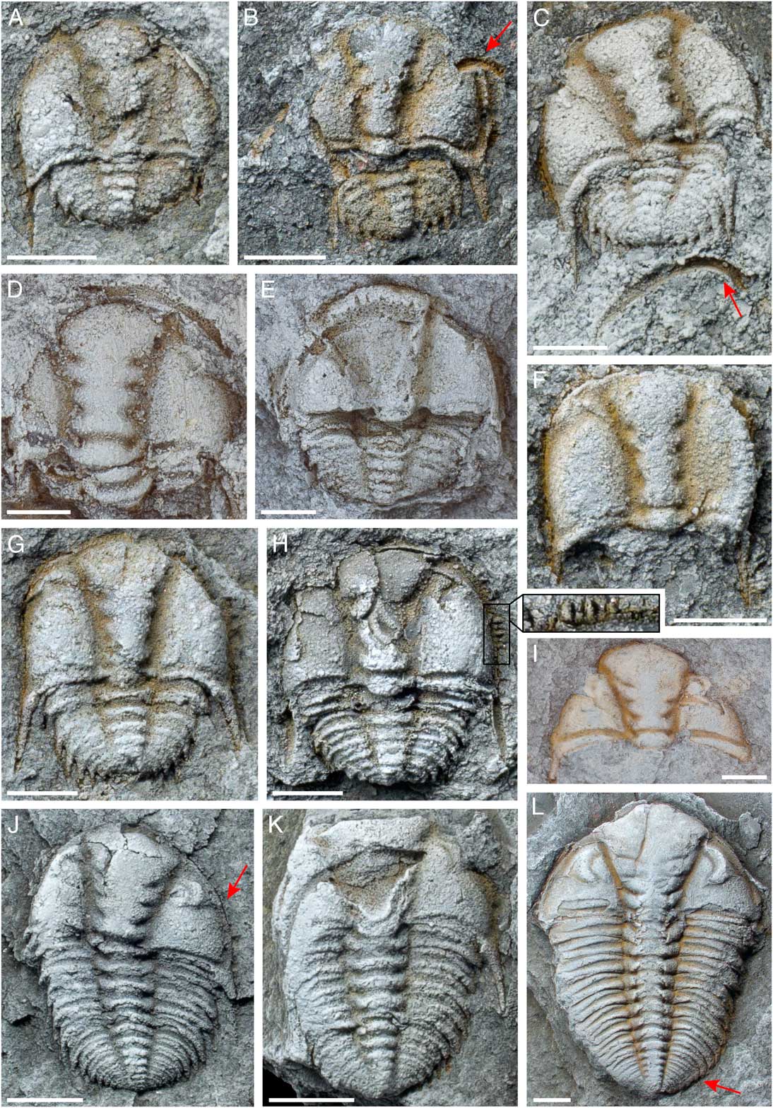
Meraspides of Dalmanitina proaeva elfrida from the Vinice Formation (Sandbian to Katian) of the Prague Basin. Red arrow in B points to a disarticulated librigena, in C to a disarticulated LCU, in J to the facial suture shape, and in L to location of the thoracic–pygidial boundary. C–E and K show Harrington’s molt configuration with the LCU detached. Magnified view of H shows numerous subhorizontal spines round the cephalic margin. A, B, meraspid degree 0; C, G, H, meraspid degree 1; E, meraspid degree 2; K, meraspid degree 5; J, meraspid degree 6; L, meraspid degree 10. A, NMP L15049; B, NMP L15060a; C, NMP L15052; D, NMP L46798; E, NMP L46807; F, NMP L46694; G, NMP L15062; H, NMP L15051a; I, NMP L46796; J, NMP L15053; K, NMP L15061a; L, NMP L15064. All illustrated specimens are from Trubín (see Fig. 1, localities 12–14). Scale bars, 0.5 mm for A–H, and 1 mm for I–L.

There are some gaps in the ontogenetic sequence, particularly between meraspid degrees 5 and 10, and within the holaspid period (Fig. 2). However, the calculated GR (Table 1) suggests that only one molting event occurred for each meraspid degree from M5 to M10 (assuming a more or less constant growth increment during development, which is typical for most trilobites; cf. Fusco et al. Reference Fusco, Garland, Hunt and Hughes2012). Dalmanitina proaeva elfrida displays protarthrous development as the full complement of 11 articulated thoracic segments is reached (Fig. 6A, with only 18 trunk segments) before the total number of trunk segments are present (20, counted from the largest specimens; Fig. 6B,C,E,F).
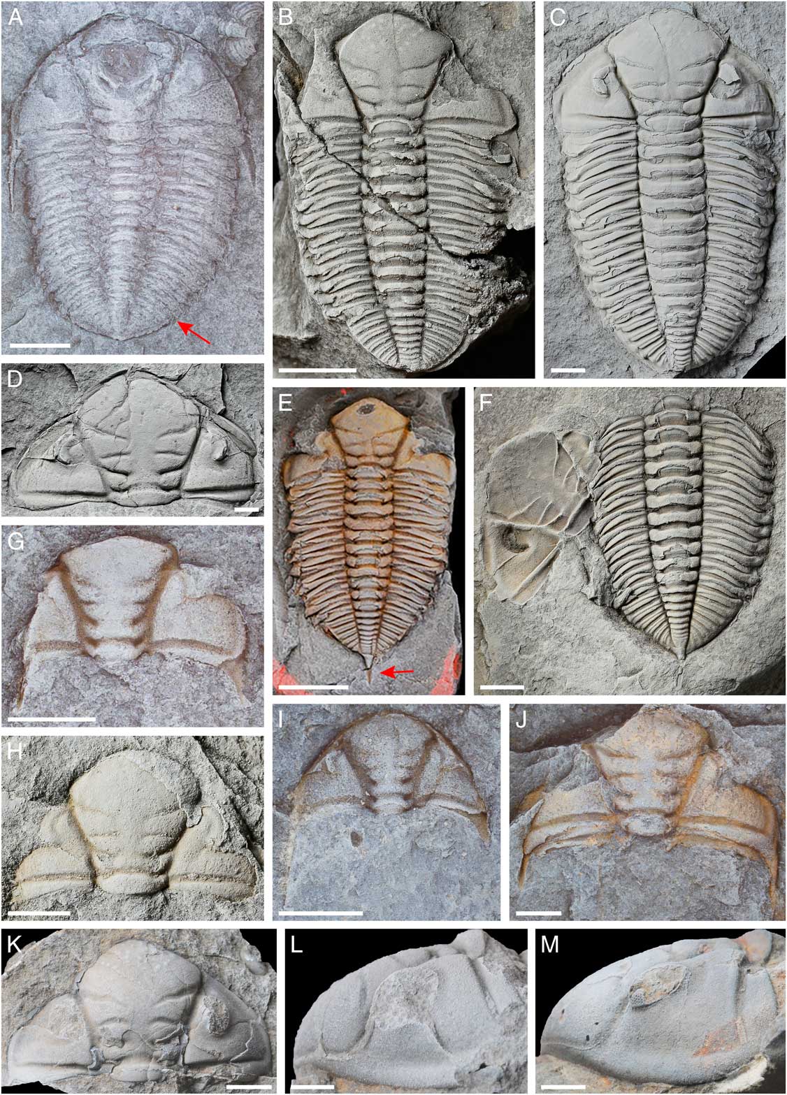
Holaspides of Dalmanitina proaeva elfrida and D. proaeva from the Vinice and Zahořany Formations, respectively (Sandbian to Katian) of the Prague Basin. Red arrow in A points to the location of the thoracic–pygidial boundary, and in E to the terminal spine. Specimens B and E are probable exuviae consisting only of the axial shields, and F is a molt in Salter’s configuration (Drage et al. Reference Drage, Holmes, García-Bellido and Daley2018). A, NMP L46804; B, NMP L46568; C, NMP L46821; D, NMP L46818; E, NMP L15055; F, NMP L46564; G, NMP L46793; H, NMP L46824; I, NMP L46708; J, NMP L46715; K, CGS MŠ2181a; L, CGS MŠ2671; M, CGS MŠ2673. All NMP specimens are from Trubín, and CGS specimens are from Zahořany (localities 12–14, and 10 and 11 respectively; Fig. 1). Scale bars, 2.5 mm for A, G–J, L, M, and 5 mm for B–F, K.

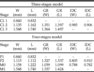
Results of developmental traits calculated for Dalmanitina proaeva elfrida from the Vinice Formation (Sandbian to Katian) of the Prague Basin. Abbreviations: AGI, average per-molt growth increment; Cl.1–Cl.3, cluster 1 to 3; GR, growth rate; IDC, index of conformity to Dyar’s rule; L, length; M0–M1, meraspid degrees; P1–P2, protaspid stages; W, width.

Protaspid Period
The earliest postembryonic stage (P1) of D. proaeva elfrida is roughly circular in outline, 0.78–0.89 mm long, and 0.74–0.92 mm wide. The cranidium is semicircular and 0.56–0.69 mm in length. The glabella expands anteriorly and has five lobes, including the occipital ring, and these are separated by deep glabellar furrows (e.g., Fig. 4D,E,I). The anterior-most lobe is subrectangular and terminates just short of the anterior cephalic margin (Fig. 4D,E). A short (sag.) anterior border is developed. Postero-lateral edges of the cranidium protrude into long spines (Fig. 4C) that are directed immediately posteriorly and extend beyond the trunk (exsag.). Based on their topological position, these seem to be homologous with the genal spines of later stages. The fixigenae are very convex, while the unfused librigenae (see Fig. 4B,F, arrows, and G) are narrow (tr.) and short (exsag.). The palpebral lobes are small and semicircular, and placed far anterolaterally, with the anterior margin created by the librigenal sutures (e.g., Fig. 4E, with missing librigenae). Both the posterior marginal furrow and posterior border furrow are well developed, running from the occipital ring and curving backward abaxially. The margins of the cranidium and librigenae are fringed by small spines likely of subhorizontal direction, which appear bifurcated on the lateral sides and singular toward the anterior (Fig. 4A,D, in particular the left margin of the latter). The trunk portion is broadly elliptical, about 60% of the maximum cranidial width, with a rounded semicircular posterior margin and two pairs of marginal spines. The axial part of the trunk is poorly differentiated but seems to contain two rings (Fig. 4A–G). The pleural parts are smooth without pleural or interpleural furrows. The hypostome, where visible (Fig. 4A inverted, in C potentially displaced to the bottom left, impressions in F,G), seems in life to have been conterminant with a suture to the cephalic doublure (Fig 4F,G). It has a circular posterior margin, an internal marginal furrow, and likely laterally and posteriorly pointing marginal spines (Fig. 4C).
The second protaspid stage (P2) is morphologically comparable to P1, differing mainly by a slightly larger overall size and a larger trunk portion with four axial rings (Fig. 4H,I).
Meraspid Period
The meraspid specimens range between 1.19 and 8.30 mm in sagittal exoskeletal length and 1.08 and 6.36 mm in maximum exoskeletal width (including the largest articulated meraspid specimen in M10). From M0 (Fig. 5A,B), the occipital ring is more defined and distinct from the remaining four lobes of the glabella. The three pairs of glabellar furrows and occipital furrow remain well defined. The general shape of the glabella in the early meraspid stages is similar to that of the protaspides, but during subsequent development the glabella widens anteriorly and becomes more pentagonal shaped in outline with a diamond-shaped anterior lobe. The posterior border furrow becomes more prominent (e.g., Fig. 5B,C), leading to the genal spines and continuing around the lateral and anterior sides of the cephalon. The margin of the entire cephalon still bears many subhorizontal spines (Fig. 5H, magnified view, also visible on left cephalic margin in F and right margin in G, on the anterior hypostome border in E and D, and on the right librigenal in B), which become less prominent during development and disappear in the late meraspid stages (present in M6 [Fig. 5J arrow], but absent by M9 [Fig. 5L]). The facial sutures migrate toward the glabella throughout the meraspid period, with the librigenae consequently becoming broader and more triangular (e.g., arrows in Fig. 5B compared with J). This also caused the semicircular eyes to move gradually away from the cephalic margin and more axially, while increasing in size and becoming more crescentic (Fig. 5L). In M0 the librigenae remain separate entities as in protaspides (Fig. 5A,B), but from M1 these structures appear to be joined together, forming a lower cephalic unit (LCU; Henningsmoen Reference Henningsmoen1975) that separates from the cranidium as a single structure (seen disarticulated in Fig. 5C–E). The hypostome of the earliest meraspides is also similar to that in the protaspid period, but later appears to become parabolic to triangular in posterior outline rather than rounded and lacks spines (Fig. 5E,K). Throughout the meraspid period, the trunk segments have small pleural spines, and five to seven axial rings can be recognized on the pygidium.
Holaspid Period
The exoskeleton of the smallest articulated holaspid specimen is 11.50 mm long and 7.26 mm wide. Holaspid individuals have 11 thoracic segments, but this condition occurs before the onset of epimorphic development, and therefore some smaller holaspid specimens exhibit 7 or 8 axial rings on the pygidium (Fig. 6A) compared with the 9 in larger individuals (Fig. 6B,C,F). The posterior of the pygidium forms a long terminal spine (Fig. 6E,F). During holaspis, the facial sutures become fused, meaning they can no longer be opened, and the librigenae become fixed to the cranidium. This appears to be flexible in timing; while the facial sutures are usually only fused in holaspid specimens based on their morphology (the full 11 thoracic segments), these are of greatly varying body sizes (as displayed in Figs. 2 and 6; see Supplementary Material 2 for more examples). For example, Figure 6B, E, G, H, and J shows a broad range of sizes, and all specimens are missing the librigenae, meaning that the facial sutures were still operable (also true for a number of additional specimens from the CGS, e.g., JK19125 with a cranidial width ~2 cm). Some specimens (e.g., Fig. 6J) are larger than or comparable to other specimens with fused sutures (e.g., Fig. 6F,I, although smaller than C and D), as are several other specimens of D. proaeva with missing librigenae from the CGS (e.g., PB130 with a cranidial width >4 cm; Supplementary Material 2).
Specimens with complete cephala, such as Figure 6C,M, are considered to have fused facial sutures because, while the lines of the sutures are still visible, they are by varying degrees much fainter and less defined (despite the excellent preservation) than sutures such as in Figure 6K, which are broader and clearly operable without the librigenae being separated from the cranidium (matrix is visible around the right librigena, and the left is displaced slightly inward; probably also true for Fig. 6A, with a broad right suture despite the poor preservation). Phacopine trilobites are defined, among other characteristics, by the proparian form of the suture lines that are ultimately fused by adulthood (Jaanusson Reference Jaanusson1975). When the librigenae are fused, these sutures remain visible in varying degrees in holaspides, more so in dalmanitids than phacopids or pterygometopids (see Henry Reference Henry1968; Eldredge Reference Eldredge1972; Hammann Reference Hammann1976: Plate 6; Holloway Reference Holloway1981, throughout; Šnajdr Reference Šnajdr1982, Reference Šnajdr1990; Budil and Bruthansová Reference Budil and Bruthansová2005; Sandford and Holloway Reference Sandford and Holloway2006; Budil et al. Reference Budil, Hörbinger and Mencl2009: Fig. 8L; Storey et al. Reference Storey, Thomas and Owens2016: Figs. 5–8). For example, the suture lines are almost invisible (although still present) on less well-preserved holaspid cephala of D. proaeva (e.g., CGS MŠ2165; Supplementary Material 2). There are many holaspid specimens from the CGS of D. proaeva of varying sizes that also have fused but faintly visible suture lines (e.g., MŠ2673, MŠ1245, MŠ2678, and MŠ2669 with cranidial widths of ~2–4 cm; see Supplementary Material 2). Further, these specimens do not show even millimeter-scale librigenal displacement (even in otherwise disarticulated cephala; e.g., Fig. 6D,M), unlike holaspides with unfused sutures (e.g., Fig. 6L and JV18212 from the CGS, which both show librigenal displacement; see Supplementary Material 2) or carcasses of other trilobite species with operable sutures that often show minor displacement through abiotic processes (e.g., Drage et al. Reference Drage, Holmes, García-Bellido and Daley2018: Fig. 3G). The fused but faintly traced facial sutures are particularly clear in extremely three-dimensional specimens of D. proaeva (such as Fig. 6M). In the aulacopleurids, another trilobite group that underwent this fusion event later in development, Hughes et al. (Reference Hughes, Kříž, MacQuaker and Huff2014) also specifically noted that, while the facial sutures were fused and Salter’s mode of molting was used, “these sutures continued to be visible throughout the holaspid phase, […], indicating that they remained expressed as vestigial structures” (p. 231). This condition (inoperable, but still visible, facial sutures) is therefore not just restricted to dalmanitoids and has wider precedence within Trilobita.
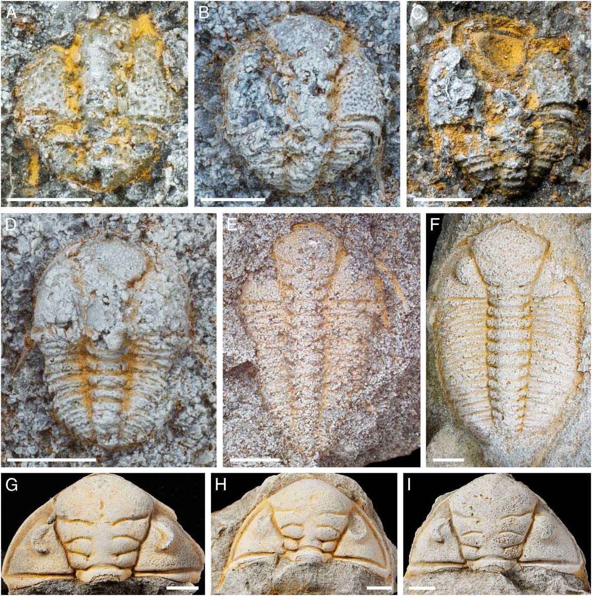
Specimens of Dalmanitina socialis from the Letná Formation (Sandbian) of the Prague Basin. A, NMP L46681; B, NMP L46725; C, NMP L46685; D, NMP L46739; E, NMP L46779; F, NMP L46785; G, NMP L46834; H, NMP L46566; I, NMP L46823. A–D are from Trubská, and E–I from Veselá (localities 12–14 and 1–7, respectively; Fig. 1). Scale bars, 0.5 mm for A–C, 1 mm for D and E, 2 mm for F, and 5 mm for G–I.

Scatter plot of cranidial or cephalic width (crw) vs. cranidial or cephalic length (crl) for Dalmanitina socialis from the Letná Formation (Sandbian) and Dalmanitina proaeva elfrida from the Vinice Formation (Sandbian to Katian) of the Prague Basin.

The developmental timing of facial suture fusion in D. proaeva elfrida does not seem to be related to the onset of epimorphic growth or of the holaspid period, as specimens such as Figure 6B,E have unfused sutures and the full 11 thoracic segments. Further, Figure 6I, based on its size (Fig. 2), may indeed represent a late-stage meraspid with fused sutures and therefore suggests an even broader range of variability for the timing of this fusion event. Budil and Bruthansová (Reference Budil and Bruthansová2005) similarly noted the occasional use of the facial sutures for molting in adult dalmanitoids and argued that this indicates facial suture fusion was not always genetically fixed in phacopines, agreeing with this interpretation of the D. proaeva specimens.
Postembryonic Developmental Sequence of D. socialis
The specimens of D. socialis (Fig. 7) provide less detailed information on the species’ ontogenetic sequence due to the coarser-grained preservation at the Letná Formation. Studied individuals of D. socialis have cranidia and cephala between 0.71 and 11.56 mm in sagittal length and 0.99 and 30.40 mm in transverse width. Most of the specimens represent meraspid and holaspid stages, and the smallest individual (Fig. 7A) may be equivalent to P2 or M0 of D. proaeva elfrida due to its similar size and overall morphology. Morphological development in D. socialis was for the most part similar to that described for D. proaeva elfrida, including the changes in general morphology, and shape of the hypostome (Fig. 7C). When compared with specimens of D. proaeva elfrida, it appears that D. socialis enters the holaspid period at a comparably smaller size (Fig. 8), with holaspides being defined from the stable thoracic number of 11 segments. The smallest recorded holaspid of D. socialis with this complete number of thoracic segments has a cranidium 3.25 mm long and 5.20 mm wide (compared with 4.20 mm long and 7.26 mm wide in D. proaeva elfrida, while the M10 of D. proaeva elfrida is 3.51 mm long and 6.36 mm wide).
Some small individuals of D. socialis show a fairly densely pitted prosopon, in particular on the fixigenae. This is clear from the best-preserved early developmental stages (Fig. 7A–C), but not visible on the internal molds of D. proaeva elfrida. Where observable, the marginal cephalic spines in meraspid individuals actually appear longer than those seen in D. proaeva elfrida and were similarly present until at least midway through the meraspid period (Fig. 7B,C, visible around the left and anterior margin, and in D, around the entire anterior). Facial suture fusion generally seemed to occur in D. socialis during early holaspid development, as far as this could be observed (e.g., Fig. 7G–I shows larger adult cephala with fused sutures, but late meraspides such as E had unfused LCUs).
Exuviae of Dalmanitina
Many of the D. proaeva elfrida and D. socialis specimens from the NMP collections, and some of those displayed in Figures 4–7, are interpreted as molted exoskeletons (exuviae). These show either the librigenae/LCU separated from the axial shield or the cephalon disarticulated from the trunk (both to create an anterior exuvial gape to exit the old exoskeleton through; see Budil and Bruthansová Reference Budil and Bruthansová2005: Fig. 3A,B1 ; Drage et al. Reference Drage, Holmes, García-Bellido and Daley2018). For the protaspid period of D. proaeva elfrida, Figure 4B shows a singular separated left librigena displaced slightly outward from the cranidium, which may also be the case for Figure 4C, and in both the right librigena appears missing as the eye ridge forms the outer margin. This is also true of Figure 4E, which is missing both librigenae. In Figure 4C, the independent hypostome may also have been displaced to the left posterior of the specimen. Figure 4F,G (and potentially 4H) shows an impressed and posteriorly displaced external mold of the two independent librigenae and doublure (i.e., the dorsal and ventral parts of the librigenae) with the hypostome (there is no visible connection between the left and right librigenae to the anterior of the hypostome). These molts are reminiscent of Maksimova’s molt configuration (described by Henningsmoen Reference Henningsmoen1975: p. 190, Fig. 8) in which both sides of the doublure, often with the hypostome, are displaced as one unit below the axial shield. However, Maksimova’s configuration was apparently not always produced, as Figures 4A–C and 5B show singular molted librigenae and/or hypostome (more comparable to the Nutcracker configuration from Drage et al. Reference Drage, Holmes, García-Bellido and Daley2018). The P2 specimen in Figure 4I may also be lacking the librigenae, much like the M0 individual in Figure 5A, but this is difficult to ascertain due to the preservation. The latter specimen does appear to have an impression of the isolated hypostome displaced posteriorly, and Figure 5B (arrow) clearly shows the separated right librigena and, potentially, the left librigena displaced outward.
From M1, several probable exuviae show a separated LCU, which is displaced backward in Figure 5C,E,K, with the hypostome attached in the latter two parts, and forward in Figure 5D. The D. socialis late meraspid in Figure 7E also appears to show a displaced LCU to the right of the cranidium. These reflect Harrington’s molt configuration (Henningsmoen Reference Henningsmoen1975; Drage et al. Reference Drage, Holmes, García-Bellido and Daley2018), although potentially with the LCU as a single structure, rather than retaining the facial sutures and joined by integument (as for Estaingia bilobata in Drage et al. Reference Drage, Holmes, García-Bellido and Daley2018). Axial shields (i.e., articulated but missing one/both the librigenae; Fig. 6B,E for D. proaeva elfrida, and F for D. socialis with the left librigena in place) are reasonably common for all but the largest Dalminitina holaspides (see Budil and Bruthansová Reference Budil and Bruthansová2005). Figure 6F presents a classic example of a molted exoskeleton in Salter’s configuration (Henningsmoen Reference Henningsmoen1975; Drage et al. Reference Drage, Holmes, García-Bellido and Daley2018), albeit with the inverted cephalon facing in an unusual direction and slightly overlapping the trunk.
Discussion
Comparative Development of Phacopina
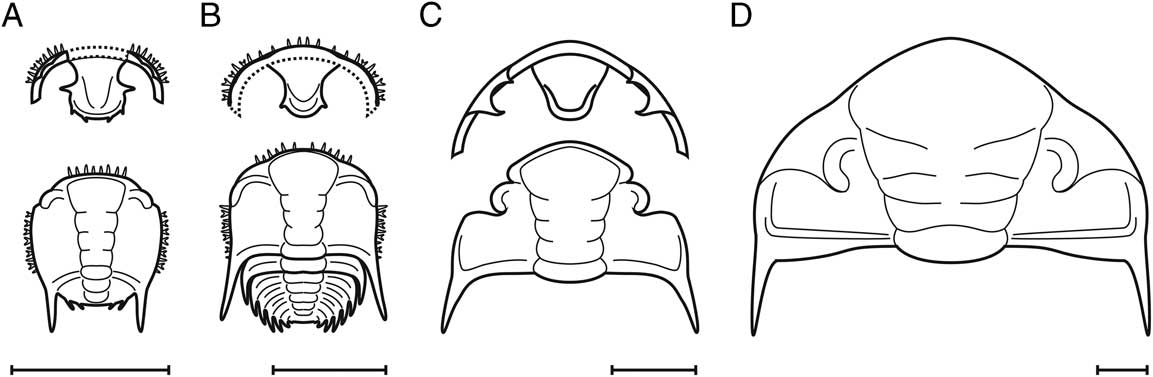
Early postembryonic stages of Dalmanitina show most (but not all) of the distinctive characteristics of Phacopina, as summarized by Chatterton and Speyer (Reference Chatterton and Speyer1997). In particular: (1) a proparian facial suture; (2) anteriorly located palpebral lobes; (3) forward expanding glabella; (4) four glabellar furrows; (5) pitted fixigenae (at least in small specimens of D. socialis); (6) short [sag.] anterior border; (7) marginal spines on the cranidium and librigenae; (8) distinct posterior marginal and border furrows; and (9) sharp spines on the trunk (e.g., Fig. 6E, arrow; see morphological reconstructions in Fig. 9).
Reconstructions of selected developmental stages of Dalmanitina proaeva elfrida from the Vinice Formation (Sandbian to Katian) of the Prague Basin. A, First protaspid stage showing librigenae and hypostome; B, meraspid degree 1 showing LCU and hypostome; C, cranidium of meraspid degree 5 showing LCU and hypostome; D, cephalon of early holaspid stage. Dotted lines represent inferred structures. Scale bars, 1 mm.

When compared with protaspides of the other phacopine families Phacopidae and Pterygometopidae, the dalmanitid protaspides lack the highly bulbous nonadult-like (pre-metamorphic) stage (see also Crônier Reference Crônier2007), instead showing only adult-like stages. All described postmetamorphic protaspides of Phacopina appear to have two dorsally expressed segments in the trunk portion, as can be inferred from the axial section and the number of marginal trunk spines preserved (cf. Temple Reference Temple1952; Whittington and Campbell Reference Whittington and Campbell1967; Chatterton and Speyer Reference Chatterton and Speyer1997; Crônier Reference Crônier2007). Protaspides and earliest meraspides of the Pterygometopidae and Dalmanitidae also share unfused, independent librigenae (Chatterton and Speyer Reference Chatterton and Speyer1997: Figs. 175.2, 175.10) and bifurcated marginal cranidial spines (Fig. 9; Chatterton and Speyer Reference Chatterton and Speyer1997: Fig. 175.8). In contrast, the earliest postembryonic stages of the Phacopidae have fused librigenae, forming an LCU (Crônier Reference Crônier2007: Fig. 2.13), and the cranidial marginal spines do not seem to be bifurcated (Crônier et al. Reference Crônier, Bartzsch, Weyer and Feist1999: Fig. 5.7; Crônier Reference Crônier2007: Figs 2.13, 3.3). Further, the protaspid and early meraspid hypostomes of Dalmanitidae, with a rounded posterior lobe, resemble those of the Pterygometopidae rather than Phacopidae (cf. Chatterton and Speyer Reference Chatterton and Speyer1997; Crônier et al. Reference Crônier, Bartzsch, Weyer and Feist1999; Crônier Reference Crônier2007). Late meraspid stages of Dalmanitidae are distinct from the Phacopidae, as the latter have an extremely inflated anterior part of the glabella (Osmólska Reference Osmólska1963; Crônier et al. Reference Crônier, Bartzsch, Weyer and Feist1999; Crônier Reference Crônier2007).
The trunk segmentation schedule described here for D. proaeva elfrida (Fig. 10) shows a protarthrous mode of segmentation, because the onset of the holaspid period precedes the onset of the epimorphic phase of development (cf. Hughes et al. Reference Hughes, Minelli and Fusco2006). This is in contrast with the schedule so far described for Phacopidae, in which only synarthromeric and hypoprotomeric modes are known (Crônier Reference Crônier2010).
Trunk segmentation schedule of Dalmanitina proaeva elfrida from the Vinice Formation (Sandbian to Katian) of the Prague Basin. Dotted lines represent estimated, but not observed stages. Abbreviations: Ac., accumulation phase; Ep., epimorphic growth; H1–Hn, holaspid stages; M0–M10, meraspid degrees; P1–P2, protaspid stages; Pr., protaspid period.

It is particularly interesting that size-based clusters of D. proaeva elfrida stages can be recognized early in ontogeny because, while these individuals are from “one locality,” they most likely represent more than one population. The specimens are all from the Vinice Formation at Trubín and therefore of Sandbian–Katian age, but as historical specimens they may have been collected from different beds and collection pits, and therefore subject to slight variations in age (see “Geological Setting” and Fig. 1). The size clustering of the D. proaeva elfrida sample may therefore suggest the species was reasonably consistent in growth increment, and this was robust to differences in regional age and location. If found to be the case for other phacopine species, this could be useful for obtaining larger sample sizes to explore broad-scale ontogenetic trends.
Variability in the Timing of First Articulation
Three clusters are apparent during the earliest postembryonic development of D. proaeva elfrida (Fig. 3). The first and third of these clusters contain individuals that can be assigned to P1 and M1, respectively. By contrast, the second cluster (Fig. 3) contains two morphotypes that differ by the presence or absence of an articulation structure, the number of postcephalic segments (four and five), and the size of the trunk (Figs. 3–5). Therefore, the second cluster contains individuals of both P2 and M0. This unusual distribution can be explained in two ways. First, the growth increment between P2 and M0 was smaller than during the rest of development, meaning the two stages greatly overlapped in their size ranges. Second, the timing of the first articulation and segment release was not developmentally constrained, producing a variability in the timing of the first articulation (and therefore variability of the protaspid–meraspid transition) and of segment number in individuals of similar sizes. Consequently, two models can be proposed to explore this distribution: the three-stages model and the four-stages model. In the former, the developmental stages are based solely on size clustering, regardless of their morphology. In the four-stages model, they are based on their morphology, irrespective of size clustering. We calculated several developmental traits under both models and compared the results with Fusco et al. (Reference Fusco, Garland, Hunt and Hughes2012) to assess which scenario better fits the traits observed more generally in Trilobita.
The GR is a premolt/postmolt size ratio known for many trilobite taxa (e.g., Palmer Reference Palmer1957; Chatterton et al. Reference Chatterton, Siveter, Edgecombe and Hunt1990; Lerosey-Aubril and Feist Reference Lerosey-Aubril and Feist2005; Laibl et al. Reference Laibl, Fatka, Crônier and Budil2014). The GR in D. proaeva elfrida ranges from 1.351 to 1.364 for width and 1.397 to 1.497 for length under the three-stages model; and from 1.039 to 1.337 for width and 1.099 to 1.424 for length under the four-stages model (see Table 1). A constant rate of size increase between molts (Dyar’s rule) is considered the null model for arthropod growth and is consistent with available evidence of the physiological mechanisms underlying growth and control of molting (Klingenberg and Zimmermann Reference Klingenberg and Zimmerman1992). Assuming Dyar’s rule, variation in GR through ontogeny for a species should be minimal. The three-stages model better fits this assumption, due to its low levels of GR variation (Table 1). This is also reflected by the values obtained for the IDC, which quantifies the level of adherence to a constant GR during development. The majority of trilobites (~80% with sufficient developmental data) apparently grew at a relatively constant rate, with IDCs >0.8 (Fusco et al. Reference Fusco, Garland, Hunt and Hughes2012). In D. proaeva elfrida, the IDC is >0.9 for the three-stages model and varies between 0.788 and 0.805, and 0.782 and 0.910 for the four-stages model for width and length respectively (Table 1). For the former, this suggests a close fit with Dyar’s rule, which may again support the three-stages model. The assumption of variation in segment number in the second size cluster under the three-stages model is not itself exceptional, because segment number flexibility within trilobite stages is well known (e.g., Hughes et al. Reference Hughes, Minelli and Fusco2006; Hou et al. Reference Hou, Hughes, Lan, Yang and Zhang2015; Dai et al. Reference Dai, Zhang, Peng and Yao2017).
However, the three-stages model is problematic, because it contradicts the general assumption that the timing of the first segment articulation is developmentally static within a species (and thereby indicates the onset of the meraspid period), although variability in the timing of first articulation within a single trilobite species has been suggested before. For example, Lee and Chatterton (Reference Lee and Chatterton2005) reported two late protaspid specimens with incomplete articulation in the Cambrian menomoniid Bolaspidella housensis (Walcott Reference Walcott1886). Based on their size, these specimens clustered with P3 and were interpreted as potential intermolt stages. These authors also described stage 2 protaspides of the lonchocephalid Glaphyraspis parva (Walcott Reference Walcott1899) that clustered with two fully articulated (i.e., not intermolt-stage) specimens of meraspid degree 0 (Lee and Chatterton Reference Lee and Chatterton2005: Fig. 6c), similar to D. proaeva elfrida (Fig. 3). There is also some support for variability in the timing of the first articulation between two closely related taxa (Park and Choi Reference Park and Choi2011). For example, Asioptychaspis subglobosa (Sun Reference Sun1924) had two protaspid stages, whereas its descendant species Quadraticephalus elongatus (Kobayashi Reference Kobayashi1935) had only one protaspid stage, but its P1 and M0 corresponded with the size and morphology of P1 and P2, respectively, in A. subglobosa (Kihm et al. Reference Kihm, Park and Choi2013).
With the data currently available on trilobite development, we cannot entirely rule out the four-stages model. Hou et al. (Reference Hou, Hughes, Lan, Yang and Zhang2015) provided a detailed discussion related to overlapping clusters of various developmental stages. They suggested that a low growth increment (and therefore GR) might have been associated with size overlap among successive stages, even during early postembryonic ontogeny (as suggested under the four-stages model). However, this is likely not the case for D. proaeva elfrida, which had a GR slightly higher than most other trilobites (cf. Fusco et al. Reference Fusco, Garland, Hunt and Hughes2012). The lower GR between P2 and M0 in the four-stages model (1.039 width and 1.099 length) may be related to the development of the articulation structure. Unfortunately, data concerning GR across the protaspid–meraspid transition that might inform on this are scarce (Fusco et al. Reference Fusco, Garland, Hunt and Hughes2012).
Based on the aforementioned evidence, it seems more likely that there existed variability in the timing of the first articulation event in D. proaeva elfrida (three-stages model). Some individuals apparently developed the first articulation (and thus entered the meraspid period) after one molt, whereas others molted without developing an articulation (and entered the P2 stage). During the subsequent molting event, the M0 individuals developed another articulation, and the individuals of P2 may have developed two articulations (this has been described in Sao hirsuta Barrande, Reference Barrande1846 and may have occurred in other trilobite taxa; Laibl et al. Reference Laibl, Fatka, Crônier and Budil2014). If the three-stages model represents the true developmental sequence, it suggests that the timing of the initial segment articulation event may have been variable even within one trilobite species. Further, it supports the view that the protaspid period is not biologically meaningful in terms of homology between developmental stages (see Park and Choi Reference Park and Choi2011) and that trilobite development reflects a complex combination of independent processes such as segmentation, articulation, and morphological changes (cf. Hughes et al. Reference Hughes, Minelli and Fusco2006).
Facial Suture Fusion in Phacopina
Molted exoskeletons were reasonably common among the specimens of D. proaeva elfrida and D. socialis in the NMP collections, including, as described earlier, protaspides and earliest meraspides with individual separated librigenae (Figs. 4B,C,F,G and 5B), meraspides with a separated LCU (Figs. 5C–E,K and 7E), holaspid axial shields missing the librigenae (Figs 6B,E and 7F; Budil and Bruthansová Reference Budil and Bruthansová2005: Fig. 4F), and large holaspides with disarticulated entire cephala (e.g., Fig. 6F). Suborder Phacopina was partially defined based on facial suture fusion during ontogeny (Jaanusson Reference Jaanusson1975), where the sutures close and the librigenae can no longer separate from the cranidium for molting. This fusion event therefore accounts for the occurrence of Dalmanitina meraspid molts with a separated LCU and holaspides with disarticulated cephala.
However, most ontogenetic descriptions for phacopine (Phacopidae) taxa have to date noted only this single fusion event. In contrast to this, we also observed a primary fusion event that occurred between stages M0 and M1 in D. proaeva elfrida, when the librigenae became joined as a single unit (the LCU). The earliest developmental stages of phacopines for which a reasonably complete developmental sequence has been described display joined librigenae (Crônier et al. Reference Crônier, Auffray and Courville2005; Crônier and Fortey Reference Crônier and Fortey2006), rather than the small individual librigenae identified in the protaspides and M0 of D. proaeva elfrida (Figs. 4B,C,F,G and 5B). Further, in discussing the evolutionary trends of Phacopina, Jaanusson (Reference Jaanusson1975) asserted that their protaspides displayed an LCU. However, both separated LCUs and independent librigenae were recognized in juveniles of Nephranops incisus incisus (Roemer Reference Roemer1866), belonging to the Phacopidae (Crônier Reference Crônier2007: Fig. 5), which also fused the facial sutures later in ontogeny, but this could not be associated with a specific fusion event during ontogeny, as was possible for D. proaeva elfrida. In addition, figured protaspides of the pterygometopid Calyptaulax annulata (Raymond Reference Raymond1905) appear to show independent librigenae retaining sutures on the doublure (Chatterton and Speyer Reference Chatterton and Speyer1997: Fig. 175.2, 175.11), and the same with the encrinuroids Physemataspis insularis (Shaw Reference Shaw1968) and Cybeloides prima (Raymond Reference Raymond1905; Chatterton and Speyer Reference Chatterton and Speyer1997: Fig. 176.2, 176.6). This indicates that the primary fusion event, with its specific ontogenetic timing in D. proaeva elfrida (Dalmanitidae), was actually widespread within the Phacopina. Interestingly, some asaphids, for example, Dikelocephalus minnesotensis (Owen Reference Owen1852), displayed a similar condition to this primary phacopine fusion event in which the median suture separating the two librigenae was fused, the timing of which varied even with single bedding planes (Hughes Reference Hughes1994). However, several ptychaspidid species (also of the Asaphida) were described as showing an opposing pattern through ontogeny, with the fused librigenae eventually becoming separated into two individual structures later in development (Kihm et al. Reference Kihm, Park and Choi2013).
Knowledge of the developmental timing of the secondary fusion event (that of the facial sutures) has been previously confined to the family Phacopidae, within which it is interspecifically variable. In Weyerites ensae (Richter and Richter Reference Richter and Richter1926), facial suture fusion takes place between stages M9 and M10 (Crônier et al. Reference Crônier, Auffray and Courville2005; Crônier and Fortey Reference Crônier and Fortey2006), and in Trimerocephalus lelievrei (Crônier and Feist Reference Crônier and Feist2000) and Plagiolaria poothaii (Kobayashi and Hamada Reference Kobayashi and Hamada1968), it occurs at the onset of holaspis at M10–H1 (Crônier et al. Reference Crônier, Auffray and Courville2005; Crônier and Fortey Reference Crônier and Fortey2006). This secondary fusion is generally known to occur in dalmanitids; for example, a number of holaspides of various species have been described with fused facial sutures in detached cephala (see Henry Reference Henry1968; Eldredge Reference Eldredge1972; Holloway Reference Holloway1981; Šnajdr, Reference Šnajdr1982, Reference Šnajdr1990; Budil and Bruthansová Reference Budil and Bruthansová2005; Sandford and Holloway Reference Sandford and Holloway2006; Budil et al. Reference Budil, Hörbinger and Mencl2009: Fig. 8L; Storey et al. Reference Storey, Thomas and Owens2016: Figs. 5–8). However, the data for Dalmanitina provide the first information on developmental timing of secondary fusion in Dalmanitidae. In comparison to these examples from the Phacopidae, the fusion of the facial sutures may occur later in development for Dalmanitina. In general, this is seemingly during the holaspid period and in individuals with a range of sizes (e.g., Fig. 6C,D,F,I,M; Supplementary Material 2; Budil and Bruthansová Reference Budil and Bruthansová2005), rather than being related to a specific developmental stage or to the meraspid–holaspid transition (Crônier et al. Reference Crônier, Auffray and Courville2005; Crônier and Fortey Reference Crônier and Fortey2006). This secondary fusion event is rare for trilobites outside of the Phacopina, but has been noted to occur during later development for some non-phacopines such as Primaspis [Taemasaspis] campbelli (Chatterton Reference Chatterton1971) and aulacopleurids (Hughes et al. Reference Hughes, Kříž, MacQuaker and Huff2014). These examples suggest that, while the secondary fusion event appeared to follow a primary fusion event (producing the LCU) in Phacopina, this need not be the case in other trilobite groups.
These changes in morphology must represent significant events in the life history of phacopine trilobites, because they necessitate alterations to exoskeletal molting behavior. Dalmanitina proaeva elfrida and D. socialis, like the Phacopidae, would have molted in their earlier developmental stages using the sutural gape mode (Henningsmoen Reference Henningsmoen1975; Daley and Drage Reference Daley and Drage2016; Drage et al. Reference Drage, Holmes, García-Bellido and Daley2018), where the facial sutures opened to create an anterior gape through which the animal would have emerged. This would have involved separation of the smaller, independent librigenae in the P1–M0 stages, producing a molt similar to the Nutcracker configuration (Drage et al. Reference Drage, Holmes, García-Bellido and Daley2018), and (following the primary fusion event) of the joined LCU, creating Harrington’s configuration (Henningsmoen Reference Henningsmoen1975; Drage et al. Reference Drage, Holmes, García-Bellido and Daley2018) from the M1 stage until near the end of the meraspid period or during holaspis. Following the secondary fusion event, the complete closure of the facial sutures, the individual could no longer have molted using the sutural gape mode, instead by necessity employing Salter’s mode, in which the entire cephalon was disarticulated from the trunk (Henningsmoen Reference Henningsmoen1975; Daley and Drage Reference Daley and Drage2016; Drage et al. Reference Drage, Holmes, García-Bellido and Daley2018), presumably creating a wider exuvial gape. This produced a Salter’s molt configuration (e.g., Fig. 6F) and would have necessitated changes in behavior, such as different patterns of movement to cause the cephalothoracic articulation to break (see Drage et al. [Reference Drage, Holmes, García-Bellido and Daley2018] for descriptions of molting mechanisms).
The interspecific differences in the timing of the secondary fusion event in the Phacopidae may relate to differing body-size proportions, and in particular, that of the relative proportions of the cephalon to thorax. However, for Dalmanitina, it is likely unrelated to body size and proportions because of the observed variation in holaspid cranidium size with fused and unfused facial sutures. This indicates that facial suture fusion was not a developmentally constrained event, unlike for the Phacopidae or the primary fusion event in D. proaeva elfrida (as also suggested by Budil and Bruthansová Reference Budil and Bruthansová2005), and instead indicates the importance of an alternative trigger. Interestingly, the two Dalmanitina species may slightly differ in this regard, D. socialis having entered the holaspid period at a smaller size than D. proaeva elfrida. This may be related to a difference in habitat, D. socialis and D. proaeva elfrida being generally found in the Letná and Vinice Formations, respectively, or a difference in the age of the two species (although these are very close, being upper Sandbian and uppermost Sandbian/earliest Katian).
The Phacopoidea are thought to represent a more derived group within Phacopina (Jaanusson Reference Jaanusson1975; Budil and Bruthansová Reference Budil and Bruthansová2005). The earliest phacopoid representatives (family Pterygometopidae) emerged almost contemporaneously with the earliest Dalmanitoidea in the Floian–Darriwillian (uppermost Lower–Middle Ordovician; see Adrain 2013; Bergström et al. Reference Bergström, Pärnaste and Zhiyi2013; Pärnaste and Bergström Reference Pärnaste and Bergström2014), but the derived family Phacopidae appear later, in the uppermost Ordovician or lowermost Silurian (Chlupáč Reference Chlupáč1977; Lespérance and Sheenan Reference Lespérance and Sheenan1987; Eldredge Reference Eldredge2001; Adrain Reference Adrain2013). Thus, it may be that the timing of the secondary fusion event became increasingly fixed during ontogeny, and therefore less intraspecifically variable, within the Phacopina throughout their evolution. This agrees with the Ordovician peak in species polymorphism and subsequent decline through the Paleozoic described by Webster (Reference Webster2007) in the Phacopida (Webster Reference Webster2007: Fig. 3F). Alternatively, this intraspecific variability might also be a characteristic of the Dalmanitidae, as the described specimens of Dalmanitina are not from the earliest origination of the Dalmanitoidea. If the timing of this secondary fusion event could be identified within additional early representatives of Phacopina, in particular the Pterygometopidae, these opposing hypotheses could be distinguished. Whether found more broadly within early phacopines, or unique to the Dalmanitidae, this developmental variability might have been an evolutionary adaptation, potentially in response to an external trigger, in addition to the inherent interspecific developmental variability well known within Trilobita (Hughes et al. Reference Hughes, Minelli and Fusco2006).
Conclusions
A large sample of protaspid to holaspid specimens of D. proaeva elfrida and D. socialis from the Letná, Vinice, and Zahořany Formations of the Prague Basin have allowed description of the first reasonably complete developmental sequence for the trilobite superfamily Dalmanitoidea. We have presented details of the morphological change in Dalmanitina from the protaspid to holaspid period, including the late disappearance of marginal cephalic spines and change in hypostome shape. Biometric data show that the specimens cluster in distinct developmental stages, with probable variability in the timing of first articulation resulting in two different morphotypes (P2 and M0) with a significant overlap in size and a broad range of body sizes in the holaspid period.
The developmental sequence for D. proaeva elfrida provides the first information on facial suture fusion for the Dalmanitoidea, which is a key characteristic of the suborder Phacopina. Between stages M0 and M1, the specimens display a fusion of the separate librigenae into an LCU, a morphological change that had not been observed as developmentally fixed in phacopines. Further, during the holaspid period (or potentially from the latest meraspid), there occurs an intraspecifically variable fusion of the facial sutures. These fusion events are related to alterations in their exoskeleton molting behavior, the secondary fusion event requiring a change from the sutural gape mode of molting to Salter’s mode (Drage et al. Reference Drage, Holmes, García-Bellido and Daley2018). This would have produced the Nutcracker (or Maksimova’s) molt configuration until M1, Harrington’s configuration until around holaspis, and Salter’s configuration when the facial sutures were inoperable (Drage et al. Reference Drage, Holmes, García-Bellido and Daley2018). Facial suture fusion is a uniting characteristic of Phacopina, but generally occurs earlier in development and with no intraspecific variation in the family Phacopidae (Crônier et al. Reference Crônier, Auffray and Courville2005; Crônier and Fortey Reference Crônier and Fortey2006), which is closely related to Dalmanitoidea but occurs later in the fossil record. This may either reflect an increasing developmental constraint on the ontogenetic timings of these events within the Phacopina, as generally supported by decreasing intraspecific variability and polymorphism after the early Ordovician (Webster Reference Webster2007), or a so-far unique condition among dalmanitids.
Acknowledgments
This research was supported by an NERC doctoral training grant (NE/L002612/1) and EU Synthesys grant (to H.B.D.), by the Grant Agency of the Czech Republic (GACR) project no.18-14575S (P.B., L.L.), and the Center for Geosphere Dynamics (UNCE/SCI/006) (L.L.). We would like to express our thanks to Š. Rak (Museum of Bohemian Karst, Beroun), who kindly provided important juvenile specimens of Dalmanitina socialis. Our thanks also to O. Fatka, N. Hughes, two anonymous reviewers, and the editor M. Hopkins for their helpful comments on this paper.
Supplementary Material
Data available from the Dryad Digital Repository: https://doi.org/10.5061/dryad.r711309