The improvement in mitral valve area is the most crucial parameter to predict the success of balloon mitral valvuloplasty procedure. Many surrogate echocardiographic parameters have been proposed to predict the severity of mitral stenosis and are validated against mitral valve area.Reference Vimal Raj, George and Jose 1
Mitral valve area can be measured using planimetry, pressure half-time, continuity equation, and proximal isovelocity surface area methods.Reference Cheitlin, Alpert and Armstrong 2 The mitral valve separation index is a simple measure of mitral valve area, and various studies have validated its use.Reference Vimal Raj, George and Jose 1 Its predictive accuracy against mitral valve area changes to define a successful balloon mitral valvuloplasty in juvenile age groups has not been studied thus far. Many echocardiographic parameters for assessing right ventricular functions are available,Reference Rudski, Lai and Afilalo 3 some of them being studied in the context of balloon mitral valvuloplasty. The role of these right ventricular function indices in envisaging the success of juvenile balloon mitral valvuloplasty is also unknown.
Objectives
We aimed to study the usefulness of a battery of echocardiographic parameters such as mitral valve separation index, left atrial volume, right ventricle function indices such as right ventricular systolic pressure, tricuspid annular plane systolic excursion, tricuspid annular systolic velocity (S wave), and right ventricular Tei index against the two-dimensional mitral valve area (planimetry) in predicting the success of juvenile balloon mitral valvuloplasty. Each of these surrogate parameters was analysed in relation to increase in valve area after valvuloplasty to predict the success of the procedure.
Design and methods
We carried out a prospective single-centre echocardiographic study at our institute from February, 2010 to March, 2014. The study was conducted after obtaining approval from the Institute’s ethics committee, and we included patients with rheumatic mitral stenosis who underwent elective balloon mitral valvuloplasty during this time period. Informed written consent was obtained from all the study patients. Exclusion criteria included patients undergoing emergency balloon mitral valvuloplasty, pre- or post-procedure mitral regurgitation of severe intensity, concomitant significant aortic valve disease – severe stenosis/regurgitation – warranting intervention, presence of congenital shunt lesions or coronary artery/myocardial disease with ventricular dysfunction, and presence of respiratory or systemic co-morbidities with potential to cause pulmonary hypertension and/or right ventricular dysfunction.
Echocardiographic parameters
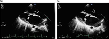
All the study details were obtained using a Philips iE33 ultrasonography machine (Philips Health Care, 5680, DA Best, Nederlands) equipped with a 4-MHz transducer. The same experienced echocardiographer performed all the measurements as per the recommendations of the American Society of Echocardiography.Reference Cheitlin, Alpert and Armstrong 2 All the patients underwent a detailed transthoracic echocardiogram, including measurement of the mitral valve area by planimetry and transmitral gradients, peak and mean. The Wilkins score of the valve was calculated and suitability for valvuloplasty was confirmed. A score of >12 was considered a contraindication as per the existing hospital protocol. The surrogate markers such as mitral valve separation index, left atrial volume, right ventricular systolic pressure, tricuspid annular plane systolic excursion, tricuspid annular systolic velocity, and right ventricular (Tei) index measurements were obtained. Patients with atrial fibrillation or in whom a left atrial thrombus was suspected underwent a transoesophageal echocardiogram. After balloon mitral valvuloplasty, the echo was repeated after 24 hours of the intervention and the same parameters were re-examined. Procedural success was defined as an increase in mitral valve area of >50% or final valve area of >1.5 cm2. The change in two-dimensional valve area was documented and changes among different surrogate parameters were compared with the valve area improvement. Mitral valve area was used as the standard parameter for defining mitral stenosis. Mitral valve area index of <2 cm2/m2 was defined as mitral stenosis. Mild mitral stenosis was defined as valve area between 1.5 and 2 cm2/m2, moderate stenosis as valve area index between 1 cm2 and 1.5 cm2/m2, and severe stenosis as valve area index of ⩽1 cm2/m2. Mitral valve separation index was estimated by measuring the maximal separation of the tip of the mitral leafets in end diastole in the parasternal long-axis view and in the apical four-chamber view during consecutive beats (Fig 1). For patients in sinus rhythm, three measurements were obtained in the long-axis and four-chamber views, and for patients in atrial fibrillation five measurements were obtained and the mean value was considered as the mitral valve separation index. Separation value of <0.70 cm was considered as severe mitral stenosis and >0.90 cm as mild stenosis. Any value in between was termed stenosis of moderate severity.Reference Vimal Raj, George and Jose 1 Left atrial volume was calculated by extrapolating area measured by Simpson’s rule at end systole in the apical four-chamber view. Right ventricle systolic pressure was determined from the tricuspid regurgitant jet using modified Bernoulli’s equation.Reference Rudski, Lai and Afilalo 3 Right atrial pressure was assumed after measuring the diameter of the inferior caval vein with sniff testing as right ventricle systolic pressure=4(tricuspid valve velocitymax)2+right atrial pressure. Tricuspid annular plane systolic excursion was assessed using M-mode, measuring the distance of tricuspid annular movement between end diastole and end systole. The values were indexed for body surface area for analysis. Tricuspid annular systolic velocity (S wave) is similar to the annular plane systolic excursion concept; the velocity of the systolic movement is measured by tissue Doppler imaging of the tricuspid annulus (normal value⩾15 cm/second).Reference Rudski, Lai and Afilalo 3 Myocardial performance index (Tei index) is an index that incorporates both systolic and diastolic time intervals in expressing global systolic and diastolic ventricular functions. We used the tissue Doppler method for calculating the Tei indexReference Rudski, Lai and Afilalo 3 (normal value⩾0.50). Right ventricle Tei index=(tricuspid valve closure to opening time−right ventricle ejection time)/right ventricle ejection time.
Measurement of mitral valve separation index in the parasternal long-axis view in a 12-year-old boy before and after balloon mitral valvuloplasty. The index increased from 0.38 to 0.87 cm. ( a ) Before valvuloplasty and ( b ) after valvuloplasty.

Valvuloplasty procedure
All the procedures were carried out under local anaesthesia and intravenous sedation. No echocardiographic guidance was used. Transseptal puncture was performed in all patients with only fluoroscopic guidance. Inoue/Accura balloon was used with sizing done according to the patient’s height (balloon size=(patient height in cm/10)+10).
Statistical analysis
Data were analysed using both descriptive and inferential statistics. The distribution of categorical data was expressed as frequencies and percentages and compared using the χ2 test. The distribution of continuous data was assessed using the Kolmogorov–Smirnov test, and the data for all the echocardiographic parameters considered for the study were expressed as mean with standard deviation. The statistical significance of the change in parameters after balloon mitral valvuloplasty was assessed using paired t-tests. The comparison of the change in values between the patients in balloon mitral valvuloplasty success and failure groups was carried out using independent student’s t-test. Receiver operative characteristic curves along with the estimation of sensitivity and specificity curves were used to assess the predictive power of the difference in parameters. The Karl–Pearson coefficient of correlation was used to assess correlation between the parameters. All the statistical analyses were carried out at 5% level of significance using SPSS version 19.0.
Results
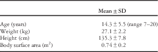
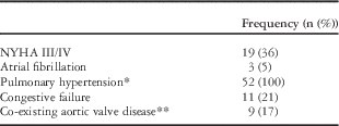
A total of 52 patients with juvenile mitral stenosis who satisfied the inclusion and exclusion criteria were included in the study. There was no evidence of cardiac perforation or significant left-to-right atrial shunting in any of the patients who participated in the study. Patient anthropometric data (Table 1) and baseline characteristics are given in the tables (Table 2). The mean age of the patients was 14.3 years (SD ±4.55), ranging from 7 to 20 years, and age was comparable between the genders (p=0.02). The mean Wilkins score of the patients was 8.1 (SD −1.2), ranging from 6 to 12.
Patient anthropometric data.

Distribution of baseline characteristics.

* Pulmonary artery systolic pressure of ⩾30 mmHg
** Mild or moderate stenosis/regurgitation
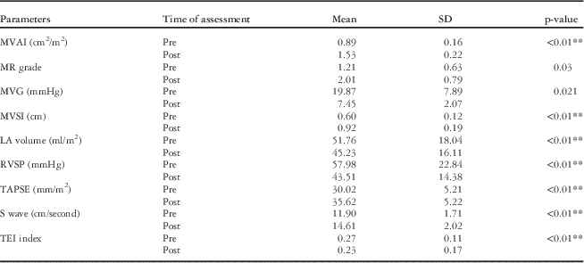
The mean mitral valve area index improved from 0.89 (SD ±0.16) to 1.73 (SD ±0.22) cm2/m2 following balloon mitral valvuloplasty (p<0.01), and the mean mitral valve gradient decreased from 19.87 (SD ±7.89) to 7.45 (SD ±2.07) (p=0.021). The details of the analysis of the change in the level of surrogate echocardiographic parameters considered for the study are given in Table 3. The surrogate echocardiographic parameters such as mitral valve separation index, tricuspid annular plane separation index, and tricuspid systolic velocity wave (S wave) significantly increased (p<0.01); however, left atrial volume, right ventricular systolic pressure, and the Tei index significantly reduced (p<0.01) after valvuloplasty. The mean grade of mitral regurgitation increased from 0.97 (SD ±0.63) to 1.31 (SD ±0.79) (p=0.08) following valvuloplasty.
Comparison of surrogate echocardiographic parameters (n=52).

LA=left atrial; MR=mitral regurgitation; MVAI=mitral valve area index; MVG=mean valve gradient; MVSI=mitral valve separation index; RVSP=right ventricular systolic pressure; S wave=tricuspid annular systolic velocity; TAPSE=tricuspid annular plane systolic excursion; TEI=Tei index
**Statistically significant at 1% level of significance
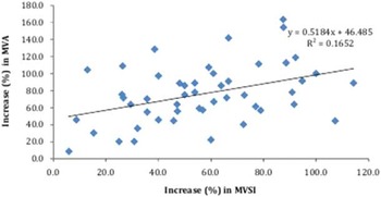
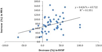
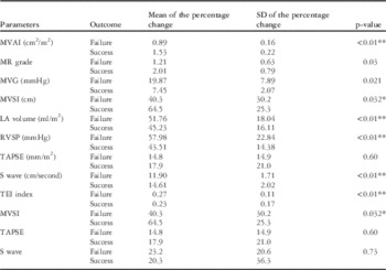
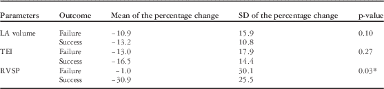
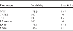
Balloon mitral valvuloplasty, by procedure definition, was successful in 49 out of 52 patients (94.2%).The change (differences) in all the surrogate parameters in relation to the outcome of balloon mitral valvuloplasty is given in Tables 4 and 5. The improvement in mitral valve separation index was significantly higher (p<0.05) among patients with successful balloon mitral valvuloplasty (64.5%) compared with patients in the failure group. The improvement in tricuspid annular plane systolic excursion and the tricuspid systolic velocity was not statistically significant (p>0.05). The mean decrease in right ventricular systolic pressure was 30% in the success group against 1% increase reported in the failure group and the difference was statistically significant (p<0.05); however, decreases in left atrial volume and the Tei index were not different between success and failure groups. The linear relationship between the difference (increase) in valve area with increase in valve separation index (r2=0.16, p<0.01) and decrease in right ventricular systolic pressure (r2=0.13, p<0.01) is given in Figs 2 and 3 and it shows that the correlations were statistically significant. Sensitivity and specificity of different parameters with respect to improvement in mitral valve area are given in Table 6.
Correlation between the difference in mitral valve area (MVA) and mitral leaflet separation index (MVSI).

Correlation between the difference in mitral valve area (MVA) and right ventricular systolic pressure (RVSP).

Comparison of the percentage change in surrogate echocardiographic parameters in relation to the treatment outcome.

LA=left atrial; MR=mitral regurgitation; MVAI=mitral valve area index; MVG=mean valve gradient; MVSI=mitral valve separation index; RVSP=right ventricular systolic pressure; S wave=tricuspid annular systolic velocity; TAPSE=tricuspid annular plane systolic excursion; TEI=Tei index
**,*Statistically significant at 1 and 5% level of significance
Comparison of the percentage decrease in surrogate echocardiographic parameters in relation to the treatment outcome.

LA volume=left atrial volume; RVSP=right ventricular systolic pressure; TEI=Tei index
*Statistically significant at 1% level of significance
Sensitivity and specificity of different parameters for predicting the success of balloon mitral valvuloplasty.

LA=left atrial; MVSI=mitral valve separation index; RVSP=right ventricular systolic pressure; S wave=tricuspid annular systolic velocity; TAPSE=tricuspid annular plane systolic excursion; TEI=Tei index
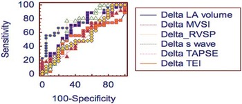
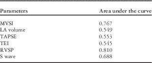
Receiver operative characteristic curves of the markers were constructed, and the area under the curves was analysed for predicting success (Fig 4). The decrease in right ventricular systolic pressure was a better indicator of the success of balloon mitral valvuloplasty followed by the increase in mitral valve separation index (Table 7). The sensitivity and the specificity were 71.1 and 87.8%, respectively, with an area under the curve of 0.810 for the cut-off value for a decrease in right ventricular systolic pressure of 16.08%. The sensitivity and specificity of the increase in mitral valve separation index as the indicator of successful balloon mitral valvuloplasty were 78.9 and 72.7%, respectively, with an area under the curve of 0.77 for the cut-off value for a decrease in mitral valve separation index of 46.4% (Table 8). Among the right ventricle function indices, right ventricle systolic pressure and S wave velocity by Doppler had better correlation with mitral valve area, whereas tricuspid annular separation index and the Tei index were poorly correlated.
Receiver operative characteristic curves of changes in mitral valve separation index (MVSI); left atrial volume (LAV); right ventricular systolic pressure (RVSP); tricuspid annular systolic plane excursion (TAPSE); tricuspid annular systolic velocity (S wave); and right ventricular Tei index (TEI) against mitral valve area (MVA). The area under the curves for RVSP and MVSI were found to be the highest (0.81 and 0.76, respectively), indicating a higher correlation with changes in MVA among the plotted variables.

Area under the receiver operative characteristic curves of parameters.

LA=left atrial; MVSI=mitral valve separation index; RVSP=right ventricular systolic pressure; S wave=tricuspid annular systolic velocity; TAPSE=tricuspid annular plane systolic excursion; TEI=Tei index
Predictive power of change in mitral valve separation index (MVSI) and right ventricular systolic pressure (RVSP) (% change) in the success of balloon mitral valvuloplasty.

Discussion
The study looked at alternative parameters for predicting the success of mitral balloon valvuloplasty. In situations where planimetry becomes difficult to assess, such as cases with irregularly thickened valves where diagnosing commissural splitting/area tracing is difficult, variables such as reduction in right ventricular pressure or improved valve separation become useful to denote success. The study results were negative in this regard with other variables of right ventricular function and left atrial volume. We used planimetry – two-dimensional tracing – as the standard measure for mitral valve area assessment, as pressure half-time method was unreliable immediately after valvuloplasty due to rapidly changing pressure dynamics.Reference Salem Omar, Tanaka and Abdel Dayem 4 Although many of the parameters tested were evaluated previously in different echocardiographic studies to measure the changes following balloon mitral valvuloplasty,Reference Vimal Raj, George and Jose 1 , Reference Fisher, Parisi and Plotnick 5 – Reference Mahfouz, Elawady and Hossein 11 we studied these in the juvenile age group. All the markers showed significant changes following balloon mitral valvuloplasty. This was comparable with the results published in earlier studies in adults.Reference Vimal Raj, George and Jose 1 , Reference Fisher, Parisi and Plotnick 5 , Reference Duggal, Bajaj and Prabhu 7 , Reference Bartunek, Vantrimpont and De Bruyne 8 Our study results indicate that mitral valve separation index is useful in such situations in predicting the success of juvenile balloon mitral valvuloplasty. A 46.4% change in mitral valve separation index predicted a final mitral valve area of 1.5 cm2 or 50% increment in mitral valve area with 78.9% sensitivity and 72.7% specificity. Fisher et alReference Fisher, Parisi and Plotnick 5 first proposed the concept of mitral valve separation index. Mitral leaflet separation index is simple to calculate and is fraught with fewer errors in measurement when compared with mitral valve area by planimetry. This has been validated in previous studies as well.Reference Vimal Raj, George and Jose 1 , Reference Fisher, Parisi and Plotnick 5 , Reference Seow, Koh and Yeo 6 We found an excellent correlation between mitral valve separation index and mitral valve area measured by planimetry. The study by Vimal Raj et alReference Vimal Raj, George and Jose 1 showed similar results earlier. A cut-off value of 0.8 cm had 92% sensitivity and specificity for predicting the severity of mitral stenosis. Our findings were similar, although with lower sensitivity and specificity. Only few studies have validated the mitral valve separation index in the immediate post-balloon mitral valvuloplasty period so far. The study by Duggal et alReference Duggal, Bajaj and Prabhu 7 showed that mitral valve separation index of >0.92 cm correlated well with mitral valve area of >1.5 cm2 immediately after balloon mitral valvuloplasty, the results of which were similar to that of our study.
The quantitative echocardiographic assessment of right ventricular function is difficult because of the ventricle’s complex trapezoidal anatomy.Reference Rudski, Lai and Afilalo 3 , Reference Bensaid, Krapf and Malanca 10 – Reference Grignola, Gines and Guzzo 13 A wide variety of techniques have been proposed, but none of them is currently considered the gold standard. S wave and annular systolic plane excursion are considered to reflect global systolic function, and the Tei index reflects both systolic and diastolic functions.Reference Dev and Shrivastava 14 , Reference Block and Palacios 15 We found that these indices poorly predicted the final valve area. Our results were similar to a study in adults by Bensaid et al,Reference Bensaid, Krapf and Malanca 10 which also did not show any significant change in echocardiographic parameters of right ventricle function after balloon mitral valvuloplasty. These poor correlations of right ventricle function require further investigation. The increase in tricuspid annular systolic excursion, S wave, and reduction in the Tei index indicate an improvement in right ventricular function. These parameters were improved in failed valvuloplasty patients as well; this could probably be reflective of a minor improvement in the valve area – mean change of 0.30 cm2 – in the failure group; however, these differences in the success and failure groups were not significant enough to predict the success of balloon mitral valvuloplasty.
Right ventricular systolic pressure reduction was robust in the juvenile patients studied, and the percentage reduction was the most specific correlate in predicting final mitral valve area. This is of paramount importance, as in juvenile patients pulmonary hypertension is highly prevalent and those who show a reduction in pulmonary artery pressure after balloon mitral valvuloplasty have better chances of successful balloon mitral valvuloplasty. A reduction of 16% in right ventricular systolic pressure was a highly specific, but relatively sensitive, marker for a successful outcome. The factors for elevated right ventricular and pulmonary artery pressures are many, starting from reactive to morphological changes in the right ventricular myocardium and pulmonary vasculature.Reference Block and Palacios 15 – Reference Levine, Weinstein and Diver 19 Our study indicates that abatement of the reactive component is predictive of successful balloon mitral valvuloplasty. Follow-up studies have indicated that there continues to be an improvement in the pulmonary vascular dynamics following balloon mitral valvuloplasty.Reference Levine, Weinstein and Diver 19
Limitations of the study
This was a short-term study involving a small number of patients. There were only three failures compared with 49 successful procedures, which decrease the value of the statistical analysis. The acute changes in left atrial and left ventricular compliance after balloon mitral valvuloplasty and their influence on the relation between mitral valve area and the six measured variables are not clearly known. All these markers vary with body surface area and heart rates, which may have an influence on the measurements. We did not perform systematic blinding, but a single physician, who did not have previous information on the procedural status, carried out the echocardiographic studies; however, there could still be a bias present, as by noting commissural split or larger valve area an indirect clue to post-procedural status might be assumed.
Conclusions
The surrogate markers studied showed favourable improvement, and right ventricular systolic pressure reduction and improved mitral valve separation index were better indicators of successful valvuloplasty. The study shows the utility of these markers in the juvenile subset of patients. In clinical practice, these parameters can be used, especially in situations where conventional echocardiographic parameters become inconclusive in assessing the success of balloon mitral valvuloplasty.
Acknowledgement
None.
Financial Support
This research received no specific grant from any funding agency, commercial or not-for-profit sectors.
Conflicts of Interest
None.
Ethical Standards
The study was approved by the Institute’s Scientific and Ethics Committee after satisfying the standards set forth by the Committee.