Primary cardiac tumours are rare in childhood, with a reported incidence of 0.027–0.08% based on postmortem studies in children.Reference Marx and Moran1 Rhabdomyoma, which is often associated with tuberous sclerosis, is the most common primary paediatric cardiac tumour, but fibroma ranks first in the surgical series.Reference Padalino, Basso and Milanesi2 Despite two-dimensional echocardiography remaining the imaging diagnostic tool of choice, cardiac magnetic resonance imaging adds important information about tumour tissue characteristics.Reference Kiaffas, Powell and Geva3 In addition, despite the benign histology of most primary paediatric cardiac tumours, there may be significant associated morbidity and mortality.Reference Stiller, Hetzer and Meyer4–Reference Stratemann, Dzurik, Fish and Parra6 We report a case of a 4-year-old boy who was symptomatic from a large left ventricular fibroma and underwent surgical excision of the tumour.
Case report
A 4-year-old boy, with a history of easy fatigability and a near-syncopal episode during exercise, was found on echocardiography to have a large mass involving the left ventricle free wall. The 24-hour Holter study showed no significant arrhythmias. However, his symptoms worsened with repeated presyncope episodes. These were characterised by dizziness, pallor, and sweating without ever losing consciousness. He was then referred to our institution, after being offered cardiac transplantation elsewhere, because the tumour was considered to be too large for safe resection.
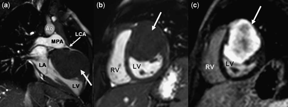
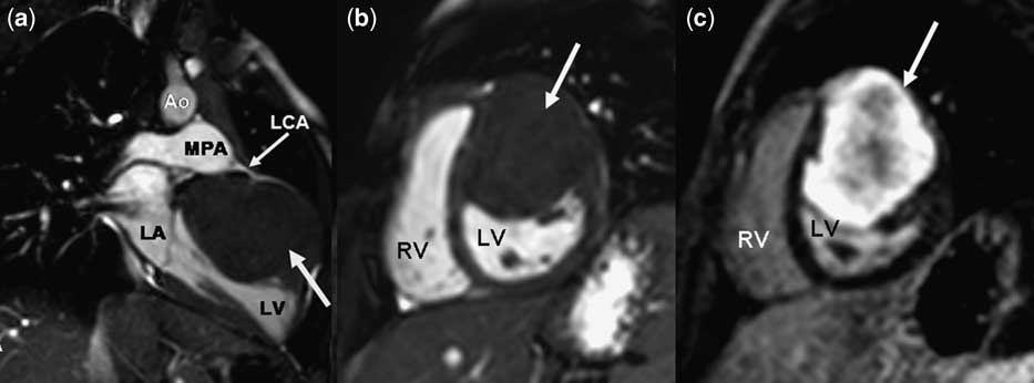
There was no family history of congenital cardiac disease, unexplained childhood deaths, or arrhythmias. The child's physical examination was unremarkable, except for a I/VI systolic murmur loudest at the left lower sternal border without radiation. Cardiomegaly and an irregular cardiac silhouette were noted on the chest radiograph. The 15-lead surface electrocardiogram showed sinus rhythm, left QRS axis deviation, and no evidence of dysrhythmias. A transthoracic echocardiogram revealed a large mass (4.6 centimetres in the antero-posterior dimension × 5.6 centimetres from apex to base) along the left ventricular antero-lateral wall, with centrally echobright regions suggestive of calcification. There was good biventricular function with no evidence of mitral inflow or aortic outflow obstruction. Cardiac magnetic resonance imaging was performed under anaesthesia and demonstrated a large solitary tumour, starting at the base of the left ventricle, involving the anterior septal, anterior, and antero-lateral segments, and extending 6 centimetres apically. These segments thickened and moved towards the centre of the ventricular cavity on cine imaging, but there was no ventricular inflow or outflow obstruction. The left main and right coronary artery origins were normal and the circumflex coronary artery coursed over the epicardial aspect of the tumour mass. The signal characteristics of this tumour were most consistent with a fibroma (Fig 1): isointense in the T1- and T2-weighted turbo spin echo sequences, minimal tumour enhancement in first pass perfusion, and hyperintense in myocardial delayed enhancement imaging.
Cardiac MRI. (a) Cine MRI image in a two-chamber left view: large solitary tumour involving the anterolateral LV wall, closely related to the left coronary artery. (b) Cine MRI image in short-axis view. (c) Myocardial delayed enhancement image in short-axis view: the tumour is hyperintense, highly suggestive of fibroma (MRI = magnetic resonance imaging; Ao = aorta; MPA = main pulmonary artery; LCA = left coronary artery; LA = left atrium; LV = left ventricle; RV = right ventricle; large arrow indicates the tumour).
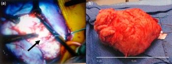
Owing to the concern that the tumour was negatively affecting left ventricle performance and possibly causing malignant arrhythmias, surgical resection was undertaken. The child underwent cardiac surgery employing cardiopulmonary bypass with aortic and bicaval cannulation. The aorta was not cross-clamped and the resection was undertaken with cold fibrillatory arrest and left ventricular venting. We have employed this technique in fibroma resection and prefer it, as it avoids cross-clamping of the aorta and facilitates differentiation of the tumour, which is relatively avascular, from the myocardium and myocardial vessels. There was sufficient myocardium between the fibroma and the left ventricular cavity to perform a complete resection without entrance into the cavity. Similarly, there was sufficient myocardial tissue between the tumour and the coronary arteries to permit complete resection and avoid major coronary injury. Subsequently, the defect in the ventricle was repaired in multiple layers of running monofilament sutures without pledgets, bringing the left ventricle free wall back together under the left anterior descending coronary artery and the circumflex. The epicardial layer was reinforced with pledgetted sutures, taking care to avoid compression of the coronary arteries. The tumour had a white glistening irregular surface (Fig 2) and the histologic examination of the surgical specimen confirmed the diagnosis of benign fibroma. His post-operative course was uneventful; he was extubated on the second post-operative day and discharged from the hospital 7 days later.
Surgery. (a) Intraoperative view: dissection of the left ventricular mass (arrow indicates the tumour). (b) Surgical specimen: the excised intramural solitary mass had a white glistening irregular surface; the histologic examination confirmed the diagnosis of benign fibroma.

At 1, 2, and 4 months following surgery, the child was asymptomatic and had no recurrence of his near-syncopal episodes. An echocardiogram revealed a thin layer of residual mass at the anterior base of the left ventricle, normal mitral valve function, unobstructed left ventricular outflow tract, and good biventricular function without regional wall motion abnormalities.
Discussion
Since the improvement and more widespread use of non-invasive imaging modalities, cardiac tumours are increasingly detected before significant symptoms develop, challenging paediatric cardiologists and surgeons to ascertain which patients need surgery and which will benefit from conservative follow-up.
Cardiac fibromas are benign tumours characteristically solitary, intramural, firm, white, and sometimes centrally calcified. They are considered neoplasms of fibroblastic origin and do not have a distinct capsule. The propensity of cardiac fibromas to involve ventricles, left more often than right, might be explained by the relative amount and distribution of myocardium and fibroblast-rich interstitium within the heart.Reference Marx and Moran1, Reference Stiller, Hetzer and Meyer4, Reference Cho, Danielson and Puga7–Reference Burke and Virmani9 The differential diagnosis of these tumours includes not only rhabdomyoma, myxoma, teratoma, lipoma, hamartoma, and haemangioma, but also other cardiac and pulmonary diseases that these tumours may mimic. There is a wide spectrum of clinical presentation from an incidental finding in an otherwise healthy patient to symptoms of congestive cardiac failure and life-threatening arrhythmias.Reference Padalino, Basso and Milanesi2, Reference Stiller, Hetzer and Meyer4, Reference Atallah, Robertson, Rebeyka, Dyck and Noga5 Sudden death is reported in 10% of patients with known cardiac fibromas.Reference Stratemann, Dzurik, Fish and Parra6
Despite two-dimensional Doppler echocardiography being the initial imaging modality of choice for evaluation of cardiac tumours,Reference Stratemann, Dzurik, Fish and Parra6, Reference Cho, Danielson and Puga7 cardiac magnetic resonance imaging has an important role due to its ability to identify the likely tissue type of the tumour, using a combination of techniques to study the tissue characteristics. In addition, cardiac magnetic resonance imaging provides important diagnostic information about tumour location, boundaries, and haemodynamic consequences.Reference Kiaffas, Powell and Geva3 Cardiac magnetic resonance imaging can also be particularly useful in visualisation of spatial relations of the tumour mass to the coronary arteries, which was helpful in guiding surgical excision.Reference Marx and Moran1
The natural course of cardiac fibromas is not well established and is unpredictable, with symptoms sometimes appearing later in life.Reference Cho, Danielson and Puga7 In addition, these tumours are not shown to regress spontaneously.Reference Atallah, Robertson, Rebeyka, Dyck and Noga5, Reference Freedom, Lee, MacDonald and Taylor8 Thus, the recommended treatment of cardiac fibromas is surgical resection, even in asymptomatic patients, to restore the normal cardiac physiology and prevent potentially lethal arrhythmias.Reference Padalino, Basso and Milanesi2, Reference Cho, Danielson and Puga7 Surgery seems to be a definitive treatment because cardiac fibromas do not usually recur after complete resection and subtotal excision has shown good late patient survival without increase in size of the residual tumour.Reference Cho, Danielson and Puga7 The only limitation to a conservative surgical approach is the extensive invasion of essential cardiac structures by the tumour mass. In these unusual cases, excision may cause irreparable injury to critical structures in the heart. In such rare circumstances, transplantation may remain the only therapeutic option.Reference Marx and Moran1, Reference Padalino, Basso and Milanesi2, Reference Cho, Danielson and Puga7, Reference Aravot, Banner and Madden10 In conclusion, prompt early diagnosis leading to a successful surgical excision may be lifesaving.