I. INTRODUCTION
The study on novel multifunctional nanomaterials which combine imaging diagnostics, sensing, and controlled drug delivery for therapy becomes a challenge in biological nanotechnology.Reference Trewyn, Giri, Slowing and Lin1–Reference Park, Lee, Kang, Kim, Choi and Kwon4 Silicon- and silica-based drug delivery systems have emerged as an alternative technique due to their high specific surface area, uniform and tunable pore structure, low cytotoxicity, and diversity in surface functionalization. The advantages above endow them with great potential applications in the fields of catalysis,Reference Maschmeyer, Rey, Sankar and Thomas5, Reference Corma6 sensing,Reference Corma, Galletero, Garcia, Palomares and Rey7 and optically active materials.Reference Nguyen, Wu, Doan, Schwartz and Tolbert8 A series of controlled release systems based on silica have been investigated. Those are responsive to distinct external stimuli, such as supramolecular nanovalves using redox,Reference Hernandez, Tseng, Wong, Stoddart and Zink9 pH,Reference Angelos, Yang, Patel, Stoddart and Zink10 competitive binding,Reference Leung, Nguyen, Stoddart and Zink11 light,Reference Ferris, Zhao, Khashab, Khatib, Stoddart and Zink12 and incorporating nanocaps.Reference Lai, Trewyn, Jeftinija, Jeftinija, Xu, Jeftinija and Lin13 The photocontrolled reversible release of drug molecules which was realized by grafting photoresponsive coumarin onto the pore outlets of MCM-41 solid to develop a photochemically controlled molecular gate was first reported by Fujiwara and his coworkers.Reference Mal, Fujiwara and Tanaka14 However, most of the release systems are based on the sensitive molecules. It is known that the strong plasmon absorption (SPA) band of Au nanorods (AuNRs) can be tuned from the visible (vis) band to the near-infrared (NIR) region where optical absorption of tissue is minimal and penetration is optimal.Reference Weissleder15 The AuNRs can convert light energy into thermal energy under laser irradiation. Highly efficient local heating can be achieved by nonradiative relaxation through electron–phonon and subsequent phonon–phonon coupling processes.Reference Link and El-Sayed16 Such an energy conversion may lead to a rise in local temperature and an increase in the small-molecule diffusion rate through the capsule walls. The photothermal effect has been used for cancer therapy.Reference Huang, El-Sayed, Qian and El-Sayed17 Although the photoregulated release can be achieved for photosensitive drugs,Reference Kim, Ghosh and Rotello18 the drug delivery vectors prepared from monolayer protected metal nanoparticles usually provide low drug-loading capacity.Reference Kim, Ghosh, Pagliuca, Zhu, Menichetti and Rotello19
In this article, we have synthesized the bifunctional nanocomposite spheres which combine the advantages of mesoporous silica and the photothermal effect of AuNRs. These bifunctional nanocomposite materials may be applied in photocontrolled release drug delivery as well as photothermal therapy.
II. EXPERIMENTAL PROCEDURE
A. Synthesis of AuNRs
The AuNRs colloid we used was prepared by seed-mediated growth procedure. First, a gold seed solution was prepared by reduction of HAuCl4 (2.0 mL, 1 mM) with NaBH4 (0.48 mL, 0.01 M) in the presence of cetrimonium bromide (CTAB) (7.52 mL, 0.13 M). After a 2-h reaction, this seed solution was used for the synthesis of AuNRs. In a flask, 8 mL of 1 mM HAuCl4 was mixed with 9.5 mL of 0.2 M CTAB and 0.12 mL of 0.01 M silver nitrate aqueous solution. After gentle mixing of the solution, 1.28 mL of 0.01 M ascorbic acid was added with continuously stirring, and then 0.3 mL of the seed solution was finally added into the mixture. A strong color change indicates the formation of the AuNRs. These nanorods were aged for 5 h, and then the excess CTAB were removed by centrifugation.
B. Synthesis of AuNR@MCM-41 NPs
First, 0.62 mL of concentrated ammonia aqueous solution (28 wt%) was added to 100 mL of the prepared AuNRs under stirring. After that, three 0.1 mL injections of 20% tetraethoxysilane (TEOS) in ethanol were added under gentle stirring at 30-min intervals. The mixture solution reacted for 6 h at 20 °C. The resultant products were washed two times with water, and then redispersed in a mixed solution of 180 mL of ethanol and 240 mL of deionized water, 0.9 g of CTAB, and 1.6 mL of concentrated ammonia aqueous solution (28 wt%). With gentle stirring, 0.4 mL of TEOS was added dropwise to the solution under vigorous stirring. After reaction for 6 h, the products were washed several times with ethanol and water. The structure-directing agent (CTAB) was subsequently removed by ultrasonication in hydrochloric acid and ethanol mixture for three times. The obtained precipitates were separated, washed with deionized water, and dried in vacuum at 30 °C for 10 h. The products were designated as AuNR@MCM-41 NPs.
C. Preparation of drug storage/release system
The loading of the drug ibuprofen (IBU) inside the AuNR@MCM-41 NPs was carried out as follows: 0.3 g of the AuNR@MCM-41 samples were added into 30 mL of IBU hexane solution (60 mg mL−1) and soaked with stirring for 24 h in a sealed vial which prevented the evaporation of hexane. The mixture was washed with hexane three times by centrifugation and dried at 30 °C for 10 h to remove the absorbed IBU on the exterior surface. The obtained samples are IBU-AuNR@MCM-41. The amount of IBU adsorbed onto the AuNR@MCM-41 was determined by ultraviolet (UV) absorption spectrum and elemental analysis. The IBU-AuNR@MCM-41 powders were compacted into 0.1 g disks (diameter and thickness, respectively, 10 × 1 mm) under a pressure of 3 MPa. The in-vitro release experiment was performed as follows: two disks were immersed in 200 mL of simulated body fluid (SBF) (pH = 7.4) at 25 °C with stirring in a beaker. One was with NIR (808 nm) laser irritation (5 W·cm−2), while the other was a reference without laser irritation. Three milliliters of the suspension was withdrawn for analysis at given time intervals, replaced with the same volume of the fresh SBF. The IBU concentration was monitored by using UV–vis spectrophotometer at 222 nm.
D. Characterization
Powder x-ray diffraction patterns (XRD) patterns were obtained on a Rigaku D/max 2550 x-ray diffractometer (Rigaku, Tokyo, Japan), using a monochromatized Cu target radiation resource (λ = 1.5045 Å).The surface morphology of the samples was inspected using a JEOL JSM-7500F field emission scanning electron microscope (FE-SEM) at an accelerating voltage of 15 kV with gold sputtered on samples. Transmission electron micrographs were carried out with a Hitachi H-8100 transmission electron microscope (TEM, Hitachi, Tokyo, Japan) operating at an acceleration voltage of 200 kV. The nitrogen isotherms at the temperature of liquid nitrogen were measured using a Micromeritics ASAP 2020M system (Micrometrics, Atlanta, Georgia). The pore size distribution for mesopores was calculated using the Barrett–Joyner–Halenda (BJH) model. Fourier transform infrared (FTIR) absorption spectra were performed on Shimadzu DT-40 model 883 IR spectrophotometer. The elemental analyses were conducted on a Perkin-Elmer 2400 element analyzer (Perkin-Elmer, Waltham, MA). UV–vis–NIR absorption spectra were measured with a Shimadzu UV-3101PC UV–vis–NIR scanning spectrophotometer (Shimadzu, Waltham, MA) ranging from 200 to 1100 nm.
III. RESULTS AND DISCUSSION
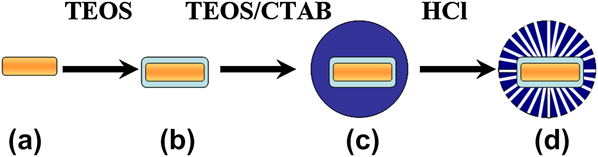
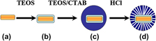
The core–shell structured AuNR@MCM-41 nanocomposites were synthesized via a modified Stöber method by a process of hydration and condensation of TEOS in a water–ethanol mixture.Reference Stober, Fink and Bohn20, Reference Gorelikov and Matsuura21 A schematic is given in Scheme 1 to simply illustrate the synthesis process of the AuNR@MCM-41 nanocomposites. All the AuNRs used here were synthesized according to the wet chemistry method invented by Murphy et al.Reference Murphy, Sau, Gole, Orendorff, Gao, Gou, Hunyadi and Li22, Reference Murphy, Gole, Hunyadi and Orendorff23 TEM images in Fig. 1(a) shows that the aspect ratio of the AuNRs is around 3.3 ± 0.5 (length: 50.0 ± 3.0 nm, width: 14.7 ± 1.5 nm). The AuNRs are easy to aggregate together or deposit in the ethyl alcohol solution. To overcome this problem and obtain uniform silica deposition, we have applied an intermediate coating to the CTAB–gold nanorods acting as a scaffold for silica deposition. The TEM image in Fig. 1(b) clearly shows that silica shells (thickness of ∼8 nm) are composed of disordered mesopores. Then, the AuNR@MCM-41 nanocomposites were synthesized by self-assembly of silica surfactant in which inorganic species simultaneously condensed, giving rise to mesoscopically ordered composites formation.
Transmission electron microscope (TEM) images of (a) Au nanorods (AuNRs) and (b) AuNR@SiO2.

The formation process of the multifunctional AuNR@MCM-41 composite nanospheres: (a) AuNR, (b) AuNR@nSiO2, (c) AuNR@nSiO2@CTAB/mSiO2, and (d) AuNR@MCM-41.

The FE-SEM, TEM images, and the XRD patterns of the AuNR@MCM-41 nanocomposites are shown in Fig. 2. Seen from the wide-angle XRD of the obtained samples [Fig. 2(a)], a broad band located at 2θ = 22° can be assigned to the characteristic diffraction peak of amorphous SiO2 (JCPDS No. 29-0085). As labeled in the figure, all of the patterns accord with the standard cards 04-0784 for cubic gold. No additional peaks appear, implying the products are Au indeed and pure in cubic phase. As for the small-angle XRD patterns [Fig. 2(b)], the strong peak (100) associated with a two-dimensional hexagonal mesostructure (p6mm) confirms their ordered mesostructure. The only one strong (100) diffraction can be associated with the low synthesis temperature.Reference Yano and Fukushima24 The size and shape of the synthesized AuNR@MCM-41 NPs were further investigated using FE-SEM and TEM. It is distinct that the as-prepared AuNR@MCM-41 with a mean particle size of ∼200 nm [see Figs. 2(c)–2(e)] are nonaggregated and with narrow size distribution. Most of the nanocomposites have a single gold core which tended to be more localized in the center of the spheres, but some have more cores. From Fig. 2(c), we can clearly see the pore channels on the surface of the mesostructure, which is in accordance with the small-angle XRD patterns.
(a) The wide-angle x-ray diffraction (XRD) patterns of as-synthesized AuNR@MCM-41 and the standard data for Au (JCPDS No. 04-0784). (b) The low-angle XRD patterns of as-synthesized AuNR@MCM-41. (d) SEM image and (c, e) TEM images of as-synthesized AuNR@MCM-41.

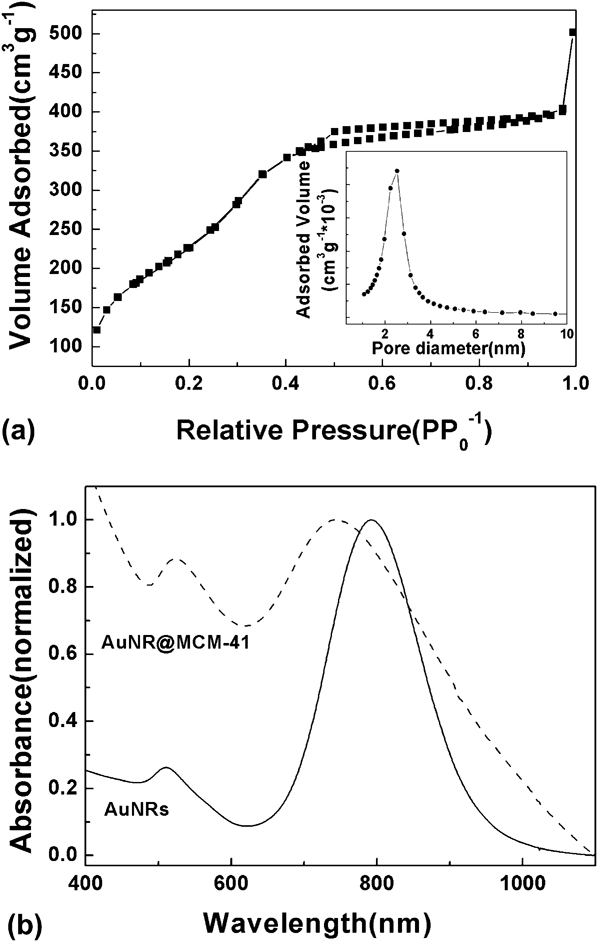
As shown in the nitrogen adsorption/desorption isotherms [Fig. 3(a)], the samples show typical IV isotherms with H3 hysteresis, together with narrow pore size distribution shown in the inset of Fig. 3(a). The surface area of the AuNRs@MCM-41 materials calculated by using the Brunauer–Emmett–Teller method is 834 m2 g−1. The pore volume is 0.769 cm3 g−1, and the pore size distribution calculated from the adsorption isotherms by using the BJH method centers in 2.5 nm. The normalized UV–vis–NIR absorbance spectra of the aqueous AuNRs (the solid line) and AuNR@MCM-41 (the dash line) solution are shown in Fig. 3(b). AuNRs possess two absorption bands, one corresponding to short-axis (transverse plasmon resonance) locates at 511 nm and the other corresponding to long-axis (longitudinal plasmon resonance) centers at 793 nm. As for the absorbance spectrum of the aqueous AuNR@MCM-41 (the dash line) solution, the transverse plasmon band at ∼520 nm is not appreciably affected by the silica coating, whereas the longitudinal plasmon band obviously blue shifts toward 750 nm relative to the plasmon absorption of pure gold nanorods due to the silica shell.
(a) Nitrogen adsorption–desorption isotherm and the inset is the pore size distribution curve of the synthesized materials. (b) The normalized ultraviolet–visible–near-infrared absorbance spectrum of the aqueous AuNRs (the solid line) and AuNR@MCM-41 (the dash line) solution.

This result is consistent with previous reports which demonstrate how the longitudinal SPA band responds to changes in the local refractive index surrounding the gold nanoparticles.Reference Cong, Toftegaard, Arnbjerg and Ogilby25, Reference Link and El-Sayed26 The novel AuNR@MCM-41 nanocomposites possess not only the property of drug carrier but also the characteristic of light-to-heat in the NIR. We chose IBU as a model drug here since it has good pharmacological activity and suitable molecule size of about 1.0 × 0.6 nm, which ensures its easy diffusion into or out of the mesoporous channels of silica.Reference Andersson, Rosenholm, Areva and Linden27 The drug storage and release system was prepared in accordance with the previous report with some modification.Reference Vallet-Regi, Ramila, Del Real and Perez-Pariente28
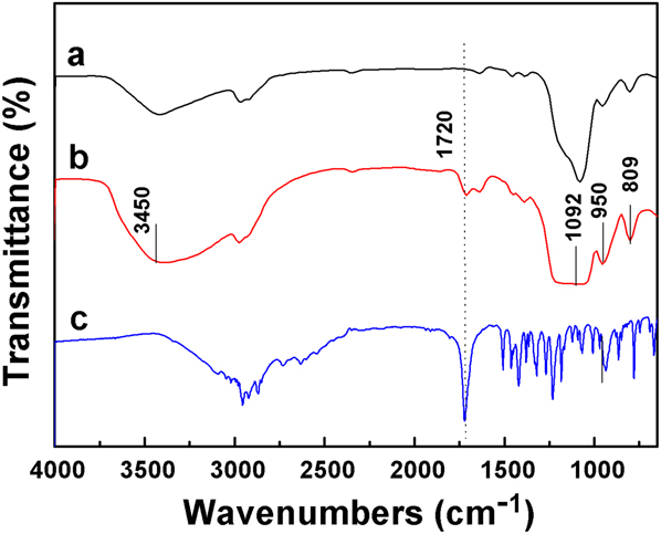
FTIR spectra of (a) AuNRs@MCM-41, (b) IBU-AuNRs@MCM-41, and (c) IBU are presented in Fig. 4. The absorption bands are ascribed to –OH (3450 and 1630 cm−1), Si–O–Si (1092 and 809 cm−1), and Si–OH (950 cm−1). For IBU-loaded IBU-AuNRs@MCM-41, the band assigned to –COOH (1720 cm−1) is apparent except for a slight intensity decrease compared with pure IBU, which proves that a large number of IBU molecules have been stored in the cavities without any interaction with Si–OH groups on the pore surface, and the storage of the IBU molecules in AuNRs@MCM-41 is physical.
Fourier transform infrared spectra of (a) AuNR@MCM-41, (b) IBU-AuNR@MCM-41, and (c) ibuprofen (IBU).

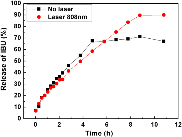
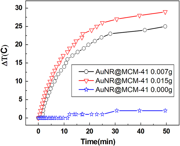
Figure 5 shows the IBU release curves from the AuNR@MCM-41 profile (diameter and thickness, respectively, 10 × 1 mm) in the SBF release medium (pH 7.4). The solid squared and circled dot curves describe the release processes with and without the continuous laser irradiation of 808 nm, respectively (the loading amount of IBU: 13.4 wt%). At the first 3 h, the release rates in different situation exhibited almost the same trend due to the dominant effect of the concentration diffusion. The profile without the laser irradiation reaches the equilibrium at 6 h with 69% release fraction, while the one under irradiation exhibited 90% release fraction at 10 h. We believe the photothermal property of gold nanorods make the difference in release fraction. It is known that the compression of the drug–matrix mixture leads to a decrease of pore size and eventually to a narrowing or closing of the pore cavity. So, this could also induce the incomplete release of IBU due to diffusion constraints.Reference Vallet-Regi, Ramila, Del Real and Perez-Pariente28 In this system, 69% should be the equilibrium point. However, high temperature can break this release balance. To investigate whether AuNR@MCM-41 NPs can increase local temperature by laser irradiation, we have examined the temperature changes under the 50-min irradiation of the laser with 0.545 w. As seen from Fig. 6, the changes of the temperature are dramatical. The temperatures of AuNR@MCM-41 NPs at 0.007 g and 0.015 g in 3mL-aqueous solution increased by 25 and 30 °C respectively. However, in the absence of AuNR@MCM-41 NPs aqueous solution, the temperature only increased by 1–2 °C. We can conclude that the photothermal effect of the AuNR@MCM-41 NPs promote the diffusion of the IBU into the solution. The release fraction of the IBU can be controlled by the AuNR@MCM-41 NPs with laser irradiation.
IBU release processes from the AuNR@MCM-41 system in the simulated body fluid release medium (pH 7.4) on the irradiation time under the laser irradiation at 808 nm (■) and no laser irradiation (●).

Plots of temperature increase for suspensions of AuNR@MCM-41 at 0.007, 0.015, and 0 g in 3 mL aqueous solution as a function of irradiation time using the diode laser at power of 0.545 w.

IV. CONCLUSIONS
In summary, to the best of our knowledge, we reported for the first time a novel kind of AuNRs@MCM-41 NPs, which combines advantages of light energy to heat and the pore channels for controlled release of drugs. The results provide new and exciting insight into developing light-driven release of drug for practical applications. Future studies of AuNR@MCM-41 NPs on biology will be extended to the synergy of chemotherapeutic drugs and thermotherapy for cancer treatments.
ACKNOWLEDGMENTS
The authors are thankful to the National Science Fund for Distinguished Young Scholars of China (Grant No. 60925018) and the National Natural Science Foundation of China (Grant Nos. 20971051 and 51002062).